. 2018 Jun 26;115(26):E5934-E5943.
doi: 10.1073/pnas.1803907115. Epub 2018 Jun 11.

Suppression of connexin 43 phosphorylation promotes astrocyte survival and vascular regeneration in proliferative retinopathy

Affiliations

Suppression of connexin 43 phosphorylation promotes astrocyte survival and vascular regeneration in proliferative retinopathy

Nefeli Slavi et al. Proc Natl Acad Sci U S A. .

Abstract

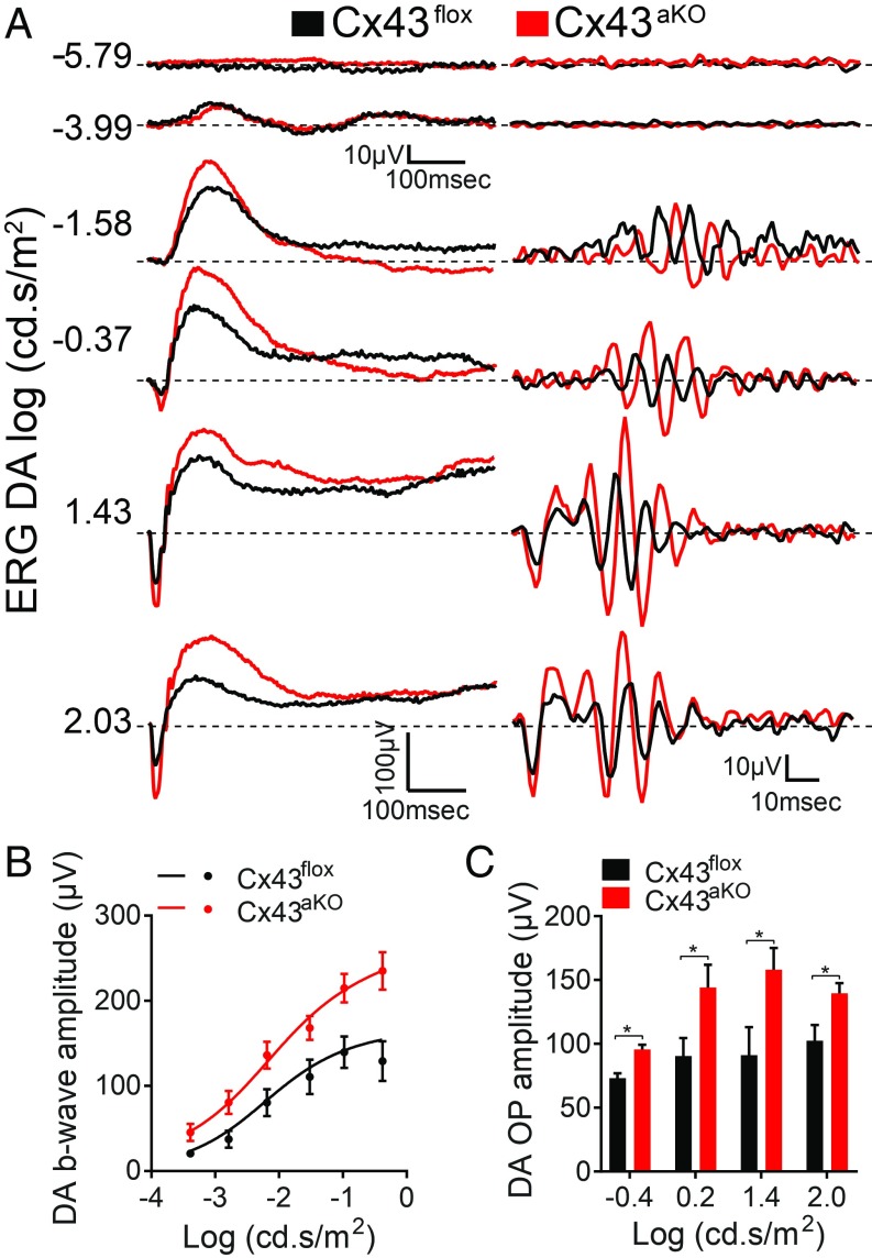
Degeneration of retinal astrocytes precedes hypoxia-driven pathologic neovascularization and vascular leakage in ischemic retinopathies. However, the molecular events that underlie astrocyte loss remain unclear. Astrocytes abundantly express connexin 43 (Cx43), a transmembrane protein that forms gap junction (GJ) channels and hemichannels. Cx channels can transfer toxic signals from dying cells to healthy neighbors under pathologic conditions. Here we show that Cx43 plays a critical role in astrocyte apoptosis and the resulting preretinal neovascularization in a mouse model of oxygen-induced retinopathy. Opening of Cx43 hemichannels was not observed following hypoxia. In contrast, GJ coupling between astrocytes increased, which could lead to amplification of injury. Accordingly, conditional deletion of Cx43 maintained a higher density of astrocytes in the hypoxic retina. We also identify a role for Cx43 phosphorylation in mediating these processes. Increased coupling in response to hypoxia is due to phosphorylation of Cx43 by casein kinase 1δ (CK1δ). Suppression of this phosphorylation using an inhibitor of CK1δ or in site-specific phosphorylation-deficient mice similarly protected astrocytes from hypoxic damage. Rescue of astrocytes led to restoration of a functional retinal vasculature and lowered the hypoxic burden, thereby curtailing neovascularization and neuroretinal dysfunction. We also find that absence of astrocytic Cx43 does not affect developmental angiogenesis or neuronal function in normoxic retinas. Our in vivo work directly links phosphorylation of Cx43 to astrocytic coupling and apoptosis and ultimately to vascular regeneration in retinal ischemia. This study reveals that targeting Cx43 phosphorylation in astrocytes is a potential direction for the treatment of proliferative retinopathies.

Keywords: astrocytes; gap junctions; ischemia; neurovascular; retina.


Conflict of interest statement

The authors declare no conflict of interest.

Figures

Fig. 1.
Fig. 1.
Conditional deletion of Cx43 preserves the astrocytic network in OIR. (A) Schematic of the mouse model of OIR. Neonatal mice are exposed to 75% O2 from P7 to P12. Hyperoxia results in vasoobliteration in the central retina (see Fig. 2 for images). Mice return to room air (21% O2) at P12, and the retina becomes immediately hypoxic due to the absence of central vasculature. The hypoxic phase (between P12 and P17) is characterized by partial revascularization and neovascular tuft formation, which commences at P14 and peaks at P17. Decrease in the density of astrocytes is also observed during hypoxia, between P12 and P14–P15. (B) (Top) Representative images of whole-mount Cx43flox and Cx43aKO retinas in OIR stained with isolectin (green) and anti-GFAP (red) at P14. (Scale bars: 200 μm.) (Middle and Bottom) Higher-magnification images from the boxed areas, corresponding to the central and peripheral retinal regions, respectively. (Scale bars: 50 μm.) (C) Quantification of astrocytes present in the avascular areas of Cx43flox and Cx43aKO retinas in OIR at P12 (n = 5–9) and P14 (n = 6–8). At P14, deletion of Cx43 led to ∼1.8-fold increase in the GFAP-labeled astrocytes in the avascular retinal regions compared with control. Data are presented as mean ± SEM. Student’s t test, ns P > 0.05, ***P < 0.001.
Fig. 2.
Fig. 2.
Deletion of astrocytic Cx43 promotes reparative angiogenesis and reduces hypoxia in OIR. Representative images of whole-mount Cx43flox and Cx43aKO retinas in OIR stained with isolectin (green) at (A) P12, (C) P14, and (E) P17. White lines indicate the border of the avascular area in each retina. Images of neovascular tufts at P17 are illustrated in E (Bottom). Quantification of the avascular areas of Cx43flox and Cx43aKO retinas in (B) P12 (n = 7–7), (D) P14 (n = 7–9), and (F) P17 (n = 18–21). Cx43aKO retinas exhibited accelerated revascularization compared with Cx43flox. (G) Quantification of the retinal areas covered with neovascular tufts at P17 revealed a reduction in neovascularization of Cx43aKO (n = 21) retinas compared with Cx43flox (n = 18). (H and I) Representative images of whole-mount Cx43flox (n = 6) and Cx43aKO (n = 6) retinas in OIR stained with isolectin (green) and Hypoxyprobe (red) at P17 and quantification of the hypoxic areas. Cx43aKO retinas were significantly less hypoxic compared with Cx43flox. Data are presented as mean ± SEM. Student’s t test, ns P > 0.05, *P < 0.05, **P < 0.01, ****P < 0.0001. (Scale bars: 250 μm.)
Fig. 3.
Fig. 3.
Deletion of astrocytic Cx43 improves neuroretinal function in OIR. (A) Recordings of (Left) a and b waves and (Right) OPs of scotopic ERGs in response to different stimulus intensities from Cx43flox and Cx43aKO mice exposed to OIR. Light intensities in log scot. cd s/m2 are shown to the left of each trace. (B and C) Cx43aKO mice showed a significant improvement in function of the neural retina. (B) Quantification of the amplitudes of the scotopic b wave and (C) summed OPs at different stimulus intensities. Lines in B represent the fit of the Naka Rushton equation to the data. Data are presented as mean ± SEM (n = 5 each of Cx43flox and Cx43aKO mice). Student’s t test, *P < 0.05.
Fig. 4.
Fig. 4.
Pharmacological inhibition of channels formed by Cx43 early in the hypoxic phase of OIR promotes reparative angiogenesis. (A) Representative images of isolectin-stained WT retinas at P17 injected intravitreally with PBS (SHAM) or with 20 µM SBO15 at P12. The borders of the avascular areas are outlined by white lines, and the neovascular tufts are marked by red lines. Injection of SBO15 reduced both the avascular and neovascular areas. (Scale bars: 250 μm.) The reduction in (B) avascular and (C) neovascular areas relative to the total retinal area was measured between P12 and P17 during the hypoxic phase of OIR after a single intravitreal injection of SBO15 at P12 (n = 3–9). SBO15 accelerated revascularization as early as P14 and reduced neovascularization between P14 and P17. (D) Representative images of isolectin- and GFAP-stained whole-mount WT retinas at P14, injected intravitreally with PBS or with 20 µM SBO15 at P12. High magnification images from the central and peripheral retinal regions are shown in SI Appendix, Fig. S5B. (Scale bars: 100 μm.) (E) Quantification of astrocytes present in the avascular areas of SHAM (n = 9) and SBO15-treated retinas (n = 9) in OIR at P14. SBO15 led to ∼1.7-fold increase in the GFAP-labeled astrocytes in the avascular retinal regions compared with control. Data are presented as mean ± SEM. Student’s t test, ns P > 0.05, *P < 0.05, **P < 0.01, ***P < 0.001, ****P < 0.0001.
Fig. 5.
Fig. 5.
Cx43 GJ coupling but not hemichannel activity in astrocytes is elevated during hypoxia in OIR. (A) Hemichannel activity as measured by ethidium bromide (EtBr) uptake in astrocytes. Images of whole-mount retinas incubated with EtBr (red) and labeled with anti-GFAP (green) after 0 and 24 h during the hypoxic phase of OIR. Arrows indicate the uptake of EtBr into cell bodies of astrocytes. (Scale bars: 50 μm.) (B) The number of cells with tracer uptake normalized to retinal area was measured in the absence (control) or in the presence of Cx43 hemichannel-selective inhibitor, Gap19 (300 μM) at 0 (n = 5–8), 6 (n = 3–4), and 24 h (n = 4–9) of hypoxia. Tracer uptake was similar at all three time points and was not blocked by Gap19. (C) GJ coupling between astrocytes of tdTomatoGFAP mice (red) was visualized by diffusion of Neurobiotin (green) introduced into a single astrocyte at 0 and 6 h in hypoxia. (Left) Images at 4× magnification at each time point. (Scale bars: 500 µm.) (Right) Higher magnification of the boxed areas. (Scale bars: 250 µm.) (D and E) Quantification of astrocytes labeled with Neurobiotin after 0, 6, and 24 h of hypoxia in the central (n = 6–8) and peripheral (n = 3) retina showed an increase in astrocytic coupling in hypoxia. Data are presented as mean ± SEM. Two-way ANOVA is used in B. One-way ANOVA is used in D and E. ns P > 0.05, *P < 0.05, **P < 0.01.
Fig. 6.
Fig. 6.
Cx43 becomes phosphorylated at Ser325/328/330 by CK1δ during hypoxia. (A) Western blot of Cx43 in WT retinas at 0, 6, and 24 h hypoxia, showing a large increase in the P2 phosphorylated form of Cx43 within 6 h. (B) Quantification of (Top) total Cx43 or (Bottom) the P2 form normalized to tubulin at 6 (n = 3), 9 (n = 7), and 24 h (n = 7) relative to 0 h (n = 7). (C and D) Western blot and quantification of total Cx43 in control (Cx43flox) (n = 11), astrocyte- (Cx43aKO) (n = 7), and endothelial cell- (Cx43eKO) (n = 6) specific knockout retinas at 6 h posthypoxia. Majority of retinal Cx43 is present in astrocytes. (E and F) Western blot and quantification of Ser325/328/330-phosphorylated Cx43 at 0 and 6 h in hypoxia. The phosphospecific antibody recognized a signal corresponding to the P2_Cx43 (∼43 kDa) (n = 3). (G) Western blot of Cx43 in Cx43WT and Cx43S3A retinas at 6 h in hypoxia. (H) Quantification of P2_Cx43 levels in Cx43WT (n = 6) and Cx43S3A retinas (n = 6). (I) Western blot of Cx43 at different time points in hypoxia following intravitreal injection of PBS (−) or the CK1δ inhibitor PF670462 (+). PF670462 reduced P2_Cx43. (J) Quantification of P2_Cx43 in PBS- and PF670462-injected retinas at 0 (n = 7), 6 (n = 3), 9 (n = 6), and 24 h (n = 7) in hypoxia. Samples from two separate animals are shown at each time point in A, E, and G. Data are presented as mean ± SEM. One-way ANOVA is used in BD. Student’s t test is used in F, H, and J. ns P > 0.05, *P < 0.05, **P < 0.01, ***P < 0.001, ****P < 0.0001.
Fig. 7.
Fig. 7.
Inhibition of CK1δ-mediated Cx43 phosphorylation rescues astrocytes, promotes reparative angiogenesis, and improves neuronal function. (A) Representative images of whole-mount Cx43WT and Cx43S3A retinas in OIR stained with isolectin (green) and anti-GFAP (red) at P14. (Scale bars: 100 μm.) (B) Quantification of astrocytes present in the avascular areas showed an ∼1.5-fold increase in their density in Cx43S3A retinas (n = 8) compared with Cx43WT (n = 8). (C) Representative images of whole-mount isolectin-stained Cx43WT and Cx43S3A retinas in OIR at P17. The borders of the avascular areas are outlined by white lines, and the neovascular tufts are marked by red lines. (Scale bars: 250 μm.) (D and E) Quantification of the avascular and neovascular areas at P17 in Cx43WT (n = 23), Cx43S3A (n = 15), and PF670462-treated (n = 8) mice. (F) Recordings of the a and b waves of scotopic ERGs at different stimulus intensities from Cx43WT and Cx43S3A mice at P28. Light intensities in log scot. cd s/m2 are shown to the left of each trace. (G) Quantification of the scotopic b-wave amplitudes at different stimulus intensities (n = 5) showed significant improvement in neuronal function of Cx43S3A mice compared with Cx43WT. Symbols represent the means ± SEM of the measured b-wave amplitudes. Lines represent the fit of the Naka Rushton equation to the data. (H) Quantification of the maximum response amplitude (Vmax) in Cx43WT and Cx43S3A mice, estimated from the fitted curves in G. Data are presented as mean ± SEM. One-way ANOVA is used in D and E, and Student’s t test is used in B and H. ns P > 0.05, *P < 0.05, **P < 0.01, ***P < 0.001, ****P < 0.0001.

References

    1. Gilbert C, Awan H. Blindness in children. BMJ. 2003;327:760–761. - PMC - PubMed
    1. Kempen JH, et al. Eye Diseases Prevalence Research Group The prevalence of diabetic retinopathy among adults in the United States. Arch Ophthalmol. 2004;122:552–563. - PubMed
    1. Cheung N, Mitchell P, Wong TY. Diabetic retinopathy. Lancet. 2010;376:124–136. - PubMed
    1. Hartnett ME. Pathophysiology and mechanisms of severe retinopathy of prematurity. Ophthalmology. 2015;122:200–210. - PMC - PubMed
    1. Sapieha P. Eyeing central neurons in vascular growth and reparative angiogenesis. Blood. 2012;120:2182–2194. - PubMed

Publication types