. 2018 Jun 26;115(26):E5944-E5953.
doi: 10.1073/pnas.1806973115. Epub 2018 Jun 11.

Regulation of a distinct activated RIPK1 intermediate bridging complex I and complex II in TNFα-mediated apoptosis

Affiliations

Regulation of a distinct activated RIPK1 intermediate bridging complex I and complex II in TNFα-mediated apoptosis

Palak Amin et al. Proc Natl Acad Sci U S A. .

Abstract

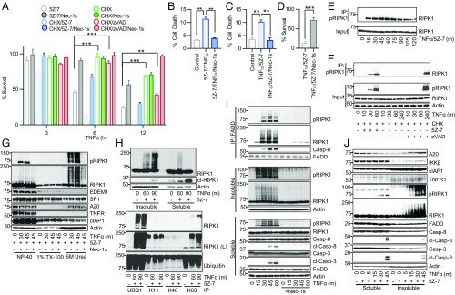
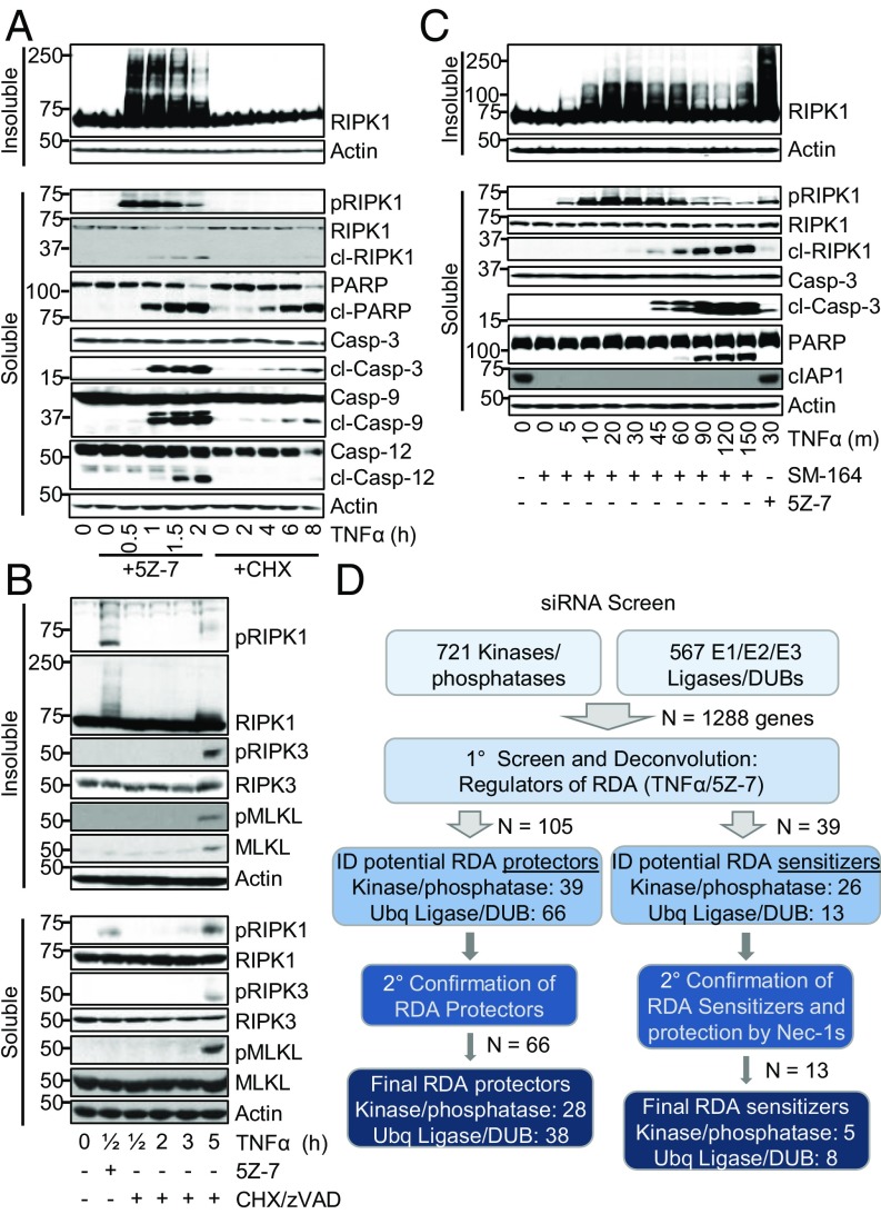
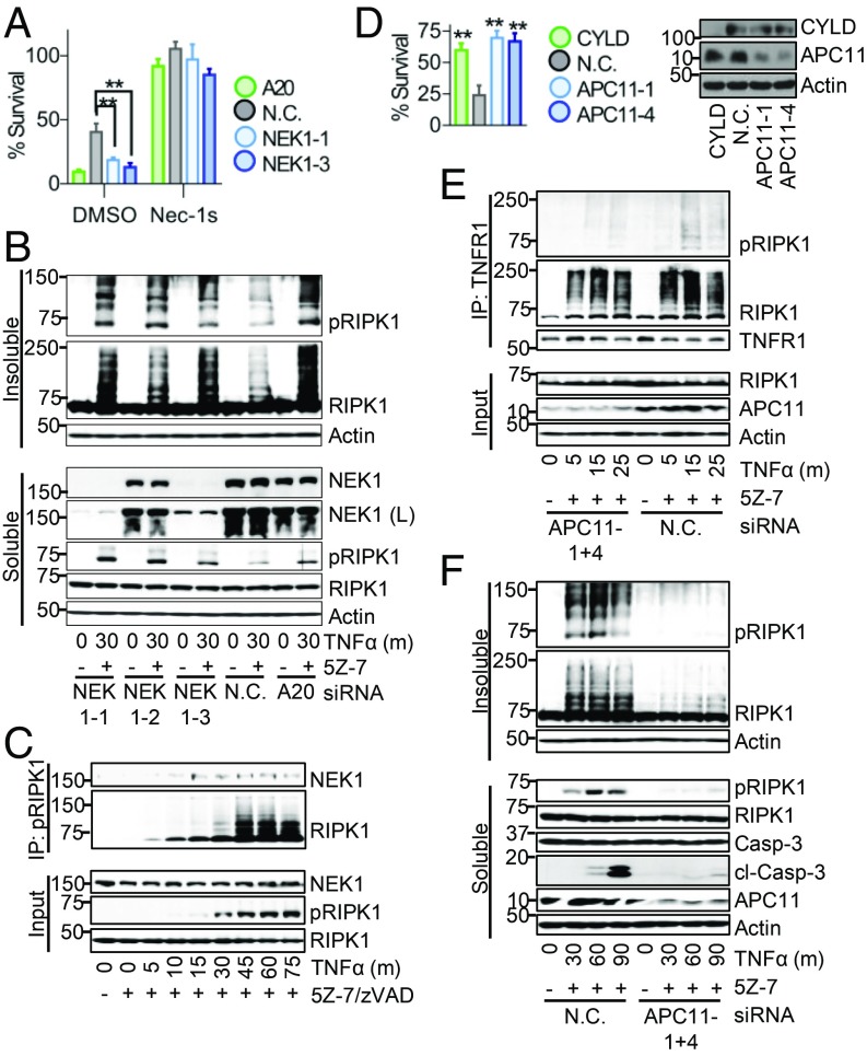
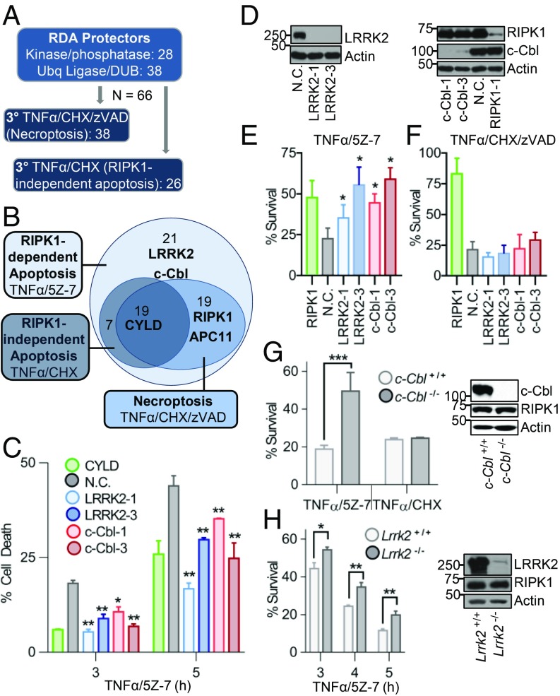
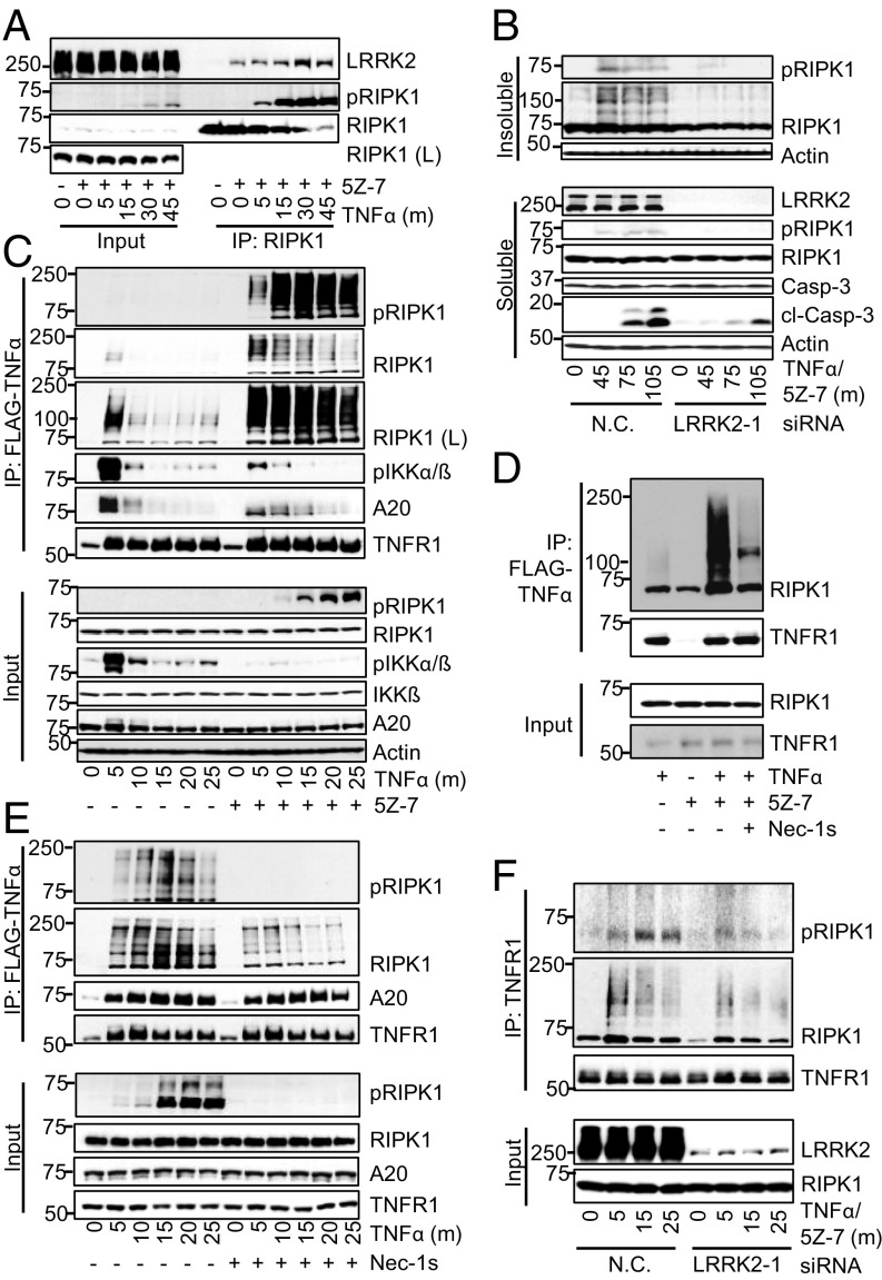
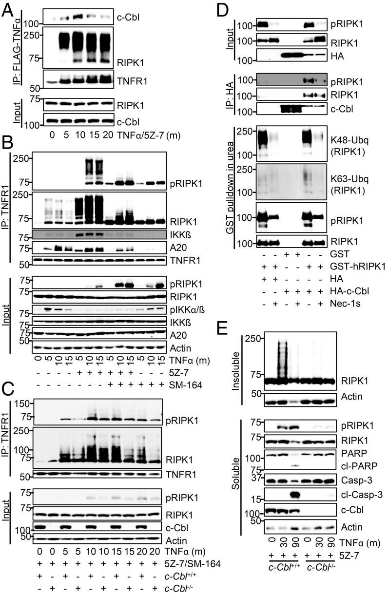
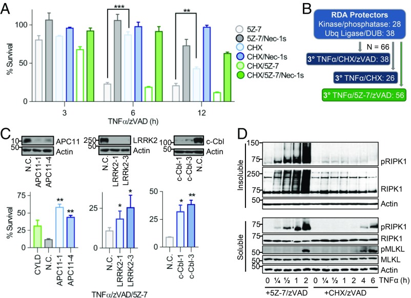
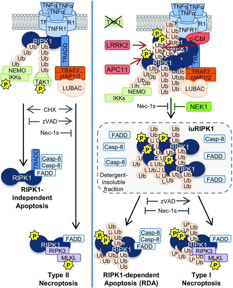
Stimulation of cells with TNFα can promote distinct cell death pathways, including RIPK1-independent apoptosis, necroptosis, and RIPK1-dependent apoptosis (RDA)-the latter of which we still know little about. Here we show that RDA involves the rapid formation of a distinct detergent-insoluble, highly ubiquitinated, and activated RIPK1 pool, termed "iuRIPK1." iuRIPK1 forms after RIPK1 activation in TNF-receptor-associated complex I, and before cytosolic complex II formation and caspase activation. To identify regulators of iuRIPK1 formation and RIPK1 activation in RDA, we conducted a targeted siRNA screen of 1,288 genes. We found that NEK1, whose loss-of-function mutations have been identified in 3% of ALS patients, binds to activated RIPK1 and restricts RDA by negatively regulating formation of iuRIPK1, while LRRK2, a kinase implicated in Parkinson's disease, promotes RIPK1 activation and association with complex I in RDA. Further, the E3 ligases APC11 and c-Cbl promote RDA, and c-Cbl is recruited to complex I in RDA, where it promotes prodeath K63-ubiquitination of RIPK1 to lead to iuRIPK1 formation. Finally, we show that two different modes of necroptosis induction by TNFα exist which are differentially regulated by iuRIPK1 formation. Overall, this work reveals a distinct mechanism of RIPK1 activation that mediates the signaling mechanism of RDA as well as a type of necroptosis.

Keywords: RIPK1; TNF; apoptosis; necroptosis; ubiquitination.


Conflict of interest statement

The authors declare no conflict of interest.

Figures

Fig. 1.
Fig. 1.
Induction of RDA by TNFα leads to rapid and elevated activation of RIPK1 in a detergent-insoluble cellular compartment before formation of complex II. (A) The 661W cells treated with TNFα/5Z-7 to induce RDA, TNFα/CHX to induce RIPK1-independent apoptosis, or TNFα/CHX/zVAD to induce necroptosis in the presence or absence of Nec-1s. Cell viability determined by CellTiter-Glo. (B) Primary neuronal cultures treated with TNFα/5Z-7 to induce RDA for 8 h and cell death measured by Toxilight. (C) Primary astrocytes treated for 5 h and cell death measured by Toxilight. (D) Primary microglia treated for 5 h and cell survival measured by CellTiter-Glo. (EJ) Cells treated as indicated and samples analyzed by Western blotting using indicated antibodies; pRIPK1 is RIPK1-pS166. (E) The 661W cells treated with TNFα/5Z-7 to induce RDA. Lysates immunoprecipitated with RIPK1-pS166 ab. (F) WT MEFs lysates immunoprecipitated with RIPK1-pS166 ab. (G) The 661W cells treated with TNFα/5Z-7 to induce RDA, harvested with Nonidet P-40 buffer, and cleared by centrifugation to yield the Nonidet P-40 fraction. Pellet resuspended in Triton X-100 buffer and cleared by centrifugation to yield the TX-100 fraction. Resulting pellet was resuspended in buffer with 6 M urea and cleared by centrifugation to yield the urea fraction. (H) The 661W cells harvested as in G; Nonidet P-40 fraction marked as detergent “soluble” fraction, 6 M urea fraction marked as detergent “insoluble” fraction (Top). Urea fraction used for immunoprecipitation with anti-linear ubiquitin (UBQ1) or anti-K11, -K48, or -K63 ubiquitin antibodies (Bottom). (I) Tak1−/− MEFs treated with TNFα to induce RDA and harvested as in H. Nonidet P-40 soluble lysate used for complex II immunoprecipitation with anti-FADD (M-19). (J) The 661W cells harvested as in H. Concentrations of reagents: TNFα, 10 ng/mL (AG, I, and J), 1 ng/mL (H); CHX, 1–2 μg/mL; 5Z-7, 500 nM; Nec-1s, 10 μM; zVAD, 20 μM; SM-164, 500 nM. Same concentrations of these compounds were used in subsequent experiments unless noted. All data shown are mean ± SD of three or more independent experiments. **P ≤ 0.01 and ***P ≤ 0.001 by one-way ANOVA (AC) or Student’s t test (D).
Fig. 2.
Fig. 2.
A siRNA screen identifies potential regulators of RDA and iuRIPK1 formation. (AC) Cells treated as indicated and samples analyzed by Western blotting using indicated antibodies; pRIPK1 is RIPK1-pS166, pRIPK3 is RIPK3-pT231/S232, and pMLKL is MLKL-pS345. (A) WT MEFs treated with TNFα/5Z-7 to induce RDA or TNFα/CHX to induce RIPK1-independent apoptosis and fractionated to yield the detergent-soluble and -insoluble fractions. (B) WT MEFs treated with TNFα/5Z-7 to induce RDA or TNFα/CHX/zVAD to induce necroptosis and analyzed as in A. (C) The 661W cells pretreated with SM-164, RDA induced by TNFα, and cells fractionated to yield the detergent-soluble and -insoluble fractions. (D) Scheme and summary of siRNA screen for regulators of RDA in 661W cells. Evaluation of hits was performed by normalizing values to nontargeting control siRNA and averaging three replicates.
Fig. 3.
Fig. 3.
NEK1 restricts while APC11 promotes RIPK1 activation and iuRIPK1 formation. (A) The 661W cells transfected with siRNAs targeting NEK1, A20 (positive control), or nontargeting control (N.C.) siRNA for 72 h and treated with TNFα/5Z-7 for 6 h to induce RDA. Cell survival determined using CellTiter-Glo. Knockdown efficiency shown in B. (BF) Cells transfected and/or treated as indicated and samples analyzed by Western blotting using indicated antibodies; pRIPK1 is RIPK1-pS166. (B) The 661W cells fractionated using lysis buffer with Nonidet P-40/TX-100 followed by 6 M urea. (C) Activated RIPK1 isolated from 661W cells treated with TNFα/5Z-7/zVAD by immunoprecipitation with RIPK1-pS166 ab. (DF) The 661W cells transfected with siRNAs against APC11, CYLD (positive control) or N.C. siRNA for 72 h. (D) Cells treated with TNFα/5Z-7 for 8 h to induce RDA. Cell survival determined using CellTiter-Glo. (E) Immunoprecipitation of complex I with anti-TNFR1 from soluble lysates. (F) Cells fractionated using lysis buffer with Nonidet P-40/TX-100 followed by 6 M urea. Concentrations of reagents: TNFα, 1 ng/mL (A, B, and DF), 10 ng/mL (C). All data shown are mean ± SD of three or more independent experiments. **P ≤ 0.01 by one-way ANOVA.
Fig. 4.
Fig. 4.
Tertiary screens identify LRRK2 and c-Cbl as specific positive regulators of RDA. (A) Scheme of tertiary siRNA screens for regulators of RIPK1-independent apoptosis induced by TNFα/CHX and necroptosis induced by TNFα/CHX/zVAD in 661W cells. Evaluation of hits was performed by normalizing values to nontargeting control (N.C.) and averaging three replicates. (B) Euler diagram of screen results. (CF) The 661W cells transfected with siRNAs against LRRK2, c-Cbl, CYLD, RIPK1 (positive control), or N.C. siRNA for 72 h. (C) Cells treated with TNFα/5Z-7 and cell death determined by Toxilight. (D) Knockdown efficiency for C, E, and F determined by Western blotting of total lysates. (E and F) The 661W cells treated as indicated for 8 h and cell survival determined by CellTiter-Glo. (G and H) Matching (G) c-Cbl WT or KO or (H) Lrrk2 WT or KO MEFs treated as indicated and cell survival determined using CellTiter-Glo. Western blots show loss of protein in mutant MEFs. Concentrations of reagents: TNFα, 1 ng/mL for TNFα/5Z-7; 10 ng/mL for TNFα/CHX and TNFα/CHX/zVAD. All data shown are mean ± SD of three or more independent experiments. *P ≤ 0.05, **P ≤ 0.01, and ***P ≤ 0.001 by one-way ANOVA.
Fig. 5.
Fig. 5.
LRRK2 promotes RDA-specific up-regulation of RIPK1 activity and association with complex I. Cells transfected and/or treated as indicated and samples analyzed by Western blotting using indicated antibodies; pRIPK1 is RIPK1-pS166. (A) The 661W cells treated with TNFα/5Z-7 to induce RDA. RIPK1 complexes immunoprecipitated using anti-RIPK1. (B) The 661W cells transfected with siRNAs against LRRK2 or nontargeting control (N.C.) siRNA for 72 h and fractionated using lysis buffer with Nonidet P-40/TX-100 followed by 6 M urea. (C) The 661W cells stimulated with FLAG-TNFα. Immunoprecipitation of complex I with FLAG-M2 beads. (D) The 661W cells stimulated with FLAG-TNFα for 15 min and complex I analyzed as in C. (E) Tak1−/− MEFs treated with FLAG-TNFα to induce RDA with and without Nec-1s and complex I analyzed as in C. (F) The 661W cells transfected as in B and complex I analyzed by TNFR1 immunoprecipitation following induction of RDA with TNFα/5Z-7. Concentrations of reagents: TNFα, 1 ng/mL (B and F), 10 ng/mL (A); FLAG- TNFα, 50 ng/mL.
Fig. 6.
Fig. 6.
c-Cbl promotes K63-ubiquitination of RIPK1 in complex I and subsequent iuRIPK1 formation in RDA. (AE) Cells transfected and/or treated as indicated and samples analyzed by Western blotting using indicated antibodies; pRIPK1 is RIPK1-pS166. (A) The 661W cells treated with FLAG-TNFα/5Z-7 to induce RDA and complex I immunoprecipitated with FLAG-M2 affinity beads. (B) Immunoprecipitation of complex I from WT MEFs with anti-TNFR1. (C) Immunoprecipitation of complex I from c-Cbl+/+ and c-Cbl−/− MEFs with anti-TNFR1. (D) The 293T cells transfected with indicated plasmids and c-Cbl immunoprecipitated with HA-affinity beads, or RIPK1 purified by GST pulldown in 1 M urea. (E) c-Cbl+/+ and c-Cbl−/− MEFs treated with TNFα/5Z-7 to induce RDA and fractionated using lysis buffer with Nonidet P-40/TX-100 followed by 6 M urea. Concentrations of reagents: TNFα, 1 ng/mL (E), 10 ng/mL (C), 50 ng/mL (B); FLAG- TNFα, 50 ng/mL.
Fig. 7.
Fig. 7.
Two distinct pathways of necroptosis initiation by TNFα. (A) The 661W cells treated with TNFα/zVAD plus either 5Z-7 or CHX to induce necroptosis. Cell viability determined by CellTiter-Glo. (B) Scheme for additional tertiary siRNA screen for regulators of necroptosis induced by TNFα/5Z-7/zVAD. (C) The 661W cells transfected with siRNAs against APC11, LRRK2, or c-Cbl or nontargeting control (N.C.) siRNA for 72 h and treated with TNFα/5Z-7/zVAD for 8 h to induce necroptosis under RDA conditions. Cell survival assayed by CellTiter-Glo. Knockdown efficiency determined by Western blotting. (D) The 661W cells treated with TNFα/5Z-7/zVAD or TNFα/CHX/zVAD to induce necroptosis. Cells fractionated using lysis buffer with Nonidet P-40/TX-100 followed by 6 M urea and analyzed by Western blotting with indicated antibodies; pRIPK1 is RIPK1-pS166, pMLKL is MLKL-pS345. Concentrations of reagents: TNFα, 1 ng/mL (C), 10 ng/mL (A and D). All data shown are mean ± SD of three or more independent experiments. *P ≤ 0.05, **P ≤ 0.01, and ***P ≤ 0.001 by one-way ANOVA.
Fig. 8.
Fig. 8.
Model for different modes of TNFα-induced apoptosis and necroptosis. When protein synthesis is blocked, TNFα can lead to RIPK1-independent apoptosis or type II necroptosis, in which active RIPK1 slowly accumulates in the cytosol or in association with complex II. When TAK1 is deficient, TNFα can lead to RDA or type I necroptosis via the accumulation of activated RIPK1 in complex I, which subsequently transitions into an insoluble pool of highly ubiquitinated and activated RIPK1 (iuRIPK1). LRRK2, c-Cbl, and APC11 cooperate to promote the formation of iuRIPK1 to mediate complex II formation, while NEK1 inhibits iuRIPK1 formation.

References

    1. Aggarwal BB. Signalling pathways of the TNF superfamily: A double-edged sword. Nat Rev Immunol. 2003;3:745–756. - PubMed
    1. Varfolomeev E, Vucic D. Intracellular regulation of TNF activity in health and disease. Cytokine. 2018;101:26–32. - PubMed
    1. Vassalli P. The pathophysiology of tumor necrosis factors. Annu Rev Immunol. 1992;10:411–452. - PubMed
    1. Micheau O, Tschopp J. Induction of TNF receptor I-mediated apoptosis via two sequential signaling complexes. Cell. 2003;114:181–190. - PubMed
    1. Ting AT, Pimentel-Muiños FX, Seed B. RIP mediates tumor necrosis factor receptor 1 activation of NF-kappaB but not Fas/APO-1-initiated apoptosis. EMBO J. 1996;15:6189–6196. - PMC - PubMed

Publication types

MeSH terms

Substances