. 2018 Oct 1;24(19):4763-4770.
doi: 10.1158/1078-0432.CCR-17-3374. Epub 2018 Jun 11.

The Spatiotemporal Evolution of Lymph Node Spread in Early Breast Cancer

Affiliations

The Spatiotemporal Evolution of Lymph Node Spread in Early Breast Cancer

Peter Barry et al. Clin Cancer Res. .

Abstract

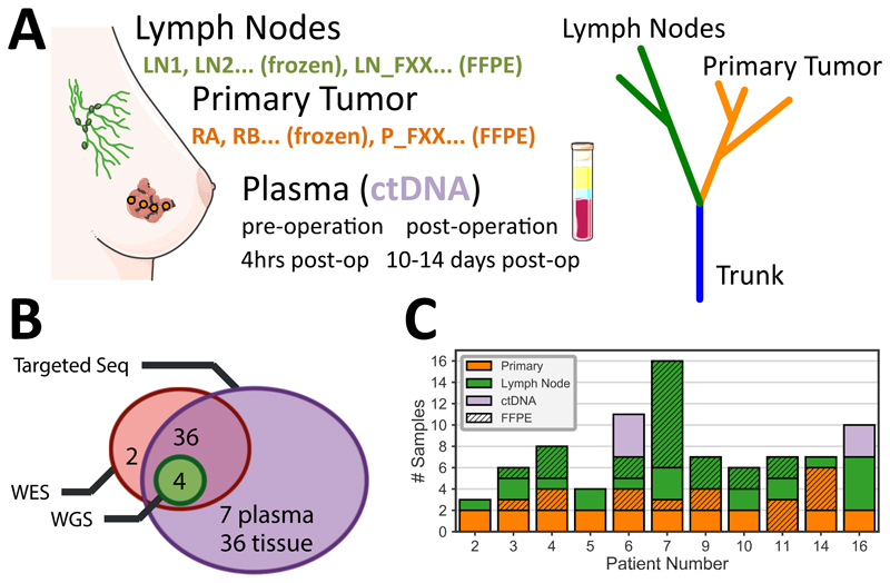
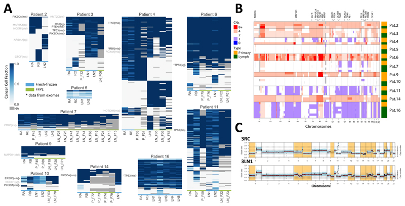
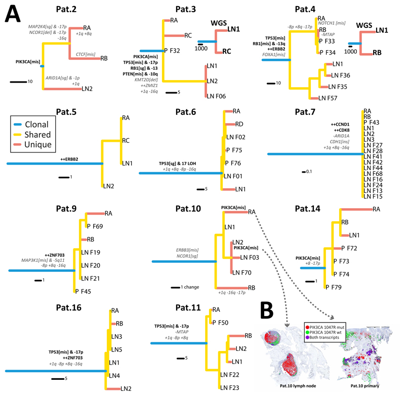
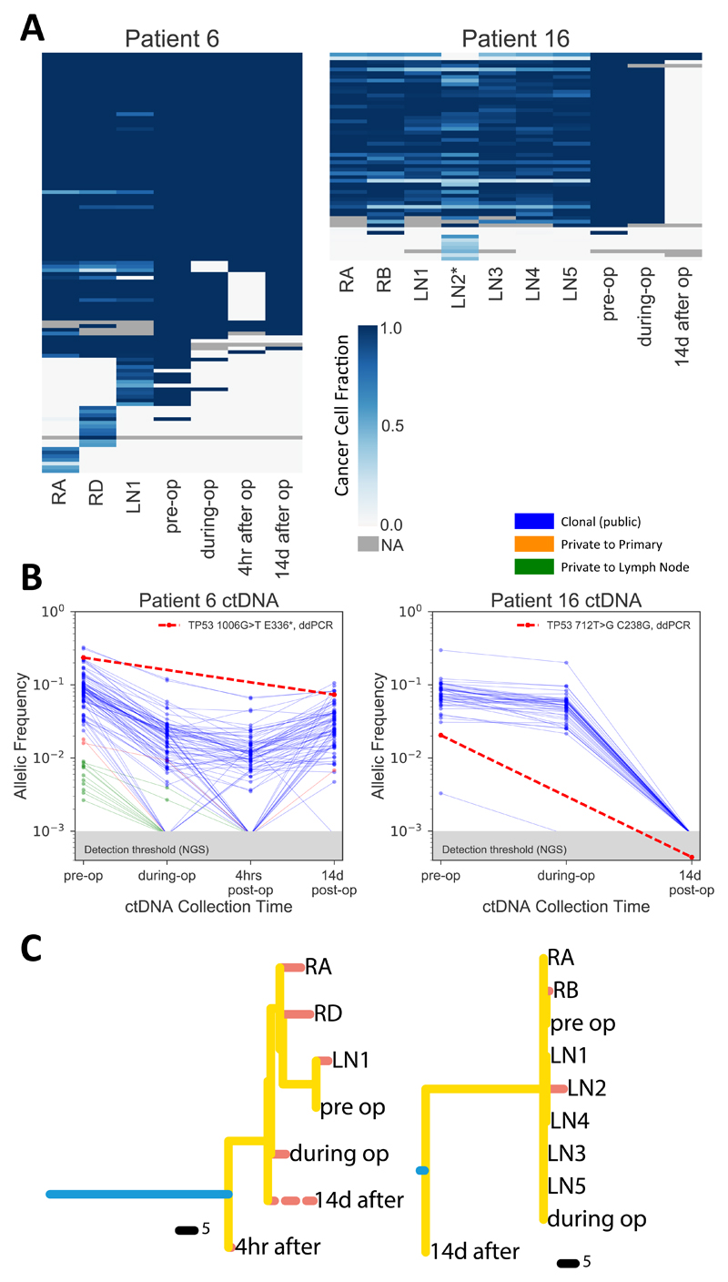
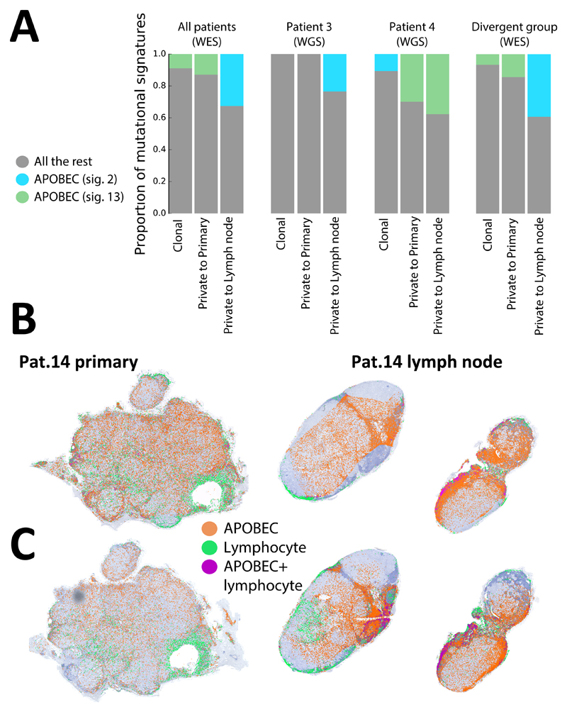
Purpose: The most significant prognostic factor in early breast cancer is lymph node involvement. This stage between localized and systemic disease is key to understanding breast cancer progression; however, our knowledge of the evolution of lymph node malignant invasion remains limited, as most currently available data are derived from primary tumors.Experimental Design: In 11 patients with treatment-naïve node-positive early breast cancer without clinical evidence of distant metastasis, we investigated lymph node evolution using spatial multiregion sequencing (n = 78 samples) of primary and lymph node deposits and genomic profiling of matched longitudinal circulating tumor DNA (ctDNA).Results: Linear evolution from primary to lymph node was rare (1/11), whereas the majority of cases displayed either early divergence between primary and nodes (4/11) or no detectable divergence (6/11), where both primary and nodal cells belonged to a single recent expansion of a metastatic clone. Divergence of metastatic subclones was driven in part by APOBEC. Longitudinal ctDNA samples from 2 of 7 subjects with evaluable plasma taken perioperatively reflected the two major evolutionary patterns and demonstrate that private mutations can be detected even from early metastatic nodal deposits. Moreover, node removal resulted in disappearance of private lymph node mutations in ctDNA.Conclusions: This study sheds new light on a crucial evolutionary step in the natural history of breast cancer, demonstrating early establishment of axillary lymph node metastasis in a substantial proportion of patients. Clin Cancer Res; 24(19); 4763-70. ©2018 AACR.


Figures

Figure 1
Figure 1. Spatio-temporal genomic profiling of lymph node evolution in breast cancer.
(A) Multi-region sampling and genomic profiling of primary (regions RA, RB, …, 1-6cm apart) and lymph node (LN1, LN2, …) samples (total samples n=78) from a selected cohort of 11 early breast cancer patients with lymph node involvement without distant metastases. Longitudinal ctDNA samples were taken before and after surgery from 2 patients (n=7 samples). (B) Sequencing analysis was performed with whole-exome sequencing (n=42), whole-genome sequencing (n=4) and targeted deep sequencing of a cohort-specific panel (n=76) to identify mutational and copy number profiles. (C) The number of samples per location for each patient in the cohort (solid = fresh frozen, stripes = FFPE).
Figure 2
Figure 2. Distinct modes of lymph node evolution.
(A) Deep targeted sequencing of a cohort-specific panel derived from whole-exome sequencing. Heatmaps indicate cancer cell fraction (CCF) of a mutation or indel in different samples from the same patient (grey=NA, not enough coverage or variant does not overlap with a copy number segment). For FFPE samples (marked in green at the bottom) CCFs were not available and presence/absence is reported (CCF=1 or CCF=0). Tier 1 cancer genes, most likely drivers are annotated in black, Tier 2 possible drivers with uncertain pathogenicity are annotated in grey. For samples/variants marked with (*) we report exome data as targeted was not available. (B) Copy number aberrations in all samples showing differences in copy number status between primary and lymph nodes in the divergent subgroup. (C) Whole-genome sequencing for a tumour and lymph node sample of Pat. 3; divergent copy number regions shown in orange.
Figure 3
Figure 3. Evolutionary trajectories during lymph node invasion.
(A) Phylogenetic trees reconstructed with maximum parsimony for each patient illustrate the patterns of lymph node spread. For Pat. 3 and 4 results were validated with whole-genome sequencing. When multiple lymph node samples were available for a patient, those clustered together in a single clade, indicating a recent common ancestor that led to lymph node colonisation. See Figure S4 for tree bootstrap values. Putative driver genes and recurrent copy number alterations in breast cancers are annotated in the trees (Tier 1 likely driver genes in black-bold, recurrent copy number alterations and Tier 2 possible drivers of uncertain pathogenicity in grey-italic). (B) We assessed the spatial heterogeneity of subclonal mutation PIK3CA 1047R in Pat.10 at single cell resolution using in situ hybridisation of mutant vs wildtype transcripts, revealing spatial segregation of mutant and wildtype subclones (only signals from cancer cells are represented).
Figure 4
Figure 4. Longitudinal ctDNA analysis recapitulates tissue evolution.
(A) The cohort-specific targeted panel was applied to ctDNA for two patients, heatmaps show presence (blue) and absence (yellow) of variants at four time points (pre-operation, immediately post-resection, 4 hours after operation and 14 days after operation) compared to the corresponding primary and lymph node samples per patient. (B) Variant allele frequency (VAF) changes of all mutations at different time points. (C) Phylogenetic trees reconstructed with both tissue and ctDNA data confirm these patterns. Post-operative ctDNA appears early in the tree in Pat.6, suggesting early disseminated micrometastatic disease (dashed line indicates possible additional variants not detectable with a targeted approach).
Figure 5
Figure 5. APOBEC signature is increased in lymph nodes.
(A) Mutational signature analysis applied to whole-exome sequencing mutations from the whole cohort and whole-genome sequencing for Pat.3 and Pat. 4 showed an increase in APOBEC signatures in mutations private to the lymph nodes. Spatial heterogeneity in expression of APOBEC3A (B) and APOBEC3B (C) was assessed at single-cell level using in situ hybridisation for Pat.14, revealing higher expression of both APOBEC3A and APOBEC3B in the lymph node lesion (only signals from cancer cells are represented).

References

    1. Curtis C, Shah SP, Chin S-F, Turashvili G, Rueda OM, Dunning MJ, et al. The genomic and transcriptomic architecture of 2,000 breast tumours reveals novel subgroups. Nature. 2012;486:346–52. - PMC - PubMed
    1. Shah SP, Roth A, Goya R, Oloumi A, Ha G, Zhao Y, et al. The clonal and mutational evolution spectrum of primary triple-negative breast cancers. Nature. 2012;486:395–9. - PMC - PubMed
    1. Nik-Zainal S, Davies H, Staaf J, Ramakrishna M, Glodzik D, Zou X, et al. Landscape of somatic mutations in 560 breast cancer whole-genome sequences. Nature. 2016;534:47–54. - PMC - PubMed
    1. Nik-Zainal S, Van Loo P, Wedge DC, Alexandrov LB, Greenman CD, Lau KW, et al. The life history of 21 breast cancers. Cell. 2012;149:994–1007. - PMC - PubMed
    1. Wang Y, Waters J, Leung ML, Unruh A, Roh W, Shi X, et al. Clonal evolution in breast cancer revealed by single nucleus genome sequencing. Nature. 2014;512:155–60. - PMC - PubMed

Publication types