Comparative Study
. 2018 Jun 11;9(1):2285.
doi: 10.1038/s41467-018-04698-4.

Diversity and evolution of the emerging Pandoraviridae family

Affiliations
Comparative Study

Diversity and evolution of the emerging Pandoraviridae family

Matthieu Legendre et al. Nat Commun. .

Abstract

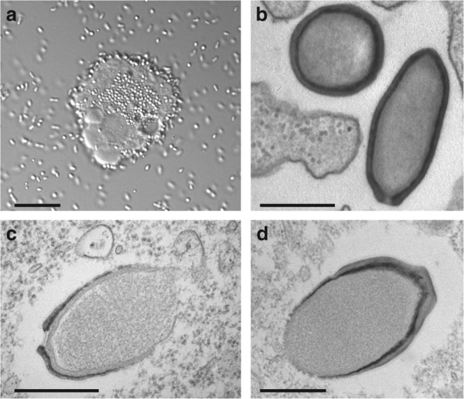
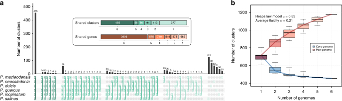
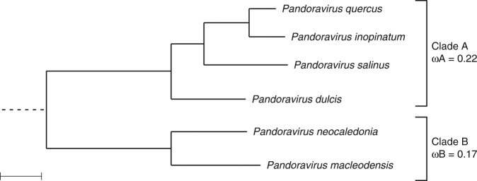
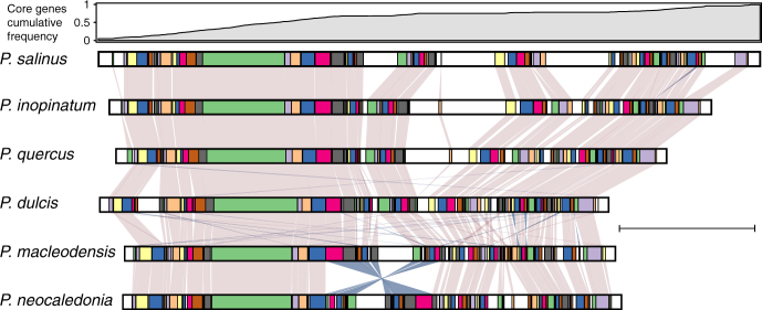
With DNA genomes reaching 2.5 Mb packed in particles of bacterium-like shape and dimension, the first two Acanthamoeba-infecting pandoraviruses remained up to now the most complex viruses since their discovery in 2013. Our isolation of three new strains from distant locations and environments is now used to perform the first comparative genomics analysis of the emerging worldwide-distributed Pandoraviridae family. Thorough annotation of the genomes combining transcriptomic, proteomic, and bioinformatic analyses reveals many non-coding transcripts and significantly reduces the former set of predicted protein-coding genes. Here we show that the pandoraviruses exhibit an open pan-genome, the enormous size of which is not adequately explained by gene duplications or horizontal transfers. As most of the strain-specific genes have no extant homolog and exhibit statistical features comparable to intergenic regions, we suggest that de novo gene creation could contribute to the evolution of the giant pandoravirus genomes.


Conflict of interest statement

The authors declare no competing interests.

Figures

Fig. 1
Fig. 1
The new pandoravirus isolates. a Overproduction by an A. castellanii cell of Pandoravirus macleodensis virions from the environmental sample prior cell lysis. Environmental bacteria can be seen in the culture medium together with P. macleodensis virions. (scale bar is 10 µm). b TEM image of an ultrathin section of A. castellanii cell during the early phase of infection by P. neocaledonia. The ameba pseudopods are ready to engulf the surrounding virions. Ten minutes pi, virions have been engulfed and are in vacuoles (scale bar is 500 nm). c TEM image of an ultrathin section of A. castellanii cell during the assembly process of a P. salinus virion (scale bar is 500 nm). d TEM image of an ultrathin section of a nascent P. quercus virion. (scale bar is 500 nm). The structures of the mature particles from the different strains do not exhibit any noticeable difference
Fig. 2
Fig. 2
Comparison of the pandoravirus gene contents. a The distribution of all the combinations of shared protein clusters is shown. The inset summarizes the number of clusters and genes shared by 6, 5, 4, 3, 2, and 1 pandoraviruses. b Core genome and pan-genome estimated from the six available pandoraviruses. The estimated heap law α parameter (α < 1) is characteristic of an open pan-genome and the fluidity parameter value characteristic of a large fraction of unique genes. Box plots show the median, the 25th, and 75th percentiles. The whiskers correspond to the extreme data points
Fig. 3
Fig. 3
Phylogenetic structure of the proposed Pandoraviridae family. Bootstrap values estimated from resampling are all equal to 1 and so were not reported. Synonymous to non-synonymous substitution rates ratios (ω) were calculated for the two separate clades and are significantly different (scale bar is 0.07 substitution/site)
Fig. 4
Fig. 4
Collinearity of the available pandoravirus genomes. Cumulative frequency of core genes is shown at the top. Conserved collinear blocks are colored in the same color in all viruses. White blocks correspond to non-conserved DNA segments (scale bar is 500 kb)
Fig. 5
Fig. 5
Functional annotations
Fig. 6
Fig. 6
Analysis of gene duplication in various giant virus families. a Distribution of single-copy versus multiple-copy genes in giant viruses. b Number of distinct gene clusters
Fig. 7
Fig. 7
Venn diagram of the particle proteomes of four different pandoravirus strains
Fig. 8
Fig. 8
Gene content-based cladistic tree of large DNA viruses. Long virus names have been replaced by acronyms (from top, clockwise). OTV1 Ostreococcus tauri virus 1, OTV2 Ostreococcus tauri virus 2, EhV86 Emiliania huxleyi virus 86, ASFAR African swine fever virus, CroV Cafeteria roenbergensis virus BV.PW1, BPSV Bovine papular stomatitis virus, ISKNV Infectious spleen and kidney necrosis virus. A maximum likelihood phylogenetic tree based on the DNA polymerase B protein sequence showing a globally similar topology was also computed (Supplementary Fig. 14)

Similar articles

Cited by

References

    1. La Scola B, et al. A giant virus in amoebae. Science. 2003;299:2033. doi: 10.1126/science.1081867. - DOI - PubMed
    1. Raoult D, et al. The 1.2-megabase genome sequence of Mimivirus. Science. 2004;306:1344–1350. doi: 10.1126/science.1101485. - DOI - PubMed
    1. Arslan D, Legendre M, Seltzer V, Abergel C, Claverie JM. Distant Mimivirus relative with a larger genome highlights the fundamental features of Megaviridae. Proc. Natl Acad. Sci. USA. 2011;108:17486–17491. doi: 10.1073/pnas.1110889108. - DOI - PMC - PubMed
    1. Yoosuf N, et al. Related giant viruses in distant locations and different habitats: Acanthamoeba polyphaga moumouvirus represents a third lineage of the Mimiviridae that is close to the megavirus lineage. Genome Biol. Evol. 2012;4:1324–1330. doi: 10.1093/gbe/evs109. - DOI - PMC - PubMed
    1. Philippe N, et al. Pandoraviruses: amoeba viruses with genomes up to 2.5 Mb reaching that of parasitic eukaryotes. Science. 2013;341:281–286. doi: 10.1126/science.1239181. - DOI - PubMed

Publication types