. 2018 Jun 11;8(1):8859.
doi: 10.1038/s41598-018-27184-9.

Interleukin-6/STAT3 signalling regulates adipocyte induced epithelial-mesenchymal transition in breast cancer cells

Affiliations

Interleukin-6/STAT3 signalling regulates adipocyte induced epithelial-mesenchymal transition in breast cancer cells

Jones Gyamfi et al. Sci Rep. .

Erratum in

Abstract

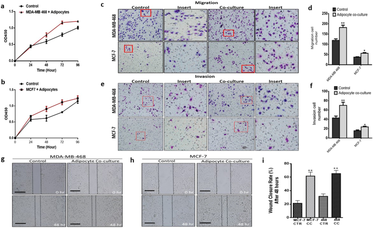
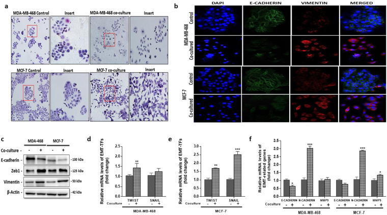
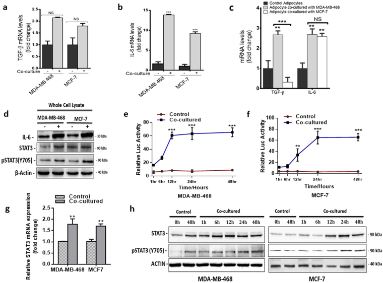
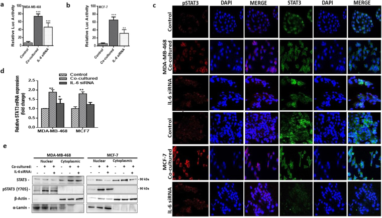
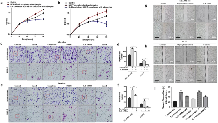
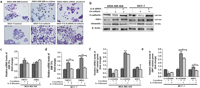
The tumour microenvironment is a key regulators of tumour progression through the secretion of growth factors that activate epithelial-mesenchymal transition (EMT). Induction of EMT is a key step for transition from a benign state to a metastatic tumour. Adipose tissue forms a bulk portion of the breast cancer microenvironment, emerging evidence indicates the potential for adipocytes to influence tumour progression through the secretion of adipokines that can induce EMT. The molecular mechanisms underlying how adipocytes enhance breast cancer progression is largely unknown. We hypothesized that paracrine signalling by adipocytes can activate EMT and results in increased migration and invasion characteristics of breast cancer cells. We found that IL-6 secreted by adipocytes induce EMT in breast cancer cells. The effect of IL-6 expression on EMT is mediated through activation of the signal transducer and activated of transcription 3 (STAT3). Blocking of IL-6 signalling in breast cancer cells and adipocytes, decreased proliferation, migration and invasion capabilities and altered the expression of genes regulating EMT. Together, our results suggest that matured human adipocytes can enhance the aggressive behaviour of breast cancer cells and induce an EMT-phenotype through paracrine IL-6/STAT3 signalling.


Conflict of interest statement

The authors declare no competing interests.

Figures

Figure 1
Figure 1
Oil Red O staining of differentiated human preadipocytes. (a) Undifferentiated human preadipocytes, 4 weeks after culturing without induction media. (b) Differentiating human preadipocytes with tiny lipid droplets 2 weeks after induction of differentiation. (c) Differentiated human preadipocytes 4 weeks after induction of differentiation with large formed lipid droplets.
Figure 2
Figure 2
Human adipocytes enhances the aggressive characteristics of breast cancer. (a,b) Proliferation capabilities of co-cultured breast cancer cells assessed with the CCK-8 assay, compared with control cells (A: MDA-MB-468 and B: MCF-7). (c) Migration rate was examined using the transwell insert without matrigel. (d) Quantification of migration capabilities ability of MDA-MB-468 and MCF-7 breast cancer cells. (e) Invasion rate was examined using the transwell insert with matrigel. (f) Quantification of invasion capabilities ability of MDA-MB-468 and MCF-7 breast cancer cells. (g,h,i) Representative images of wound healing assay. Breast cancer cell motility accessed by a wound healing assay after 48 hours (magnification, x100), MDA-MB-468 and MCF-7 co-cultured breast cancer cells had a higher motility rates. All results are representative of 3 independent experiments. (Data indicate mean ± SD; ***p < 0.001; **p < 0.01; *p < 0.05).
Figure 3
Figure 3
Human adipocytes induced EMT-phenotype with alteration in EMT-related gene expression. (a) Morphological characteristics of MDA-MB-468 and MCF-7 cells compared with cells co-cultured with human adipocytes. Co-cultured breast cancer cells were dispersed and slightly elongated. (b) Immunofluorescence staining of e-cadherin and vimentin. (c) Western blot for e-cadherin, vimentin and ZEB1 in MDA-MB-468 and MCF-7 breast cancer cells cultured with or without adipocytes. Full-length blots are presented in Supplementary Fig. 5. (d,e) Quantitative PCR (qt-PCR) comparing the expression of EMT-TFs (TWIST and SNAIL) in co-cultured breast cancer cells to control cells. (f) qt-PCR comparing the expression of EMT-related genes (MMP9, N-cadherin and E-cadherin) in co-cultured breast cancer cells to control cells. Relative mRNA expression was normalized to GADPH. All results are representative of 3 independent experiments. (Data indicate mean ± SEM; ***p < 0,001; **p < 0.01; *p < 0.05).
Figure 4
Figure 4
Analysis of STAT3 activity in breast cancer cells. (a,b) Quantitative PCR comparing the expression of TGF-B and IL-6 in co-cultured breast cancer cells to control cells. (c) qt-PCR comparing the expression of TGF-B and IL-6 in adipocytes cultured with/without breast cancer cells. (d) Comparing the expression levels of IL-6, STAT3 and pSTAT3 (Y705) in co-cultured and control breast cancer cells by immunoblotting. Full-length blots are presented in Supplementary Fig. 5. (e,f) Relative luciferase activity in breast cancer cells with STAT3 luciferase reporter plasmid and cultured with/without human adipocytes (e). MDA-MB-468 STAT3 activity, (f). MCF-7 STAT3 activity. Luciferase activity was normalized against a non-inducible luciferase construct. (i) Western blot for STAT3 and pSTAT3 (Y705) over 48 hours are regular time intervals in breast cancer cells cultured with/without adipocytes. B-actin was used as loading control. Full-length blots are presented in Supplementary Fig. 5. All results are representative of 3 independent experiments. Data is presented as mean ± SEM; ***p < 0,001; **p < 0.01; *p < 0.05.
Figure 5
Figure 5
Adipocytes induces phosphorylated STAT3 nuclear localization. Using IL-6R siRNA and IL-6 neutralizing antibodies paracrine IL-6 signaling was blocked in co-cultured cells. (a) IL-6 mRNA expression was analyzed 72 hours after IL-6 signal was blocked. (b,c) Western blot for IL-6 in breast cancer cells over 72 hours after IL-6 signal was blocked at 24, 48 and 72 hours. Full-length blots are presented in Supplementary Fig. 5. (d) IL-6 mRNA expression was analyzed at 24, 48 and 72 hours in adipocytes after IL-6 siRNA transfection. (e) Western blot for IL-6 in IL-6 siRNA transfected adipocytes at 24, 48 and 72. Full-length blots are presented in Supplementary Fig. 5. (f,g) Relative luciferase activity in breast cancer cells with STAT3 luciferase reporter plasmid, with/without human adipocytes and after IL-6 was blocked. (h) Quantitative PCR comparing the expression of STAT3 mRNA in co-cultured breast cancer cells with/without IL-6 blocking and in control cells. Relative mRNA expression was normalized to GAPDH. (i) Increased STAT3 phosphorylation and nuclear localization in co-cultured human breast cancer cells with/without IL-6 neutralization and in control cells assessed by immunofluorescence staining. (j) Representative western blot analysis of pSTAT3 expression in cytoplasmic and nuclear fraction of co-cultured breast cancer with/without IL-6 neutralization and in control breast cancer cells. α-Lamin and β-actin was used as loading control for nuclear and cytoplasmic fraction respectively. Full-length blots are presented in Supplementary Fig. 5. All results are representative of 3 independent experiments. Data is presented as mean ± SEM; ***p < 0,001; **p < 0.01; *p < 0.05.
Figure 6
Figure 6
Blocking IL-6 signals reverses human adipocytes induced aggressive characteristics of breast cancer. (a,b) Proliferation capabilities of co-cultured breast cancer cells assessed in co-cultured breast cancer cells with/without IL-6 neutralization and in control cells. (c) Migration rate was examined using the transwell insert without matrigel in co-cultured breast cancer cells with/without IL-6 neutralization and in control cells. (d) Quantification of migration capabilities ability of in co-cultured breast cancer cells with/without IL-6 neutralization and in control cells. (e) Invasion rate in co-cultured breast cancer cells with/without IL-6 neutralization and in control cells. (f) Quantification of invasion capabilities in co-cultured breast cancer cells with/without IL-6 neutralization and in control cells. (g, h) Representative images of wound healing assay. Breast cancer cell motility accessed by a wound healing assay after 48 hours (magnification, x100), MDA-MB-468 and MCF-7 co-cultured breast cancer cells had a higher motility rates and blocking IL-6 inhibited breast cancer cell motility. (i) Quantification of breast cancer cell motility in co-cultured breast cancer cells with/without IL-6 neutralization and in control cells. All results are representative of 3 independent experiments. (Data indicate mean ± SD; ***p < 0.001; **p < 0.01; *p < 0.05).
Figure 7
Figure 7
Expression of EMT-related genes was reversed after blocking IL-6 signalling. (a) Morphological characteristics of co-cultured breast cancer cell with/without IL-6 blocking and control breast cancer cells. (b) Western blot for e-cadherin, vimentin and ZEB1 in co-cultured breast cancer cells with/without IL-6 blocking and in control cells. Full-length blots are presented in Supplementary Fig. 5. (cf) Quantitative PCR (qt-PCR) comparing the expression of EMT-TFs and EMT-related genes in co-cultured breast cancer cells with/without IL-6 blocking and control cells. Relative mRNA expression was normalized to GAPDH. All results are representative of 3 independent experiments. (Data indicate mean ± SEM; ***p < 0,001; **p < 0.01; *p < 0.05).

Similar articles

Cited by

References

    1. Siegel, R. L., Miller, K. D. & Jemal, A. Cancer statistics, 2016. CA Cancer J Clin. 66, 7–30, 10.3322/caac.21332, Epub22016 Jan 21337 (2016). - PubMed
    1. Mao Y, Keller ET, Garfield DH, Shen K, Wang J. Stromal cells in tumor microenvironment and breast cancer. Cancer metastasis reviews. 2013;32:303–315. doi: 10.1007/s10555-012-9415-3. - DOI - PMC - PubMed
    1. Place AE, Jin Huh S, Polyak K. The microenvironment in breast cancer progression: biology and implications for treatment. Breast Cancer Res. 2011;13:227. doi: 10.1186/bcr2912. - DOI - PMC - PubMed
    1. Takebe, N., Warren, R. Q. & Ivy, S. P. Breast cancer growth and metastasis: interplay between cancer stem cells, embryonic signaling pathways and epithelial-to-mesenchymal transition. Breast Cancer Res. 13, 211, doi: 210.1186/bcr2876 (2011). - PMC - PubMed
    1. Yang, J. & Weinberg, R. A. Epithelial-mesenchymal transition: at the crossroads of development and tumor metastasis. Dev Cell. 14, 818–829, doi: 810.1016/j.devcel.2008.1005.1009 (2008). - PubMed

Publication types