. 2018 Jul;3(7):773-780.
doi: 10.1038/s41564-018-0174-y. Epub 2018 Jun 11.

Retraction of DNA-bound type IV competence pili initiates DNA uptake during natural transformation in Vibrio cholerae

Affiliations

Retraction of DNA-bound type IV competence pili initiates DNA uptake during natural transformation in Vibrio cholerae

Courtney K Ellison et al. Nat Microbiol. 2018 Jul.

Abstract

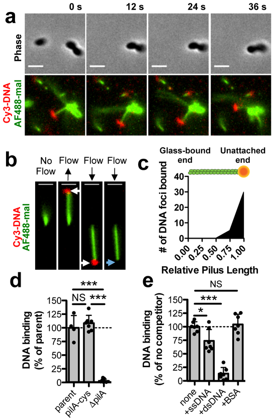
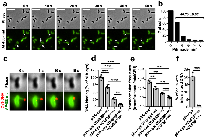
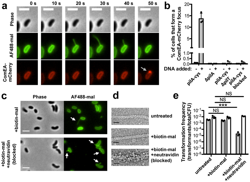
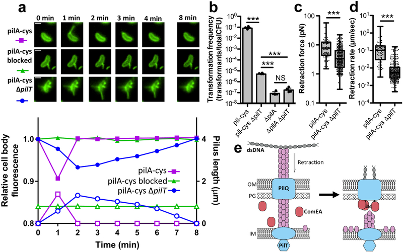
Natural transformation is a broadly conserved mechanism of horizontal gene transfer in bacterial species that can shape evolution and foster the spread of antibiotic resistance determinants, promote antigenic variation and lead to the acquisition of novel virulence factors. Surface appendages called competence pili promote DNA uptake during the first step of natural transformation 1 ; however, their mechanism of action has remained unclear owing to an absence of methods to visualize these structures in live cells. Here, using the model naturally transformable species Vibrio cholerae and a pilus-labelling method, we define the mechanism for type IV competence pilus-mediated DNA uptake during natural transformation. First, we show that type IV competence pili bind to extracellular double-stranded DNA via their tip and demonstrate that this binding is critical for DNA uptake. Next, we show that type IV competence pili are dynamic structures and that pilus retraction brings tip-bound DNA to the cell surface. Finally, we show that pilus retraction is spatiotemporally coupled to DNA internalization and that sterically obstructing pilus retraction prevents DNA uptake. Together, these results indicate that type IV competence pili directly bind to DNA via their tip and mediate DNA internalization through retraction during this conserved mechanism of horizontal gene transfer.


Conflict of interest statement

Competing interests:

The authors declare no competing interests.

Figures

Fig. 1.
Fig. 1.. The tips of type IV competence pili directly bind DNA.
Because type IV competence pili are dynamically active and relatively few pili are extended at any given time, all pilus-DNA binding studies were performed in ΔpilT backgrounds to ensure that cells contained numerous extended pili as shown in Supplementary Fig. 1c. (a) Montage of time-lapse imaging of a pilA-cys ΔpilT cell labeled with AF488-mal after mixing with Cy3-labeled DNA in a wet mount. Scale bar, 2 μm. (b) Example of a sheared pilus labeled with AF488-mal bound to glass within a microfluidic device after the addition of fluorescently labeled DNA. Before flow is applied, the relatively long pilus fiber is out of the field of view, but after flow is applied, bound DNA becomes apparent (white arrows) and moves with the direction of flow. When flow is maintained, the DNA detaches from the pilus fiber (blue arrow). Black arrows above images represent the direction of flow. Scale bar, 2 μm. Data in a-b are representative of 3 independent experiments. (c) Localization of bound DNA along sheared pili in microfluidic devices. Pili were normalized in length to 1.00 (arbitrary units), and the position of DNA foci bound along the length for each fiber was plotted, n = 51 independent DNA-bound pili analyzed. Green rod above plot is a visual representation of a labeled pilus fiber bound to orange DNA. (d) Relative DNA binding assay using a fluorescently labeled 6 kb PCR product. parent n = 4, pilA-cys n = 7, ΔpilA, n = 7. (e) Relative DNA binding as in d except with the addition of the indicated non-labeled competitors at 50-fold excess. Competitors were ssDNA (PhiX174 virion), dsDNA (PhiX174 RFII), or bovine serum albumin (BSA). n = 7 for all samples. Each data point in d-e represents an independent biological replicate and bar graphs indicate the mean ± SD. Statistical comparisons were made by two-tailed Student’s t-test. NS = not significant, *P < 0.05, ***P < 0.001.
Fig. 2.
Fig. 2.. Type IV competence pilus dynamic activity and DNA binding are critical for DNA internalization.
(a) Montage of time-lapse imaging of pilA-cys cells after labeling with AF488-mal dye. The white arrows indicate pili. Scale bar, 5 μm. (b) Number of cells making 0–5 pili within a one-minute period. Data are from three independent, biological replicates, n = 192 total pili observed. Percentage is the percent of cells within the population that make pili within the one-minute time-frame ± SD. (c) Montage of time-lapse imaging of a retracting pilA-cys strain after labeling with AF488-mal and incubation with fluorescently labeled DNA in a wet mount. Scale bar, 1 μm. Similar events were captured in three independent experiments. (d) Relative DNA binding assay of the indicated strains in ΔpilT mutant backgrounds using a fluorescently labeled 6 kb PCR product. (e) Natural transformation assays of the indicated strains. Cells were incubated with 5 ng of transforming DNA (tDNA) and DNase I was added to reactions after 10 mins to prevent additional DNA uptake. (f) DNA internalization assays of the indicated strains using a fluorescently labeled 6 kb PCR product. VC0858 and VC0859 encode minor pilins. Each data point in d-f represents an independent biological replicate (d-e, n = 4 for all samples; f, n = 3 for all samples) and bar graphs indicate the mean ± SD. All statistical comparisons were made by two-tailed Student’s t-test. **P < 0.01, ***P < 0.001.
Fig 3.
Fig 3.. Pilus retraction is required for DNA uptake.
(a) Montage of time-lapse imaging of pilA-cys ComEA-mCherry strain labeled with AF488-mal showing ComEA focus formation after pilus retraction (gray arrow). Scale bar, 2 μm. (b) Percent of cells that formed a ComEA-mCherry focus within a five min window in the presence or absence of DNA. Cells that had already formed ComEA-mCherry foci at the start of imaging were excluded from the analysis. Data are from three independent experiments (n = 3 for each condition) and shown as the mean ± SD. (c) Static images of pilA-cys cells labeled with a 1:1 ratio of biotin-mal:AF488-mal with or without added neutravidin. White arrows indicate pili. Scale bar, 2 μm. (d) Transmission electron micrographs of pilA-cys pili with the indicated treatments. While untreated and biotin-mal treated pili exhibit a similar thickness, the biotin-mal + neutravidin treated pilus is approximately twice as thick. Scale bar, 10 nm. Images in c-d are representative of two independent experiments. (e) Natural transformation assays of pilA-cys (black bars) or parent (white bars) strains after the indicated treatment. Cells were incubated with 500 ng of tDNA and DNase I was added to reactions after 10 mins to prevent additional DNA uptake. Data are from three independent, biological replicates and shown as the mean ± SD. Statistical comparisons were made by two-tailed Student’s t-test. NS = not significant. ***P < 0.001.
Fig 4.
Fig 4.. Residual retraction in ΔpilT mutants allows for low rates of transformation.
(a) Montage (top) of cells measured for corresponding plots (bottom) showing the relative cell body fluorescence (closed symbols) and correlated pilus length (open symbols) over time for pilA-cys labeled with AF488-mal (circles), pilA-cys blocked for retraction by labeling with 1:1 ratio of biotin-mal:AF488-mal and neutravidin (triangles), or pilA-cys ΔpilT labeled with AF488-mal (squares). Scale bar, 1 μm. Data are representative of 3 independent experiments. (b) Natural transformation assays of the indicated strains using 500 ng of tDNA. Data are from four independent, biological replicates and shown as the mean ± SD. Micropillar assays were performed with the indicated strains to measure the retraction (c) force (pilA-cys n = 79, pilA-cys ΔpilT n = 339) and (d) speed (pilA-cys n = 76, pilA-cys ΔpilT n = 288). Box plots indicate the median and first and third quartiles, while the whiskers denote the range. Each data point in the overlay for c-d represents an independent retraction event. Statistical comparisons in b-d were made by two-tailed Student’s t-test. NS = not significant. ***P < 0.001. (e) Model of pilus retraction-mediated DNA uptake. Retraction of DNA-bound pili threads dsDNA across the outer membrane (left) followed by ComEA-dependent molecular ratcheting (right) to promote uptake; OM = outer membrane, PG = peptidoglycan, IM = inner membrane.

Similar articles

Cited by

References

    1. Chen I & Dubnau D DNA uptake during bacterial transformation. Nat Rev Microbiol 2, 241–249 (2004). - PubMed
    1. Hepp C & Maier B Kinetics of DNA uptake during transformation provide evidence for a translocation ratchet mechanism. Proc Natl Acad Sci U S A 113, 12467–12472 (2016). - PMC - PubMed
    1. Muschiol S, Balaban M, Normark S & Henriques-Normark B Uptake of extracellular DNA: competence induced pili in natural transformation of Streptococcus pneumoniae. Bioessays 37, 426–435 (2015). - PMC - PubMed
    1. Graupner S, Weger N, Sohni M & Wackernagel W Requirement of novel competence genes pilT and pilU of Pseudomonas stutzeri for natural transformation and suppression of pilT deficiency by a hexahistidine tag on the type IV pilus protein PilAI. J Bacteriol 183, 4694–4701 (2001). - PMC - PubMed
    1. Laurenceau R et al. A type IV pilus mediates DNA binding during natural transformation in Streptococcus pneumoniae. PLoS Pathog 9, e1003473 (2013). - PMC - PubMed