. 2018 Jun 11;8(1):8858.
doi: 10.1038/s41598-018-27195-6.

D5 dopamine receptors control glutamatergic AMPA transmission between the motor cortex and subthalamic nucleus

Affiliations

D5 dopamine receptors control glutamatergic AMPA transmission between the motor cortex and subthalamic nucleus

Lionel Froux et al. Sci Rep. .

Abstract

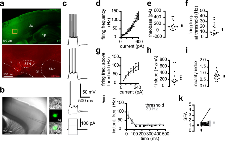
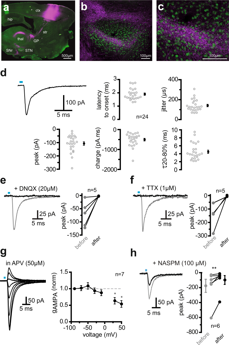
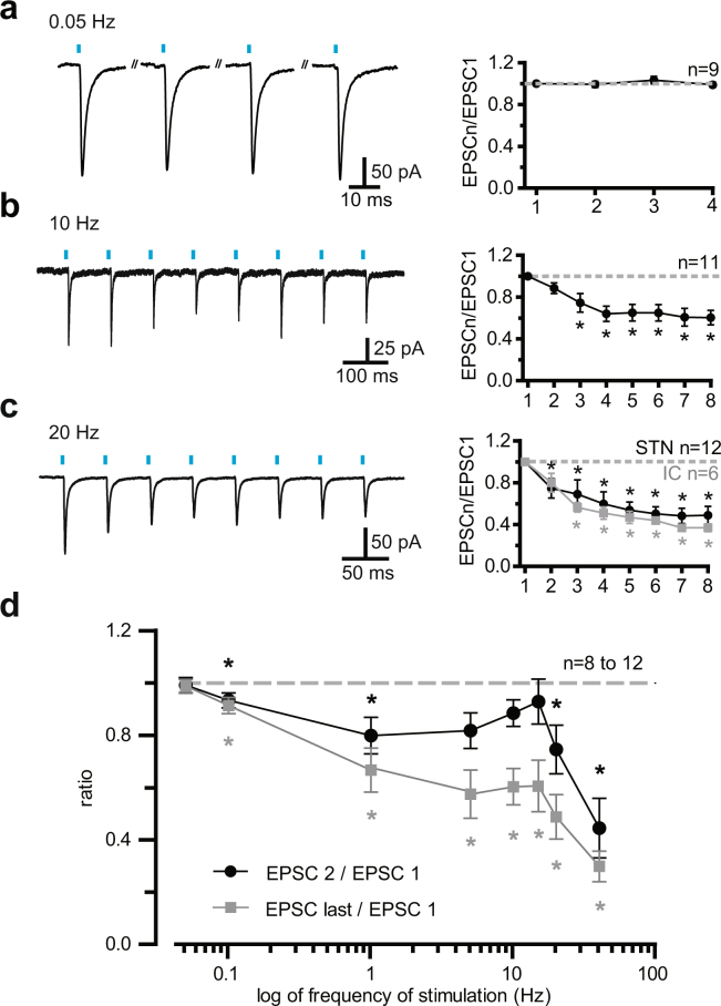
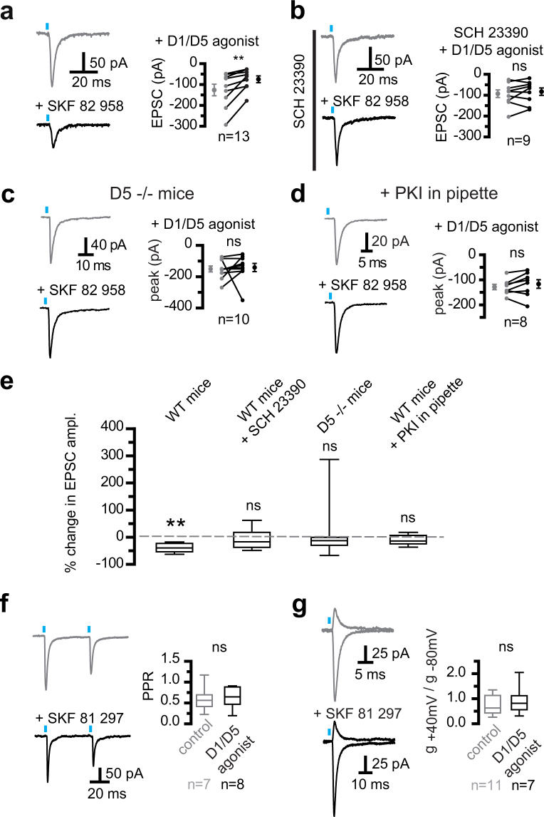
Corticofugal fibers target the subthalamic nucleus (STN), a component nucleus of the basal ganglia, in addition to the striatum, their main input. The cortico-subthalamic, or hyperdirect, pathway, is thought to supplement the cortico-striatal pathways in order to interrupt/change planned actions. To explore the previously unknown properties of the neurons that project to the STN, retrograde and anterograde tools were used to specifically identify them in the motor cortex and selectively stimulate their synapses in the STN. The cortico-subthalamic neurons exhibited very little sag and fired an initial doublet followed by non-adapting action potentials. In the STN, AMPA/kainate synaptic currents had a voltage-dependent conductance, indicative of GluA2-lacking receptors and were partly inhibited by Naspm. AMPA transmission displayed short-term depression, with the exception of a limited bandpass in the 5 to 15 Hz range. AMPA synaptic currents were negatively controlled by dopamine D5 receptors. The reduction in synaptic strength was due to postsynaptic D5 receptors, mediated by a PKA-dependent pathway, but did not involve a modified rectification index. Our data indicated that dopamine, through post-synaptic D5 receptors, limited the cortical drive onto STN neurons in the normal brain.


Conflict of interest statement

The authors declare no competing interests.

Figures

Figure 1
Figure 1
Properties of cortico-subthalamic neurons. (a) Confocal images of the cortex (top) after an injection of Fluoro-Gold (FG)retrograde tracer in the STN. To check the location of the injection site (bottom), the FG solution was mixed with a solution of anterograde virus expressing mCherry that labelled neurons at the injection site. FG and mCherry were detected by immunocytochemistry in a 350 μm slice used for electrophysiology. The orange box on the cortex indicates a high-density zone of FG-labelled neurons. Ctx, cortex; hip, hippocampus; ic, internal capsule; cp, cerebral peduncle; SNr, substantia nigra reticulata. (b) Infrared view of a living slice (left) and an FG-positive neuron at high magnification (right). (c) Example of the electrical properties of a retrograde-labelled cortico-subthalamic neuron. (di) Input-output relationship of a sample of 17 cortico-subthalamic neurons. (d) Mean firing frequency-current relationship. (e) Rheobase. (f) Firing frequency at threshold. (g) Mean firing frequency-current relationship above threshold. The current values represent the amplitude of step stimuli minus the amplitude of the first action potential-evoking step. (h) Firing frequency–current (f.i) slope. (i) Linearity index of the firing frequency–current relationship. e,f,g,h,i, left: individual values, right: mean ± sem. (j,k) Spike frequency over time in the same sample. (j) Instantaneous firing rate versus time (time was divided into 50 ms bins; times shown are the center of each bin) and (k) Spike firing adaptation, with black and grey symbols for values at threshold and 30 Hz, respectively. Three neurons with firing frequencies close to 30 Hz at threshold and those with above threshold values much higher than 30 Hz were not included in the 30 Hz data. (ck) Saline was supplemented with 20 μM DNQX, 50 μM APV, 50 μM picrotoxin, and 1 μM CGP 55845.
Figure 2
Figure 2
Optical activation of corticofugal fibers in the STN results in action potential-dependent AMPA synaptic transmission (a) Expression of mCherry following transfection of a ChR2-mCherry viral solution in the motor cortex. (b,c) mCherry-positive corticofugal fibers in the STN. The STN boundaries are delineated by the STN neuron-specific FoxP2 marker (in green). (d) Example of the current evoked by a 1 ms flash and descriptive parameters of the inward currents measured in a sample of 24 subthalamic neurons. Current peak amplitude and charge, onset latency, time required to decay from 80 to 20% of peak value, and jitter are reported as individual values (left) and mean ± sem (right). Flash duration and luminance ranges were 0.4 to 1 ms and 0.55 to 1.15 mW.mm−2. (e,f) The light-induced inward currents were fully blocked by the specific AMPA/kainate receptor antagonist, DNQX (20 µM), or the sodium channel inhibitor, TTX (1 µM). The gray and black traces illustrate currents photo-evoked in controls and in the continued presence of the drugs, respectively. The graphs to the right of each recording display the summary data of current values before and after drug application. (g) Voltage-dependence and conductance of pharmacologically-isolated AMPA receptor EPSCs. Left: Representative traces of photo-induced EPSCs in the presence of the specific NMDA receptor antagonist, APV (50 µM). Holding voltage was changed from −90 to +50 mV in 20 mV steps. Right: Plot showing chord conductance against voltage. Conductance was significantly different from 1 at +30 and +50 mV, indicating rectifying AMPA receptors. (h) Naspm reduced the EPSC magnitude, indicating Ca2+-permeant, GluA2-lacking, as well as GluA2-containing, AMPA receptors. *Significantly different from the theoretical median = 1, two-tailed Wilcoxon signed rank test, p = 0.0313 for both +30 mV and +50 mV. **Indicates significant changes at p = 0.0156, one-tailed Wilcoxon matched-pairs signed rank test.
Figure 3
Figure 3
Short-term plasticity of AMPA/kainate cortico-subthalamic transmission at stimulation frequencies between 0.05 and 40 Hz (a,c) Examples of EPSCs obtained at light frequencies of 0.05, 10, and 20 Hz (left) and group synaptic dynamics data (right). Mean response amplitudes were normalized to the first responses and plotted as a function of stimulus number within trains. The gray symbols in C depict the values obtained by flashing the internal capsule (and not the STN). The stimuli trains contained 8 photo-stimuli, except those at 0.05 Hz, which comprised only four photo-stimuli. (d) Summary graph of the filter properties of the cortico-subthalamic synapses. The black line and symbols display the ratios of paired pulse values, i.e. peak values of the second to the first EPSC in a train. The gray plot shows the ratio of the last to the first EPSC in a train (the 8th EPSC was the last, except for the tests at 0.05 and 0.1 Hz, where the ratio of the fourth to the first EPSC was calculated). As paired-pulse ratios exhibited no change in the 5 to 15 Hz range, a limited band-pass frequency was assumed. *Values significantly different from 1, α = 0.05, two-tailed Wilcoxon signed rank test.
Figure 4
Figure 4
D5 dopamine receptors reduced AMPA/kainate synaptic strength. (a) Activating D1/D5 receptors reduced cortico-subthalamic AMPA/kainate EPSCs in subthalamic neurons. Illustration of the action of the D1/D5 agonist, SKF 82958 (5 µM). D1/D5 agonists refer to SKF 82958, SKF 81297, or SKF 38393, at 2–5 µM. The 3 dopaminergic agonists were used to neutralize the differences in the potency of the drugs or their affinity for D5 or D1 receptors, as reported in the literature. (b) The continued presence of SCH 23390 (10 µM) prevented the action of SKF 82958 (5 µM). (c) Perfusion of 5 µM D1/D5 agonists had no significant effect in slices from D5−/− mice. (d) D1/D5 agonists (5 µM) were no longer active in the neuron sample tested with a peptide inhibitor (PKI, 20 µM in pipette solution) to intracellularly inhibit protein kinase A. In (a) to (d), traces show the currents recorded immediately prior to (top) and 10 min after agonist perfusion (bottom), and group data show individual paired values as well as mean ± sem. Two-tailed Wilcoxon matched-pairs signed rank tests were used; α was 0.05; **indicates significant changes at p = 0.0039; ns, not significant. (e) Box plot summary of the changes in EPSC amplitude obtained under the conditions depicted in a to d. The boxes present the distribution with the median as a central line. The hinges and edges display the 25th and 75th percentiles, whereas the “whiskers” display the minimal and maximal values. Action of D1/D5 agonists only reached significance in wild-type mice and not in any other 3 conditions. A Wilcoxon signed rank test was used to test a null hypothesis (no action of D1/D5 agonists). α = 0.05; **significant changes at p = 0.0039; ns, not significant. (f) No change in photo-induced paired pulse ratio (PPR) when D1/D5 receptors were activated. (g) The conductance ratio at −80 mV and +40 mV is the rectification index. It did not change in neurons bathed in D1/D5 agonists for 10–60 min. Left, representative traces of EPSCs photo-evoked at −80 mV and +40 mV in control (top) and 5 μM SKF 81 297 (bottom); right, group data showing the ratio of chord conductance at +40 mv to that at −80 mV. The reversal potential of the synaptic response in the presence of D1/D5 agonists was measured in 3 neurons, where a full I.V curve was established. Examples in (f) and (g) depict different neurons and group data report EPSCs obtained in saline alone or supplemented with 5 µM D1/D5 agonist for 10 to 60 min.

References

    1. Aron AR, Herz DM, Brown P, Forstmann BU, Zaghloul K. Frontosubthalamic Circuits for Control of Action and Cognition. The Journal of neuroscience: the official journal of the Society for Neuroscience. 2016;36:11489–11495. doi: 10.1523/JNEUROSCI.2348-16.2016. - DOI - PMC - PubMed
    1. Eagle DM, et al. Stop-signal reaction-time task performance: role of prefrontal cortex and subthalamic nucleus. Cereb Cortex. 2008;18:178–188. doi: 10.1093/cercor/bhm044. - DOI - PubMed
    1. Schmidt R, Leventhal DK, Mallet N, Chen F, Berke JD. Canceling actions involves a race between basal ganglia pathways. Nature neuroscience. 2013 - PMC - PubMed
    1. Mallet N, et al. Arkypallidal Cells Send a Stop Signal to Striatum. Neuron. 2016;89:308–316. doi: 10.1016/j.neuron.2015.12.017. - DOI - PMC - PubMed
    1. Nambu A, et al. Excitatory cortical inputs to pallidal neurons via the subthalamic nucleus in the monkey. Journal of neurophysiology. 2000;84:289–300. doi: 10.1152/jn.2000.84.1.289. - DOI - PubMed

Publication types

Substances

LinkOut - more resources