. 2018 Jun 11;8(1):8822.
doi: 10.1038/s41598-018-27180-z.

Extracellular vesicles with altered tetraspanin CD9 and CD151 levels confer increased prostate cell motility and invasion

Affiliations

Extracellular vesicles with altered tetraspanin CD9 and CD151 levels confer increased prostate cell motility and invasion

Joshua S Brzozowski et al. Sci Rep. .

Abstract

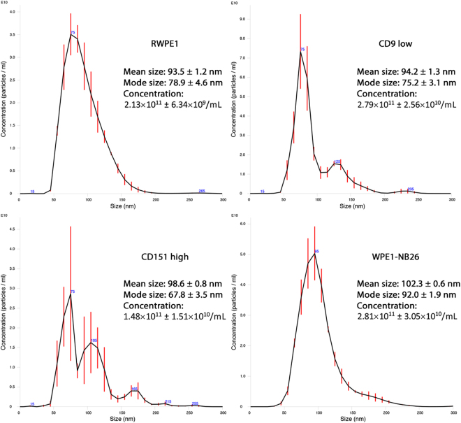
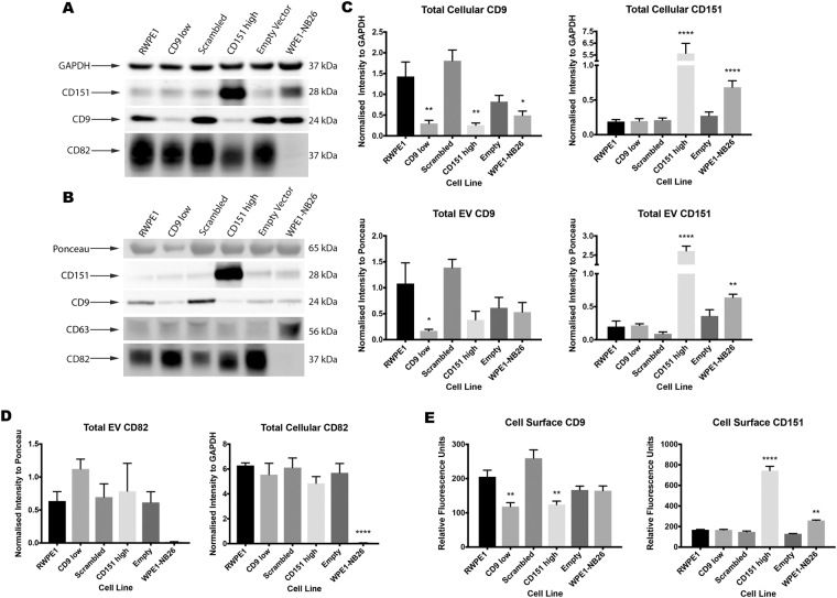
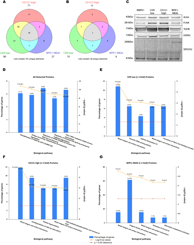
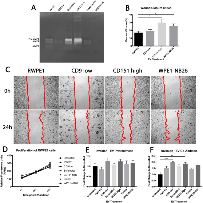
To facilitate intercellular communication, cells release nano-sized, extracellular vesicles (EVs) to transfer biological cargo to both local and distant sites. EVs are enriched in tetraspanins, two of which (CD9 and CD151) have altered expression patterns in many solid tumours, including prostate cancer, as they advance toward metastasis. We aimed to determine whether EVs from prostate cells with altered CD9 and CD151 expression could influence cellular behaviour and increase the metastatic capabilities of non-tumourigenic prostate cells. EVs were isolated by ultrafiltration and characterised for their tetraspanin expression and size distribution. iTRAQ was used to identify differences between RWPE1 and tetraspanin-modified RWPE1 EV proteomes, showing an enrichment in protein degradation pathways. Addition of EVs from RWPE1 cells with reduced CD9 or increased CD151 abundance resulted in increased invasion of RWPE1 cells, and increased migration in the case of high CD151 abundance. We have been able to show that alteration of CD9 and CD151 on prostate cells alters the proteome of their resultant EVs, and that these EVs can enhance the migratory and invasive capabilities of a non-tumourigenic prostate cellular population. This work suggests that cellular tetraspanin levels can alter EVs, potentially acting as a driver of metastasis in prostate cancer.


Conflict of interest statement

The authors declare no competing interests.

Figures

Figure 1
Figure 1
Average size distribution curve of EVs determined by NTA. Three 60 s videos were recorded for each sample, and NTA analysis settings kept constant between samples. The average concentration of vesicles was plotted against their size with error bars representing the standard deviation in concentration in 10 nm size increments. The mean and mode sizes and the average concentration are given ± standard deviation.
Figure 2
Figure 2
Cell and EV Tetraspanin Expression. (A) Cells were assessed for their total CD9, CD151 and CD82 content via western blot, with GAPDH used as a loading control. (B) EVs were assessed for their total CD9, CD151 and CD82 content via western blot, using Ponceau-S stained bands as a loading control. EVs were further assessed for the expression of CD63, a common EV marker. (C) Differences in CD9 and CD151 expression compared to RWPE1 cells and EVs were determined using one-way ANOVA with Bonferroni’s multiple comparisons test. CD9 low, CD151 high and WPE1-NB26 cells all showed a significant decrease in CD9 expression compared to RWPE1 cells, however only CD9 low EVs had a significant decrease in expression compared to RWPE1 EVs. Both CD151 high and WPE1-NB26 cells and EVs showed significant increases in CD151 expression compared to RWPE1 cells and EVs. (D) Differences in CD82 expression in cell and EV lysates were determined as above. WPE1-NB26 cells and EVs were negative for CD82 expression, whereas alterations in CD9 and CD151 expression did not significantly affect CD82 abundance in either cells or EVs. (E) Cell surface expression of CD9 and CD151 was determined using flow cytometry, and one-way ANOVA with Bonferroni’s multiple comparisons test was used to determine significance. CD9 low and CD151 high cells showed significant decreases in CD9 surface expression to RWPE1 cells. CD151 high and WPE1-NB26 cells showed significant increases in CD151 surface expression to RWPE1 cells. Data shown are representative of at least three independent experiments. *P < 0.05; **P < 0.01; ****P < 0.0001. Error bars are SEM. Western blot images are cropped, with full-length blots presented in Supplementary Fig. S7.
Figure 3
Figure 3
FunRich analysis of iTRAQ data. (A) Proteins with a ≥ 2-fold increase in expression and (B) a ≥ 2-fold decrease in expression when compared to RWPE1 EVs. Venn diagrams were created using the Public Research Centre for Health’s Venn diagram generator tool (http://www.bioinformatics.lu/venn.php). (C) Western blot analysis of Actin, Filamin-A, TGFBI, HSPA5 and SPOCK2 confirmed iTRAQ quantitated values. (C) Enriched biological pathways for all detected EV proteins. Degradation pathways were the predominant feature in the total dataset. (D) Enriched biological pathways for CD9 low EV proteins with a ≥ 2-fold increase in expression to RWPE1 EVs. (E) Enriched biological pathways for CD151 high EV proteins with a ≥ 2-fold increase in expression to RWPE1 EVs. (F) Enriched biological pathways for WPE1-NB26 EV proteins with a ≥ 2-fold increase in expression to RWPE1 EVs. Graphs were created using FunRich v2.1.2. which converts protein IDs to gene names. Western blot images are cropped, with full-length blots presented in Supplementary Fig. S8.
Figure 4
Figure 4
Migration and invasion of EV treated RWPE1 cells. (A) Gelatin zymography of EVs detected MMP2 and the pro- and active form of MMP9, with an increase in pro-MMP9 observed in Scrambled and CD151 high EVs. (B) Both CD151 high and WPE1-NB26 EV treated RWPE1 cells displayed enhanced wound closure using a Kruskal-Wallis test. (C) A wound healing assay was performed to determine the migratory capacity of RWPE1 cells treated with EVs. Red lines indicate the edge of the wound used for calculations. (D) A proliferation assay was performed to ensure that effects seen were not due to proliferation. No differences were seen in proliferation in EV treated RWPE1 cells. (E) Invasion assay of RWPE1 cells through a BME coating that was pretreated with 10 µg EVs for 24 h. No significant differences were observed compared to untreated. (F) Invasion assay of RWPE1 cells through a BME coating with co-addition of cells and EVs. CD9 low and CD151 high EVs significantly enhanced RWPE1 cell invasion when compared to untreated cells. Significance was determined using a Kruskal-Wallis test. All data are representative of three independent experiments. *P < 0.05; **P < 0.01; ***P < 0.001. Error bars are shown as SEM.

References

    1. Zöller, M. Tetraspanins and Cancer Metastasis. The Tumor Microenvironment (2010).
    1. Wang J-C, et al. Down-regulation of CD9 expression during prostate carcinoma progression is associated with CD9 mRNA modifications. Clinical Cancer Research. 2007;13:2354–2361. doi: 10.1158/1078-0432.CCR-06-1692. - DOI - PubMed
    1. Levina E, et al. Identification of novel genes that regulate androgen receptor signaling and growth of androgen-deprived prostate cancer cells. Oncotarget. 2015;6:13088–13104. doi: 10.18632/oncotarget.3743. - DOI - PMC - PubMed
    1. Ang J, Lijovic M, Ashman LK, Kan K, Frauman AG. CD151 protein expression predicts the clinical outcome of low-grade primary prostate cancer better than histologic grading: a new prognostic indicator? Cancer Epidemiol. Biomarkers Prev. 2004;13:1717–1721. - PubMed
    1. Detchokul S, Newell B, Williams ED, Frauman AG. CD151 is associated with prostate cancer cell invasion and lymphangiogenesis in vivo. Oncol. Rep. 2014;31:241–247. doi: 10.3892/or.2013.2823. - DOI - PubMed

Publication types

LinkOut - more resources