. 2018 Jun 1:(34):63-92.
doi: 10.3897/mycokeys.34.25974. eCollection 2018.

New Fusarium species from the Kruger National Park, South Africa

Affiliations

New Fusarium species from the Kruger National Park, South Africa

Marcelo Sandoval-Denis et al. MycoKeys. .

Abstract

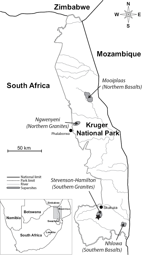
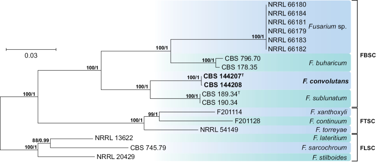
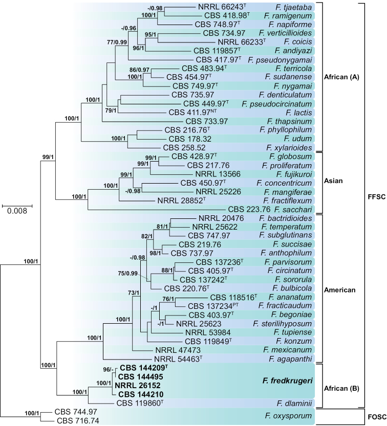
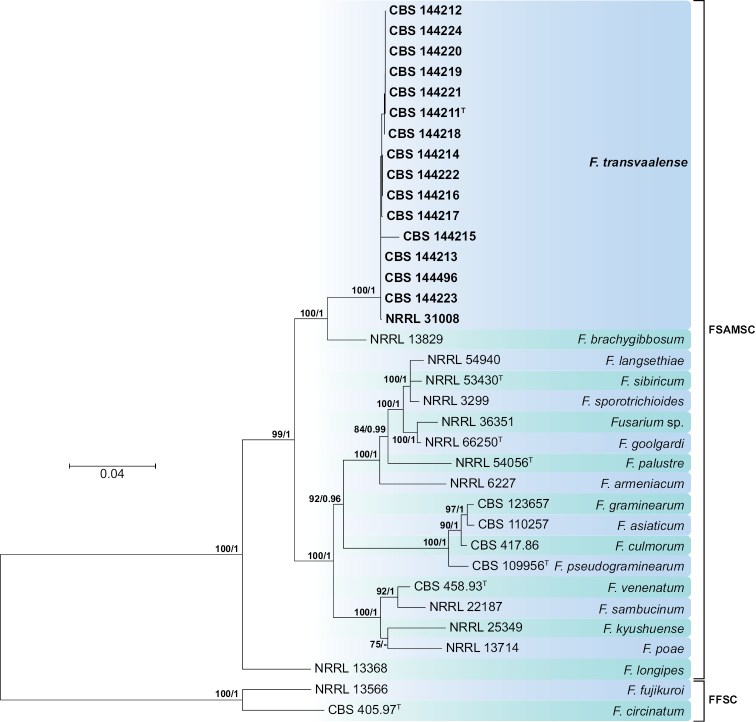
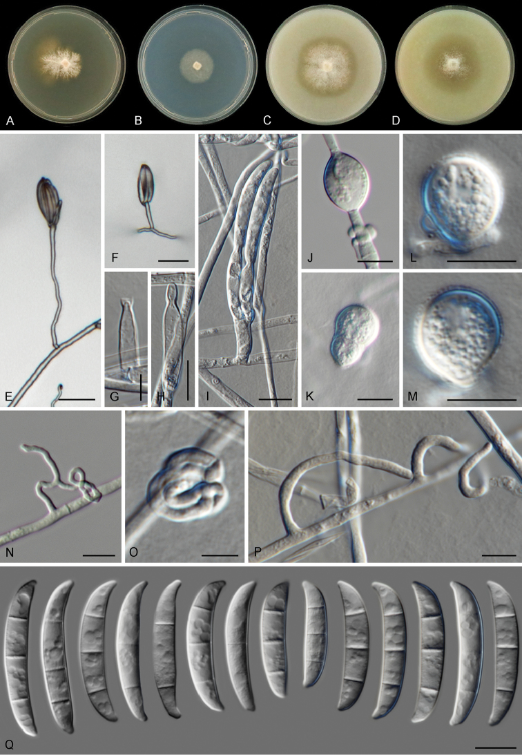
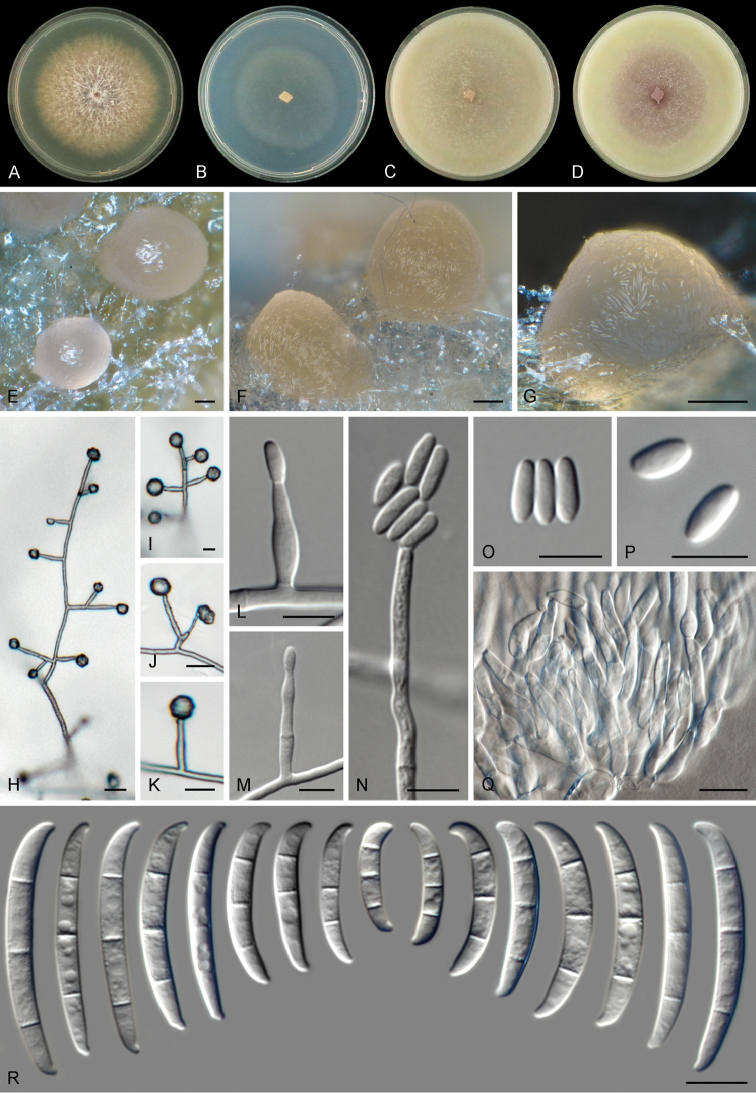
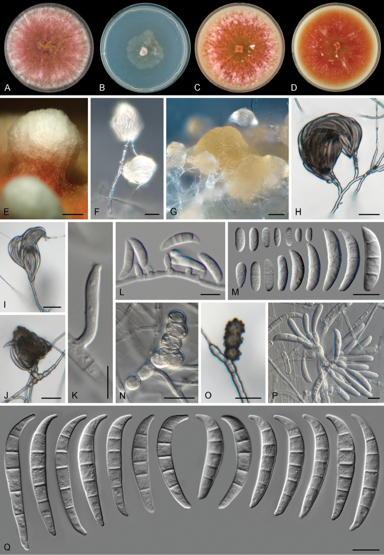
Three new Fusarium species, F. convolutans, F. fredkrugeri, and F. transvaalense (Ascomycota, Hypocreales, Nectriaceae) are described from soils collected in a catena landscape on a research supersite in the Kruger National Park, South Africa. The new taxa, isolated from the rhizosphere of three African herbaceous plants, Kyphocarpa angustifolia, Melhania acuminata, and Sida cordifolia, are described and illustrated by means of morphological and multilocus molecular analyses based on sequences from five DNA loci (CAL, EF-1 α, RPB1, RPB2 and TUB). According to phylogenetic inference based on Maximum-likelihood and Bayesian approaches, the newly discovered species are distributed in the Fusarium buharicum, F. fujikuroi, and F. sambucinum species complexes.

Keywords: Natural parks; diversity; fungi; morphology; multigene; phylogeny.


Figures

Figure 1.
Figure 1.
Map of the Kruger National Park (KNP) in South Africa. The arrows indicate the location of the four research “supersites” (adapted from Smit et al. 2013). Sampling site is indicated with a black star. The inset shows the location of the KNP within South Africa, indicated by a grey box.
Figure 2.
Figure 2.
Maximum-likelihood (ML) phylogram obtained from combined EF-1α, RPB1 and RPB2 sequences of 18 strains belonging to the Fusarium buharicum (FBSC), Fusarium tricinctum (FTSC) and Fusarium lateritium (FLSC) species complexes. Numbers on the nodes are ML bootstrap values above 70% and Bayesian posterior probability values above 0.95. Branch lengths are proportional to distance. Ex-type strains are indicated with T. Strains corresponding to new species described here are shown in bold.
Figure 3.
Figure 3.
Maximum-likelihood (ML) phylogram obtained from combined CAL, EF-1α, RPB1, RPB2 and TUB sequences of 48 strains belonging to the Fusarium fujikuroi (FFSC) and Fusarium oxysporum (FOSC) species complexes. Numbers on the nodes are ML bootstrap values above 70% and Bayesian posterior probability values above 0.95. Branch lengths are proportional to distance. Ex-type, ex-neotype and ex-paratype strains are indicated with T, NT and PT, respectively. Strains corresponding to new species described here are shown in bold.
Figure 4.
Figure 4.
Maximum-likelihood (ML) phylogram obtained from combined RPB1 and RPB2 sequences of 35 strains belonging to the Fusarium sambucinum (FSAMSC) and Fusarium fujikuroi (FFSC) species complexes. Numbers on the nodes are ML bootstrap values above 70% and Bayesian posterior probability values above 0.95. Branch lengths are proportional to distance. Ex-type strains are indicated with T. Strains corresponding to new species described here are shown in bold.
Figure 5.
Figure 5.
Fusarium convolutans sp. nov. A–D Colonies on PDA, SNA, OA and CMA, respectively, after 7 d at 24 °C in the dark E–I Conidiophores, phialides and conidia J–M Chlamydospores N–P Sterile hyphal projections Q Conidia. Scale bars: 20 μm (E, F); 5 μm (G–I); 10 μm (J–Q).
Figure 6.
Figure 6.
Fusarium fredkrugeri sp. nov. A–D Colonies on PDA, SNA, OA and CMA, respectively, after 7 d at 24 °C in the dark E–G Sporodochia formed on the surface of carnation leaves H–N Aerial conidiophores, phialides and conidia O, P Aerial conidia Q Sporodochial conidiophores and phialides R Sporodochial conidia. Scale bars: 100 μm (E–G); 10 μm (H–R).
Figure 7.
Figure 7.
Fusarium transvaalense sp. nov. A–D Colonies on PDA, SNA, OA and CMA, respectively, after 7 d at 24 °C in the dark E Pustule-like growth on OA F, G Sporodochia formed on the surface of carnation leaves H–L Aerial conidiophores phialides and conidia M Aerial conidia N, O Chlamydospores P Sporodochial conidiophores and phialides Q Sporodochial conidia. Scale bars: 2 mm (E); 20 μm (F–J); 5 μm (K); 10 μm (L–Q).

References

    1. Aoki T, Smith JA, Mount LL, Geiser DM, O’Donnell K. (2013) Fusarium torreyae sp. nov., a pathogen causing canker disease of Florida torreya (Torreya taxifolia), a critically endangered conifer restricted to northern Florida and southwestern Georgia. Mycologia 105: 312–319. https://doi.org/10.3852/12-262 - DOI - PubMed
    1. Aydogdu H, Asan A. (2008) Airborne fungi in child day care centers in Edirne City, Turkey. Environmental Monitoring and Assessment 147: 423–444. https://doi.org/10.1007/s10661-007-0130-4 - DOI - PubMed
    1. Bent E, Kiekel P, Brenton R, Taylor DL. (2011) Root-associated ectomycorrhizal fungi shared by various boreal forest seedlings naturally regenerating after a fire in interior Alaska and correlation of different fungi with host growth responses. Applied and Environmental Microbiology 77: 3351–3359. https://doi.org/10.1128/AEM.02575-10 - DOI - PMC - PubMed
    1. Brown DJ, Clayton MK, McSweeney K. (2004) Potential terrain controls on soil color, texture contrast and grain-size deposition for the original catena landscape in Uganda. Geoderma 122: 51–72. http://doi.org/10.1016/j.geoderma.2003.12.004 - DOI
    1. Burgess LW. (2014) 2011 McAlpine Memorial Lecture – A love affair with Fusarium. Australasian Plant Pathology 43: 359–368. https://doi.org/10.1007/s13313-013-0261-8 - DOI

LinkOut - more resources