. 2018 May 28:9:501.
doi: 10.3389/fphys.2018.00501. eCollection 2018.

Blood Shift During Cough: Negligible or Significant?

Affiliations

Blood Shift During Cough: Negligible or Significant?

Antonella LoMauro et al. Front Physiol. .

Abstract

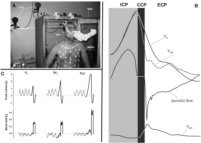
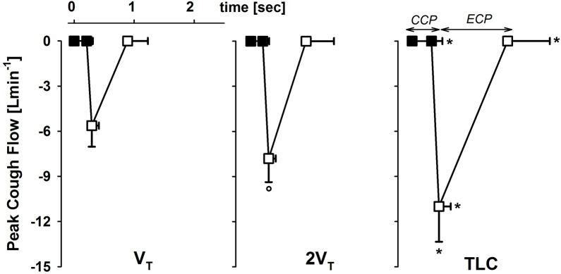
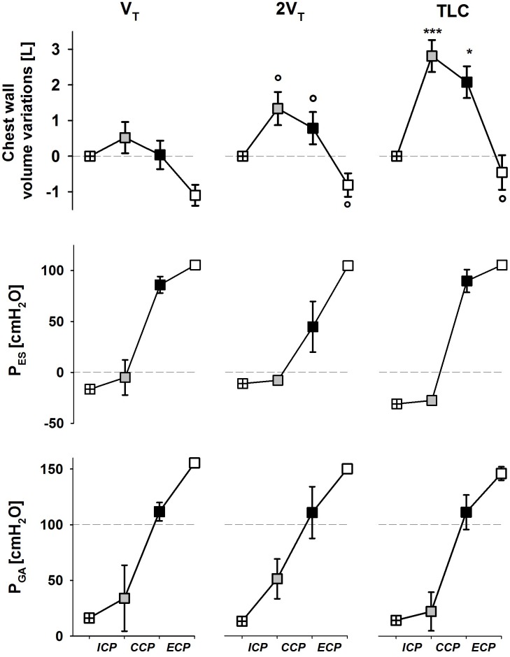
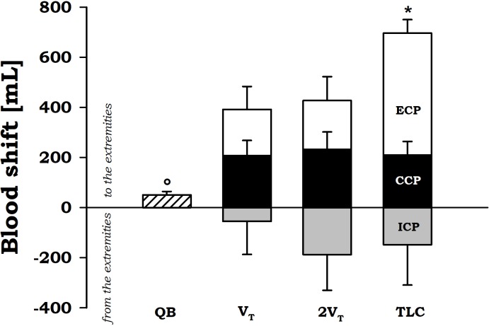
Rationale: It was reported how forceful rhythmic coughing can provide effective blood flow during ventricular fibrillation without direct chest compression. This mechanism of cough-assisted cardiopulmonary resuscitation constitutes a form of "cardiac massage" secondary to the intrathoracic and intra-abdominal pressure changes during cough. We have previously shown that significant blood shifts (BSs) occurs from the thorax to the extremities during expulsive maneuvers and that abdominal pressure controls the outflow of blood from the splanchnic vasculature. This mechanism was called abdominal circulatory pump. BS was quantified by using double body plethysmography (DBP), which combines total body plethysmography and opto-electronic plethysmography. Aim: We hypothesized that coughing activates also the abdominal circulatory pump, being an additional mechanism that displaces a circulatory output sufficient to maintain consciousness in a patient with a non-beating heart. Methods and Results: We studied seven healthy subjects (age: 28.6 ± 2.5 years) during series of voluntary coughs at three different operating volumes: after a spontaneous tidal volume, at total lung capacity (TLC) and at an intermediate volume. BS from the thorax to the extremities were measured by DBP during quiet breathing and during cough at each operating lung volume. BS during cough resulted significantly higher than during quiet breathing (p < 0.05). During the compressive phase, the blood outflow is around 200 ml, whereas during the expulsive phase BS increased (p < 0.05) with increasing operating volume, being almost 700 ml at TLC. At lower operating volume it is almost 400 ml. Conclusion: Deep, vigorous coughing and the consequent fluctuations in intra-thoracic and intra-abdominal pressure activate both the thoracic and the abdominal pump mechanism. The former leads the low-resistance pulmonary veins to empty into the left heart. The latter can generate a circulatory output from the splanchnic region, which acts as a blood reservoir, to other body tissues. These findings might help to better understand the cardiopulmonary interactions during cough, particularly in patients with unstable cardiac function, and the mechanism by which coughing during unstable cardiac rhythms can maintain consciousness in human subjects.

Keywords: blood flow; cardiopulmonary resuscitation; cough; double body plethysmography; physiology.


Figures

FIGURE 1
FIGURE 1
Double plethysmograph, traces and protocol. (A) Experimental set-up: the transparent whole body plethysmograph (WBP), the infra-red TV cameras (TVC), and the markers (MRKs) of optoelectronic plethysmography, and the pneumotacograph (PNT) to measure the flow at the mouth. (B) Time courses of body (VB) and trunk (VTR) volumes, flow measured at the mouth, and blood shift (VBS) during a single maximal voluntary cough. Gray area: inspiratory cough phase (ICP); black area: compressive cough phase (CCP); white area: expulsive cough phase (ECP). (C) Representative case of trunk volume changes (top panels) and blood shift (bottom panels) during spontaneous breathing (gray) and a single cough maneuver (black) at each operating lung volume: a tidal volume above functional residual capacity (VT, left panels), total lung capacity (TLC, right panels), and an intermediate volume (2VT, middle panels). VBS was calculated as the difference between trunk and body volume variations. Positive values of VBS indicate blood shifts occurring from the thorax to the extremities and vice versa. Written informed consent was obtained for the publication of the (A).
FIGURE 2
FIGURE 2
Average values ± standard deviation of peak cough flow at each operating lung volume: one tidal volume (VT) above functional residual capacity, left panel), total lung capacity (TLC, right panel), and an intermediate volume (2VT, middle panel). The x-axis displays the average values ± standard deviation of the duration of the compressive (CCP, close symbols) and the expulsive (ECP, open symbol) cough phases. p < 0.05 vs. both VT and 2VT; °p < 0.05 vs. VT.
FIGURE 3
FIGURE 3
Average values ± standard deviation of chest wall (trunk) volume (upper panels), esophageal (middle panels) and gastric (bottom panels) pressures at start inspiration (crossed symbols), end of the ICP (gray symbols), end of the CCP (black symbols), and end of the ECP (white symbols) at each operating lung volume: a tidal volume above functional residual capacity (VT, left panels), total lung capacity (TLC, right panels), and an intermediate volume (2VT, middle panels). ICP, inspiratory cough phase; CCP, compressive cough phase; ECP, expulsive cough phase. p < 0.05, ∗∗∗p < 0.001 vs. both VT and 2VT; °p < 0.05 vs. VT.
FIGURE 4
FIGURE 4
Average values ± standard error of blood shift during quiet breathing (QB) (measured during expiration), inspiratory cough phase (ICP, gray bars), compressive cough phase (CCP, black bars), and expiratory cough phase (ECP, white bars) at each operating lung volumes: a tidal volume above functional residual capacity (VT), total lung capacity (TLC), and an intermediate volume (2VT). p < 0.05 vs. both VT and 2VT; °p < 0.05 vs. both CCP and ECP.

References

    1. Aliverti A., Bovio D., Fullin I., Dellaca R. L., Lo Mauro A., Pedotti A., et al. (2009). The abdominal circulatory pump. PLoS One 4:e5550. 10.1371/journal.pone.0005550 - DOI - PMC - PubMed
    1. Aliverti A., Uva B., Laviola M., Bovio D., Lo Mauro A., Tarperi C., et al. (2010). Concomitant ventilatory and circulatory functions of the diaphragm and abdominal muscles. J. Appl. Physiol. 109 1432–1440. 10.1152/japplphysiol.00576.2010 - DOI - PubMed
    1. American Heart Association (2018). Cough CPR. Available at: http://www.heart.org/HEARTORG/Conditions/More/CardiacArrest/Cough-CPR_UC... [Accessed March 20, 2018].
    1. American Thoracic Society [ATS]/European Respiratory Society [ERS] (2002). ATS/ERS statement on respiratory muscle testing. Am. J. Respir. Crit. Care Med. 166 518–624. 10.1164/rccm.166.4.518 - DOI - PubMed
    1. Bergmann H. (1992). The scientific basis of cardiopulmonary and cerebral resuscitation. Anästhesiol. Intensivmed. Notfallmed. Schmerzther. 27 196–204. 10.1055/s-2007-1000281 - DOI - PubMed

LinkOut - more resources