. 2018 May 28:9:624.
doi: 10.3389/fphys.2018.00624. eCollection 2018.

Biochemical Comparison of dsRNA Degrading Nucleases in Four Different Insects

Affiliations

Biochemical Comparison of dsRNA Degrading Nucleases in Four Different Insects

Yingchuan Peng et al. Front Physiol. .

Abstract

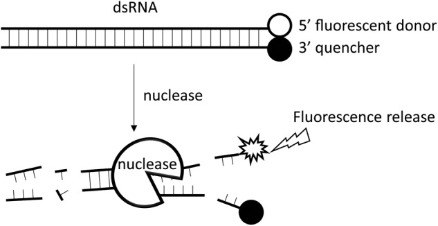
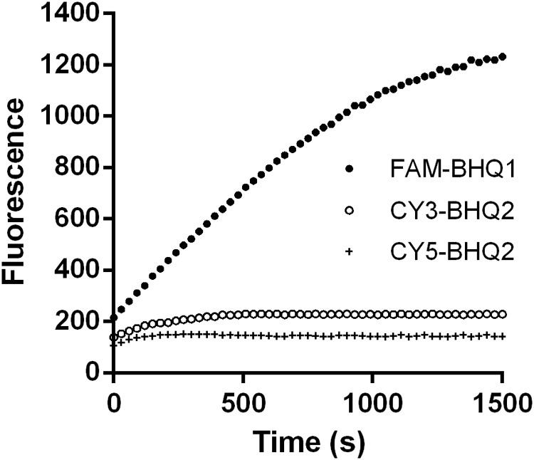
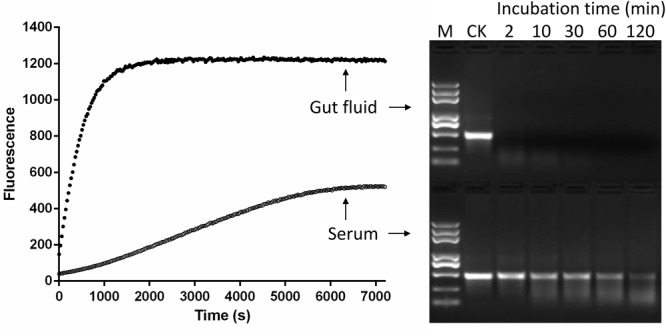
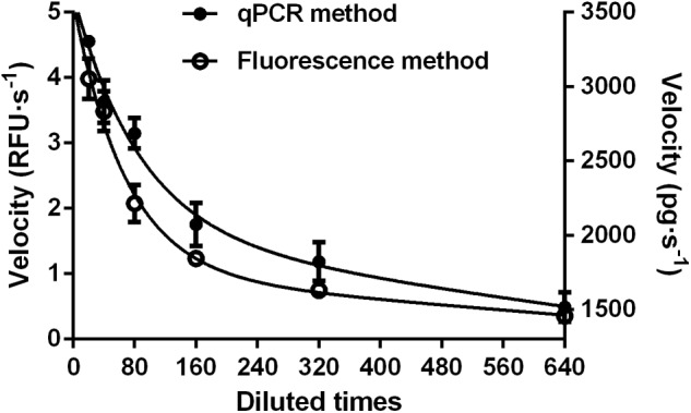
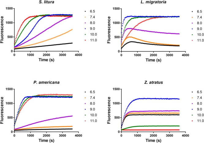
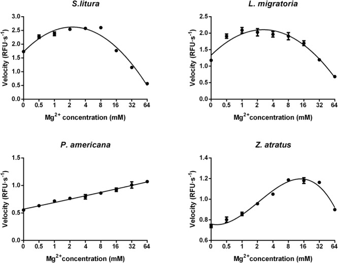
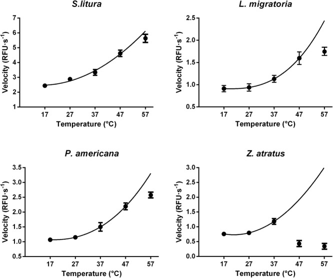
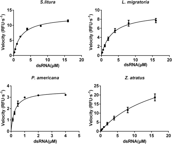
Double stranded RNAs (dsRNA) degrading nuclease is responsible for the rapid degradation of dsRNA molecules, and thus accounts for variations in RNA interference (RNAi) efficacy among insect species. Here, the biochemical properties and tissue-specific activities of dsRNA degrading nucleases in four insects (Spodoptera litura, Locusta migratoria, Periplaneta americana, and Zophobas atratus) from different orders were characterized using a modified assay method. The results revealed that all insect dsRNA degrading nucleases tested showed high activity in alkaline environments at optimal Mg2+ concentrations and elevated temperatures. We also found that enzymes from different insects varied in terms of their optimal reaction conditions and kinetic parameters. Whole body enzyme activity differed dramatically between insect species, although enzymes with higher substrate affinities (lower Km) were usually balanced by a smaller Vmax to maintain a proper level of degradative capacity. Furthermore, enzyme activities varied significantly between the four tested tissues (whole body, gut, hemolymph, and carcass) of the insect species. All the insects tested showed several hundred-fold higher dsRNA degrading activity in their gut than in other tissues. Reaction environment analysis demonstrated that physiological conditions in the prepared gut fluid and serum of different insects were not necessarily optimal for dsRNA degrading nuclease activity. Our data describe the biochemical characteristics and tissue distributions of dsRNA degrading activities in various insects, not only explaining why oral delivery of dsRNA often produces lower RNAi effects than injection of dsRNA, but also suggesting that dsRNA-degrading activities are regulated by physiological conditions. These results allow for a better understanding of the properties of dsRNA degrading nucleases, and will aid in the development of successful RNAi strategies in insects.

Keywords: RNA interference; dsRNA degrading enzyme; dsRNase; fluorescence; nuclease.


Figures

FIGURE 1
FIGURE 1
The working principle of the fluorescence method. A 24-bp synthesized dsRNA with a fluorophore group attached to the 3′ end of its sense strand and a quencher group to the 5′ end of its antisense strand is used as the substrate for analysis of dsRNA nucleases. No fluorescence is emitted when the substrate remain intact as the quencher group inhibits the fluorophore group. When dsRNA nuclease is added to the reaction tube, the dsRNA substrate will be gradually degraded, leading to separation of the fluorophore group from the quencher group, and thus the emission of the corresponding fluorescence light. The more the dsRNA degraded, the stronger the fluorescence signal.
FIGURE 2
FIGURE 2
Degradation time course of a 24-bp EGFP dsRNA substrate labeled with different fluorophores and quenchers. Three different combinations of fluorophore and quencher types were considered: 5-FAM fluorescent donor and 3-BHQ1 quencher-labeled strands (5-FAM/3-BHQ1), 5-Cy3 fluorescent donor and 3-BHQ2 quencher-labeled strands (5-Cy3/3-BHQ2), and 5-Cy5 fluorescent donor and 3-BHQ2 quencher-labeled strands (5-Cy5/3-BHQ2). The gut fluid from S. litura was 20-fold diluted (1 μL fluid + 19 μL nuclease-free water) and incubated with 1 μL of a 24-bp EGFP dsRNA labeled with different fluorophore/quencher combination (final concentration 0.5 μM) at 37°C. The florescence signal resulted from degradation of the dsRNA was monitored at 30 s intervals from 0 to 1500 s.
FIGURE 3
FIGURE 3
Comparison of fluorescent (Left) and gel electrophoresis (Right) methods for analyzing dsRNA degrading nucleases in S. litura serum and gut fluid. Both serum and gut fluid were diluted 20 times with nuclease free water, and the fluorescence reaction was incubated with 24 bp fluorescence labeled dsEGFP in the final concentration 0.5 μM. The gel electrophoresis reaction was incubated with 414 bp naked dsEGFP in the final concentration 0.05 μg/μL. CK: Control without enzymes. M: Trans 2K Plus DNA marker. Numbers 2–120 were the minutes indicating sample incubation time.
FIGURE 4
FIGURE 4
Comparison of fluorescent (Left) and qPCR (Right) methods for analyzing dsRNA degrading nucleases in S. litura gut fluid. Serial dilutions of the gut fluid (20, 40, 80, 160, 320, and 640 times) were incubated with 1 μL 24 bp fluorescence labeled dsEGFP or 1 μL 414 bp naked dsEGFP in a total volume of 20 μL at 37°C. The final substrate concentration was 0.5 μM for fluorescence method and 0.05 μg/μL for qPCR method. Values are mean ± SE; n = 3.
FIGURE 5
FIGURE 5
Distinct impacts of pH on the dsRNA degrading nucleases of four insects. The homogenates were prepared from whole body of different insects. The reaction buffer containing 0.1 M Glycine, 0.1 M NaCl, 1 mM MgCl2, 1 mM PTU, 1 mM DTT, 1 mM PMSF and 10% Glycerol was adjusted by KOH to different pH (6.5, 7.4, 8.0, 9.0, 10.0, and 11.0). Each reaction containing 19 μL enzyme solution and 1 μL fluorescence labeled dsRNA substrate at a final concentration of 0.5 μM. The fluorescence intensity in different reactions were continuously monitored at 37°C. Values are mean of fluorescence intensity; n = 3.
FIGURE 6
FIGURE 6
Distinct impacts of Mg2+ concentrations on the dsRNA degrading nucleases of four insects. The homogenates were prepared from whole body of different insects. Tested conditions were conducted under their optimal pH (pH 10.0 for S. litura and pH 9.0 for the other three species). Each reaction containing 19 μL enzyme solution and 1 μL fluorescence labeled dsRNA substrate at a final concentration of 0.5 μM. The fluorescence intensity in different reactions were continuously monitored at 37°C. Values are mean ± SE; n = 3.
FIGURE 7
FIGURE 7
Distinct impacts of temperature on the dsRNA degrading nucleases of four insects. The homogenates were prepared from whole body of different insects. The test reaction used the buffer with optimal pH and Mg2+ concentrations for different insect species (pH 10.0 and 8 mM Mg2+ for S. litura, pH 9.0 and 8 mM Mg2+ for the other three species). Each reaction containing 19 μL enzyme solution and 1 μL fluorescence labeled dsRNA substrate at a final concentration of 0.5 μM. The fluorescence intensity in different reactions were continuously monitored at different temperatures. Values are mean ± SE; n = 3.
FIGURE 8
FIGURE 8
Comparison of the saturation curves of the dsRNA degrading nuclease of four insects. The homogenates were prepared from whole body of different insects. The test reaction used the buffer with optimal pH and Mg2+ concentrations for different insect species (pH 10.0 and 8 mM Mg2+ for S. litura, pH 9.0 and 8 mM Mg2+ for the other three species). Different concentration of dsRNA substrates were incubated with 19 μL enzyme solutions in a total volume of 20 μL. The fluorescence intensity in different reactions were continuously monitored at 37°C. Values are mean ± SE; n = 3.

References

    1. Allen M. L., Walker W. B. (2012). Saliva of Lygus lineolaris digests double stranded ribonucleic acids. J. Insect Physiol. 58 391–396. 10.1016/j.jinsphys.2011.12.014 - DOI - PubMed
    1. Almeida Garcia R., Lima Pepino Macedo L., Cabral do Nascimento D., Gillet F.-X., Moreira-Pinto C. E., Faheem M., et al. (2017). Nucleases as a barrier to gene silencing in the cotton boll weevil, Anthonomus grandis. PLoS One 12:e0189600. 10.1371/journal.pone.0189600 - DOI - PMC - PubMed
    1. Arimatsu Y., Furuno T., Sugimura Y., Togoh M., Ishihara R., Tokizane M., et al. (2007a). Purification and properties of double-stranded RNA-degrading nuclease, dsRNase, from the digestive juice of the silkworm, Bombyx mori. J. Insect Biotechnol. Sericol. 76 57–62.
    1. Arimatsu Y., Kotani E., Sugimura Y., Furusawa T. (2007b). Molecular characterization of a cDNA encoding extracellular dsRNase and its expression in the silkworm, Bombyx mori. Insect Biochem. Mol. Biol. 37 176–183. 10.1016/j.ibmb.2006.11.004 - DOI - PubMed
    1. Baum J. A., Bogaert T., Clinton W., Heck G. R., Feldmann P., Ilagan O., et al. (2007). Control of coleopteran insect pests through RNA interference. Nat. Biotechnol. 25 1322–1326. 10.1038/nbt1359 - DOI - PubMed

LinkOut - more resources