Review
. 2018 May 28:9:368.
doi: 10.3389/fneur.2018.00368. eCollection 2018.

Abnormalities in Skeletal Muscle Myogenesis, Growth, and Regeneration in Myotonic Dystrophy

Affiliations
Review

Abnormalities in Skeletal Muscle Myogenesis, Growth, and Regeneration in Myotonic Dystrophy

Laurène M André et al. Front Neurol. .

Abstract

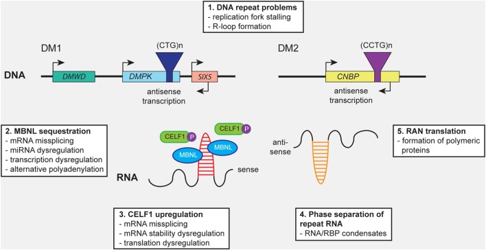
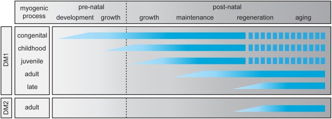
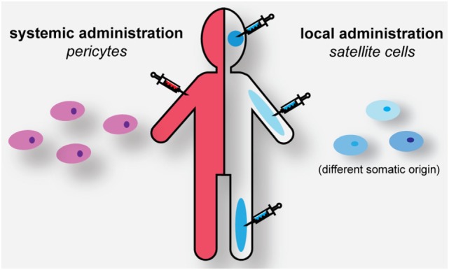
Myotonic dystrophy type 1 (DM1) and 2 (DM2) are autosomal dominant degenerative neuromuscular disorders characterized by progressive skeletal muscle weakness, atrophy, and myotonia with progeroid features. Although both DM1 and DM2 are characterized by skeletal muscle dysfunction and also share other clinical features, the diseases differ in the muscle groups that are affected. In DM1, distal muscles are mainly affected, whereas in DM2 problems are mostly found in proximal muscles. In addition, manifestation in DM1 is generally more severe, with possible congenital or childhood-onset of disease and prominent CNS involvement. DM1 and DM2 are caused by expansion of (CTG•CAG)n and (CCTG•CAGG)n repeats in the 3' non-coding region of DMPK and in intron 1 of CNBP, respectively, and in overlapping antisense genes. This critical review will focus on the pleiotropic problems that occur during development, growth, regeneration, and aging of skeletal muscle in patients who inherited these expansions. The current best-accepted idea is that most muscle symptoms can be explained by pathomechanistic effects of repeat expansion on RNA-mediated pathways. However, aberrations in DNA replication and transcription of the DM loci or in protein translation and proteome homeostasis could also affect the control of proliferation and differentiation of muscle progenitor cells or the maintenance and physiological integrity of muscle fibers during a patient's lifetime. Here, we will discuss these molecular and cellular processes and summarize current knowledge about the role of embryonic and adult muscle-resident stem cells in growth, homeostasis, regeneration, and premature aging of healthy and diseased muscle tissue. Of particular interest is that also progenitor cells from extramuscular sources, such as pericytes and mesoangioblasts, can participate in myogenic differentiation. We will examine the potential of all these types of cells in the application of regenerative medicine for muscular dystrophies and evaluate new possibilities for their use in future therapy of DM.

Keywords: RNA toxicity; mesoangioblast; muscle stem cell; myoblast; myogenesis; myotonic dystrophy; pericyte; proteotoxicity.


Figures

Figure 1
Figure 1
Distinct molecular mechanisms contribute to pathology in myotonic dystrophy type 1 (DM1) and myotonic dystrophy type 2 (DM2). (1) Expanded (CTG)n and (CCTG)n repeats in DMPK and CNBP, or the complementary repeats in the antisense genes (not shown), can cause cellular stress by (1) promoting DNA replication fork stalling and R-loop formation. Expression of repeat-containing sense and antisense RNAs results in (2) sequestration of members of the MBNL protein family, leading to mRNA missplicing, alternative polyadenylation, microRNA (miRNA) deregulation, and transcription deregulation. In addition, (3) CELF1 gets hyperphosphorylated and stabilized, resulting in mRNA missplicing and dysregulation of mRNA stability and translation. (4) Formation of abnormal RNA-protein condensates by repeat RNA and RNA-binding proteins (RBPs) may alter the intracellular distribution fate and biological activity of RBPs. (5) Repeat-associated non-ATG (RAN) translation of the repeats may result in the production of toxic polymeric polypeptides, which perturb cellular proteostasis.
Figure 2
Figure 2
Abnormalities in skeletal muscle myogenesis in myotonic dystrophy (DM). For myotonic dystrophy type 1 (DM1), five clinical subtypes have been identified (19), while for myotonic dystrophy type 2 (DM2) only the adult-onset manifestation is known. The myogenic process in skeletal muscle is divided in two prenatal and four postnatal stages. The graphic summarizes which stages of the pre- and postnatal myogenic process are affected in each clinical DM (sub)type. A discontinuous bar indicates decreased life expectancy.
Figure 3
Figure 3
Skeletal muscle growth, maintenance, and repair by different myogenic progenitor cells. Satellite cells from the basal lamina of the myofiber are activated and undergo asymmetric and symmetric division to generate heterogeneous progeny. Some cells undergo self-renewal and return to quiescence, others become myoblast that will proliferate and differentiate to become myocytes, which fuse to myofibers, enabling repair and/or growth. Mesoangioblasts (MABs) can contribute to muscle regeneration during embryonic growth, while pericytes (PCs) are involved in postnatal muscle growth by repopulating the quiescent stem cell population or maybe by transforming into a myoblast. Participation in growth and/or repair or direct fusion with the myofiber probably occurs along the same pathways as given for satellite cells. Uncertainties in cell fate are indicated by dashed arrows. PW1+ interstitial cells (PICs) are mostly involved in perinatal growth. Expression signatures of differentiation markers in all different cell types are listed at the bottom.
Figure 4
Figure 4
Strategies for cell-based muscle therapy in myotonic dystrophy. Genome-edited autologous or HLA-matched pericytes (PCs) can be administered systemically for muscle healing. Genome-edited or HLA-matched satellite cells need to be engrafted locally in the corresponding muscles to have a regenerative effect.

Similar articles

Cited by

References

    1. Meola G, Cardani R. Myotonic dystrophy type 2 and modifier genes: an update on clinical and pathomolecular aspects. Neurol Sci (2017) 38:535–46.10.1007/s10072-016-2805-5 - DOI - PubMed
    1. Gourdon G, Meola G. Myotonic dystrophies: state of the art of new therapeutic developments for the CNS. Front Cell Neurosci (2017) 11:101.10.3389/fncel.2017.00101 - DOI - PMC - PubMed
    1. Yum K, Wang ET, Kalsotra A. Myotonic dystrophy: disease repeat range, penetrance, age of onset, and relationship between repeat size and phenotypes. Curr Opin Genet Dev (2017) 44:30–7.10.1016/j.gde.2017.01.007 - DOI - PMC - PubMed
    1. Sicot G, Gourdon G, Gomes-Pereira M. Myotonic dystrophy, when simple repeats reveal complex pathogenic entities: new findings and future challenges. Hum Mol Genet (2011) 20:116–23.10.1093/hmg/ddr343 - DOI - PubMed
    1. Mateos-Aierdi AJ, Goicoechea M, Aiastui A, Fernández-Torrón R, Garcia-Puga M, Matheu A, et al. Muscle wasting in myotonic dystrophies: a model of premature aging. Front Aging Neurosci (2015) 7:125.10.3389/fnagi.2015.00125 - DOI - PMC - PubMed

LinkOut - more resources