

Reduced graphene oxide-loaded nanocomposite scaffolds for enhancing angiogenesis in tissue engineering applications
- PMID: 29892387
- PMCID: PMC5990794
- DOI: 10.1098/rsos.172017
Reduced graphene oxide-loaded nanocomposite scaffolds for enhancing angiogenesis in tissue engineering applications
Abstract
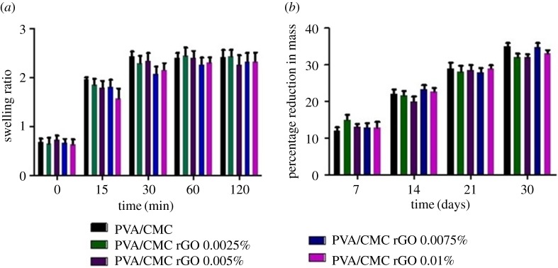
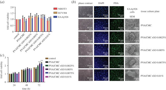
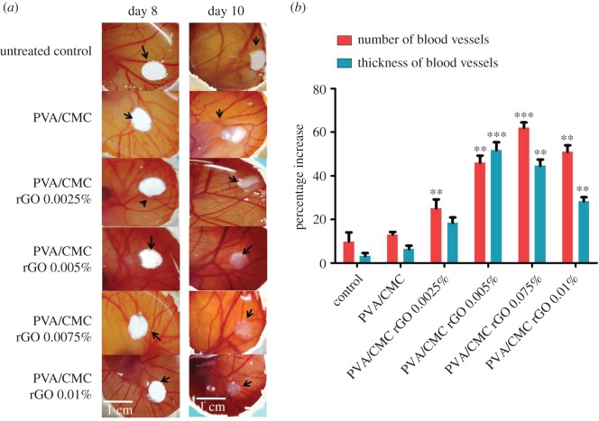
Tissue engineering combines cells, scaffolds and signalling molecules to synthesize tissues in vitro. However, the lack of a functioning vascular network severely limits the effective size of a tissue-engineered construct. In this work, we have assessed the potential of reduced graphene oxide (rGO), a non-protein pro-angiogenic moiety, for enhancing angiogenesis in tissue engineering applications. Polyvinyl alcohol/carboxymethyl cellulose (PVA/CMC) scaffolds loaded with different concentrations of rGO nanoparticles were synthesized via lyophilization. Characterization of these scaffolds showed that the rGO-loaded scaffolds retained the thermal and physical properties (swelling, porosity and in vitro biodegradation) of pure PVA/CMC scaffolds. In vitro cytotoxicity studies, using three different cell lines, confirmed that the scaffolds are biocompatible. The scaffolds containing 0.005 and 0.0075% rGO enhanced the proliferation of endothelial cells (EA.hy926) in vitro. In vivo studies using the chick chorioallantoic membrane model showed that the presence of rGO in the PVA/CMC scaffolds significantly enhanced angiogenesis and arteriogenesis.
Keywords: angiogenesis; nanocomposite scaffolds; reduced graphene oxide; tissue engineering; vascularization.
Conflict of interest statement
The authors have no competing interests.
Figures
References
-
- Bae H, Puranik AS, Gauvin R, Edalat F, Carrillo-Conde B, Peppas NA, Khademhosseini A. 2012. Building vascular networks. Sci. Transl. Med. 4, 160ps123 (doi:10.1126/scitranslmed.3003688) - DOI - PMC - PubMed
-
- Lovett M, Lee K, Edwards A, Kaplan DL. 2009. Vascularization strategies for tissue engineering. Tissue Eng. Part B Rev. 15, 353–370. (doi:10.1089/ten.TEB.2009.0085) - DOI - PMC - PubMed
-
- Hunziker E, et al. 2006. Translation from research to applications. Tissue Eng. 12, 3341–3364. (doi:10.1089/ten.2006.12.3341) - DOI - PubMed
-
- Malda J, Klein TJ, Upton Z. 2007. The roles of hypoxia in the in vitro engineering of tissues. Tissue Eng. 13, 2153–2162. (doi:10.1089/ten.2006.0417) - DOI - PubMed
-
- Griffith CK, Miller C, Sainson RC, Calvert JW, Jeon NL, Hughes CC, George SC. 2005. Diffusion limits of an in vitro thick prevascularized tissue. Tissue Eng. 11, 257–266. (doi:10.1089/ten.2005.11.257) - DOI - PubMed
Associated data
LinkOut - more resources
Full Text Sources
Other Literature Sources
Miscellaneous