. 2018 May 16;5(5):172293.
doi: 10.1098/rsos.172293. eCollection 2018 May.

Enamel formation and growth in non-mammalian cynodonts

Affiliations

Enamel formation and growth in non-mammalian cynodonts

Rachel N O'Meara et al. R Soc Open Sci. .

Abstract

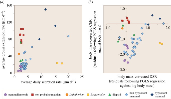
The early evolution of mammals is associated with the linked evolutionary origin of diphyodont tooth replacement, rapid juvenile growth and determinate adult growth. However, specific relationships among these characters during non-mammalian cynodont evolution require further exploration. Here, polarized light microscopy revealed incremental lines, resembling daily laminations of extant mammals, in histological sections of enamel in eight non-mammalian cynodont species. In the more basal non-probainognathian group, enamel extends extremely rapidly from cusp to cervix. By contrast, the enamel of mammaliamorphs is gradually accreted, with slow rates of crown extension, more typical of the majority of non-hypsodont crown mammals. These results are consistent with the reduction in dental replacement rate across the non-mammalian cynodont lineage, with greater rates of crown extension required in most non-probainognathians, and slower crown extension rates permitted in mammaliamorphs, which have reduced patterns of dental replacement in comparison with many non-probainognathians. The evolution of mammal-like growth patterns, with faster juvenile growth and more abruptly terminating adult growth, is linked with this reduction in dental replacement rates and may provide an additional explanation for the observed pattern in enamel growth rates. It is possible that the reduction in enamel extension rates in mammaliamorphs reflects an underlying reduction in skeletal growth rates at the time of postcanine formation, due to a more abruptly terminating pattern of adult growth in these more mammal-like, crownward species.

Keywords: cynodont; dental histology; enamel development; enamel increment; mammaliaform; mammaliamorph.


Conflict of interest statement

The authors declare no competing interests.

Figures

Figure 1.
Figure 1.
The relationships between non-mammalian cynodonts investigated in this study, and the acquisition of characteristics related to growth and dentition in these animals. Teeth were sectioned from all species with the exception of Sinoconodon. In statistical tests, non-mammalian cynodonts were assigned to two different groups, non-probainognathians (red) and mammaliamorphs (purple) and compared with diapsids, non-hypsodont crown mammals and hypsodont mammals. Irajatherium was not assigned to any group as its enamel showed a pattern of growth intermediate between non-probainognathians and mammaliamorphs. Exaeretodon was excluded from statistical analyses due to its extremely unusual pattern of enamel deposition. Phylogeny after Ruta et al. [31].
Figure 2.
Figure 2.
The periodic increments of mammalian enamel and their development. Striae of Retzius (green) represent the forming front of enamel as it is deposited from cusp to cervix along the EDJ. Cross-striations and laminations both have a daily periodicity, but laminations follow a course parallel to the striae of Retzius, while cross-striations are formed perpendicular to enamel prisms, and thus may be oblique to striae of Retzius. Enamel is secreted by ameloblasts moving progressively further from the EDJ. Prismatic enamel is produced by the distal Tomes' process (red), and interprismatic enamel by more proximal portions of the Tomes' process (blue). In extant mammals, the distal Tomes' process is initially absent during early enamel development and also regresses towards the end of development. Thus, during these stages of development, enamel is produced across a relatively flat, ‘proximal', secretory surface, forming regions of interprismatic enamel close to the EDJ and outer enamel surface. Laminations are most frequently observed in these interprismatic regions in extant mammals, forming in line with the flat secretory surface of the ameloblasts. Lower frames of figure follow Smith [3].
Figure 3.
Figure 3.
Quantifying CER. Black lines indicate long-period incremental lines of the enamel. The distance along a prism, A, from the EDJ to a particular accentuated line, B, was divided by the average cross-striation length (DSR) along that prism to yield x days to form that length of prism. By following the accentuated line B (which represents the forming front of enamel at a particular time) back to its intersection with the EDJ, the length of EDJ, y, from its intersection with stria B and its intersection with prism A can be measured. Length y therefore represents the increase in EDJ length after x days. Daily extension rate is, therefore, y/x. Selecting a new prism, intersecting the EDJ at the same point as stria B, allows the process to be repeated in an additive manner along the entire length of the EDJ.
Figure 4.
Figure 4.
Polarized light microscope images of incremental lines in the enamel of postcanines of non-mammalian cynodonts. Red arrows indicate long-period incremental lines, and white (a,c,e) or black arrows (b,d) indicate short-period incremental lines. In all species of non-mammalian cynodont, there appears to be one short-period line between any two adjacent long-period lines.(a) Diademodon sp. (UMZC T.486), (b) Massetognathus pascuali (CRILAR-C 162.2), (c) Irajatherium hernandezi (UFRGS-PV-1348-T-S1), (d) Oligokyphus sp. (UMZC 1236a), (e) Morganucodon watsoni (UMZC 2017.4.1). Scale bars = 10 µm. Outer enamel surface to the top. Cervix to the left. Image (e) slightly out of focus to visualize both long- and short-period increments in the same image as they are not in precisely the same plane within the section.
Figure 5.
Figure 5.
Polarized light microscope image of incremental lines in the enamel (top) and dentine (bottom) of a postcanine of the non-mammalian cynodont Diademodon sp. Coloured arrows indicate daily lines of von Ebner in the dentine. Incremental lines in both the enamel and the dentine can be seen along the same length of EDJ in this specimen, and lines of the same colour indicate equivalent daily lines in the dentine and enamel. Scale bar, 100 µm. Cervix to the right.
Figure 6.
Figure 6.
Enamel growth rates from different individual teeth belonging to different amniote groups, assuming a 2-day periodicity between long-period lines in non-mammalian cynodonts. (a) Raw growth rates: average DSR (μm d−1) versus average CER (μm d−1). (b) Body mass-corrected growth rates: the residuals of log DSR (µm d−1) and log CER (µm d−1) following phylogenetic generalized least-squares regression against log body mass. The growth rates of individual teeth were included as separate data points, rather than using the species average.
Figure 7.
Figure 7.
Polarized light microscope images of incremental lines in the enamel of postcanines of the mammaliamorph M. watsoni UMZC 2017.4.1 (a) and the non-probainognathian Diademodon sp. UMZC T.486 (b). Red arrows indicate prominent long-period incremental lines, with the paths of some traced for emphasis. White lines indicate the EDJ. Long-period lines of Morganucodon approach the EDJ at a steeper angle of inclination, indicating more gradual accretion of enamel than in Diademodon, in which enamel is rapidly extended, causing long-period lines to approach the EDJ at a very small angle. Scale bars, 50 µm. Cervix to the left in both images.

Similar articles

Cited by

References

    1. Dean MC. 1987. Growth layers and incremental markings in hard tissues: a review of the literature and some preliminary observations about enamel structure in Paranthropus boisei. J. Hum. Evol. 16, 157–172. (doi:10.1016/0047-2484(87)90074-1) - DOI
    1. Dean MC. 1995. The nature and periodicity of incremental lines in primate dentine and their relationship to periradicular bands in OH 16 (Homo habilis). In Aspects of dental biology: paleontology, anthropology and evolution (ed. Moggi-Cecchi J.), pp. 239–265. Florence, Italy: International Institute for the Study of Man.
    1. Smith TM. 2006. Experimental determination of the periodicity of incremental features in enamel. J. Anat. 208, 99–113. (doi:10.1111/j.1469-7580.2006.00499.x) - DOI - PMC - PubMed
    1. Smith TM. 2008. Incremental dental development: methods and applications in hominoid evolutionary studies. J. Hum. Evol. 54, 205–224. (doi:10.1016/j.jhevol.2007.09.020) - DOI - PubMed
    1. Shellis RP. 1998. Utilization of periodic markings in enamel to obtain information on tooth growth. J. Hum. Evol. 35, 387–400. (doi:10.1006/jhev.1998.0260) - DOI - PubMed

LinkOut - more resources