. 2018 Jun 7;5(4):e466.
doi: 10.1212/NXI.0000000000000466. eCollection 2018 Jul.

Molecular signature of Epstein-Barr virus infection in MS brain lesions

Affiliations

Molecular signature of Epstein-Barr virus infection in MS brain lesions

Monica A Moreno et al. Neurol Neuroimmunol Neuroinflamm. .

Abstract

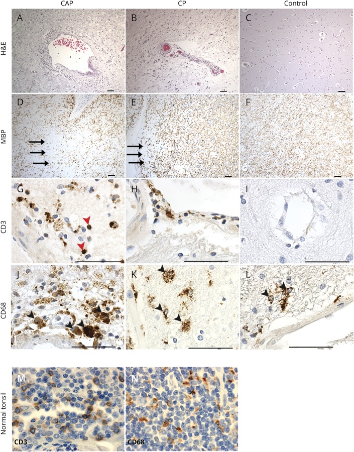
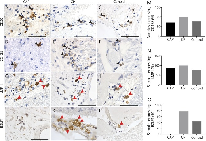
Objective: We sought to confirm the presence and frequency of B cells and Epstein-Barr virus (EBV) (latent and lytic phase) antigens in archived MS and non-MS brain tissue by immunohistochemistry.

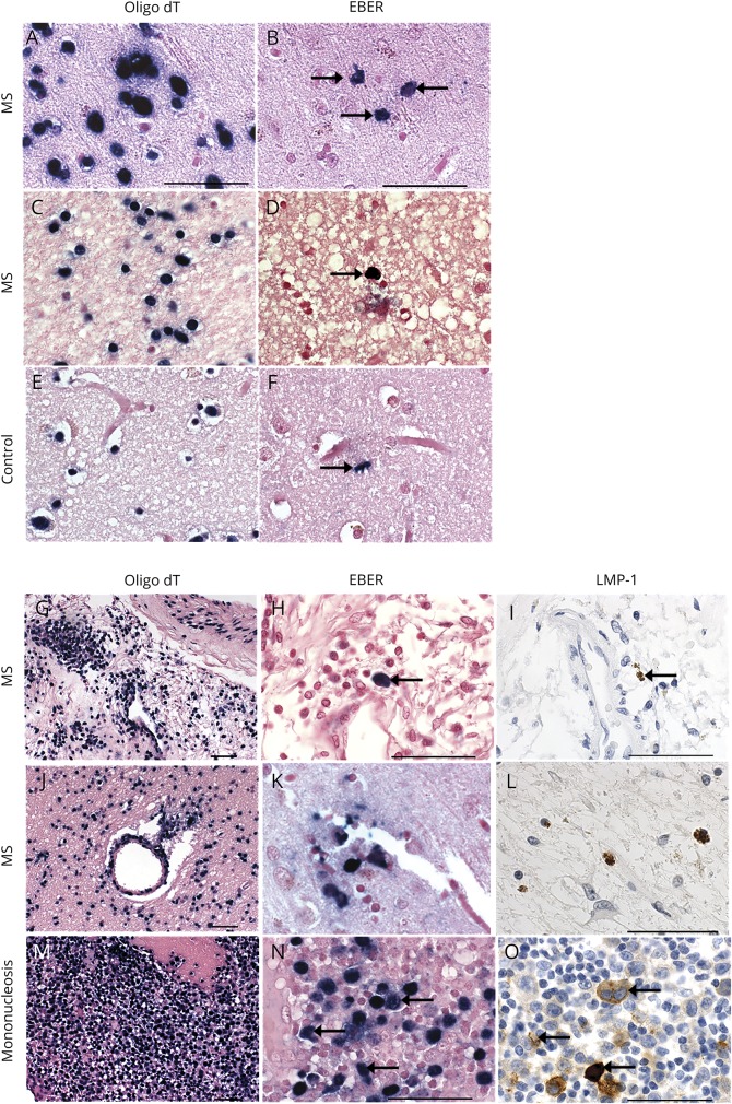
Methods: We quantified the type and location of B-cell subsets within active and chronic MS brain lesions in relation to viral antigen expression. The presence of EBV-infected cells was further confirmed by in situ hybridization to detect the EBV RNA transcript, EBV-encoded RNA-1 (EBER-1).

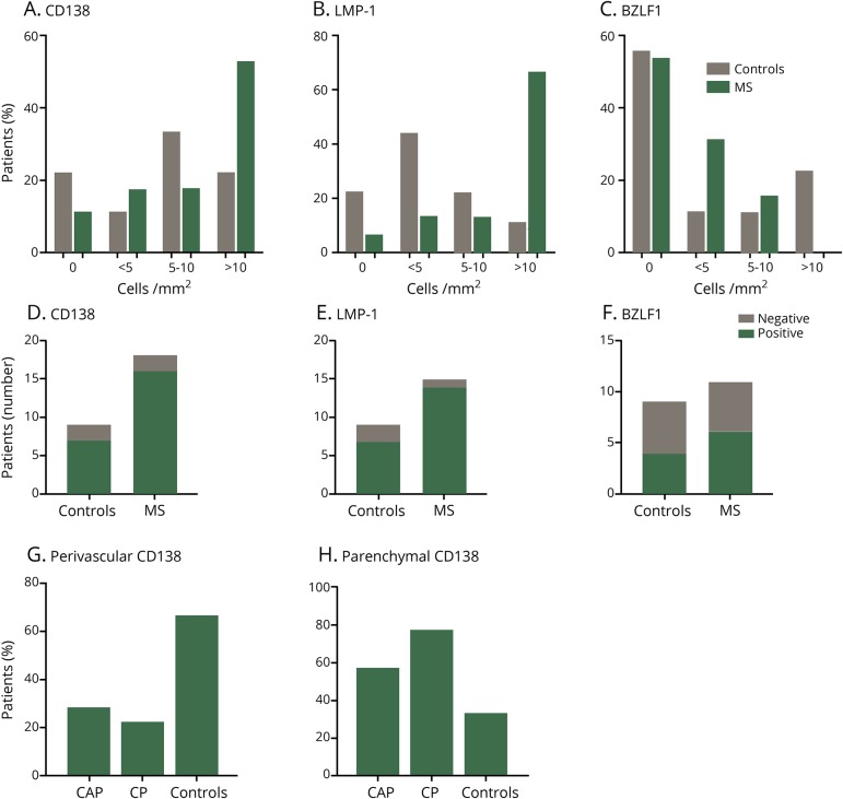
Results: We report the presence of EBV latent membrane protein 1 (LMP-1) in 93% of MS and 78% of control brains, with a greater percentage of MS brains containing CD138+ plasma cells and LMP-1-rich populations. Notably, 78% of chronic MS lesions and 33.3% of non-MS brains contained parenchymal CD138+ plasma cells. EBV early lytic protein, EBV immediate-early lytic gene (BZLF1), was also observed in 46% of MS, primarily in association with chronic lesions and 44% of non-MS brain tissue. Furthermore, 85% of MS brains revealed frequent EBER-positive cells, whereas non-MS brains seldom contained EBER-positive cells. EBV infection was detectable, by immunohistochemistry and by in situ hybridization, in both MS and non-MS brains, although latent virus was more prevalent in MS brains, while lytic virus was restricted to chronic MS lesions.

Conclusions: Together, our observations suggest an uncharacterized link between the EBV virus life cycle and MS pathogenesis.


Figures

Figure 1
Figure 1. Histopathologic features of a chronic active and a chronic plaque in the MS brain
Representative hematoxylin & eosin (H&E) staining (A–C) and immunohistochemistry (D–L) of active MS (A, D, G, J), chronic MS (B, E, H K), and healthy control (C, F, I, L) brain samples. Perivascular (PV) inflammation (A and B), demyelination, as indicated by the loss of MBP staining (black arrows) (D and E), presence of inflammatory cells, CD3+ T lymphocytes (red arrowheads) were prominent within active MS lesions (G) and present to a lesser extent in chronic MS lesions (H). Numerous macrophages/microglia (black arrowheads) were observed in and around PV cuffs in active MS (J) and were also observed chronic MS lesions, although to a lesser extent (K). Healthy controls, without neurologic disease, showed little or no CD3+ immunoreactivity (I) and positive CD68+ immunostaining in the parenchyma resembling resident microglia (black arrowheads) (L). Normal tonsils were used as a positive control and show CD3+ (M) and CD68+ (N) immunostaining. CAP = chronic active plaque, CP = chronic plaque. Scale bars = 50 μm.
Figure 2
Figure 2. Immunohistological detection of EBV latent and early lytic proteins in MS and control brains
CD20+ B lymphocytes (A–C) and CD138+ plasma cells (black arrowheads) (D–F) in the parenchyma and vasculature in a chronic active plaque (CAP) (A and D), in a chronic plaque (CP) (B and E), and in the vasculature of a healthy control brain sample (C and F). Percentage of CAP, CP, and control brain samples expressing CD138 protein detectable by immunohistochemistry are shown (M). We observed latent membrane protein-1 (LMP-1) expression (red arrowheads) in CAP, CP, and control brain samples (G–I). Percentage of CAP, CP, and control brain samples expressing LMP-1 protein detectable by immunohistochemistry are shown (N). Cells expressing LMP-1 (red arrowheads) were found in the vasculature of CAPs (G) and control brains (I), and within the parenchyma in a CP (H). The expression of the viral immediate-early protein BZLF1 was not observed in a CAP (J) and was observed in and around the vasculature in tissue from a CP (red arrowheads) (K). BZLF1 was also observed to a lesser extent in healthy controls (red arrowheads) (L). Percentage of CAP, CP, and control brain samples expressing BZLF1 protein detectable by immunohistochemistry are shown (O). Scale bar = 50 μm. Pictures are representative of analysis from 17 MS samples and 9 healthy controls. EBV = Epstein-Barr virus.
Figure 3
Figure 3. Immunohistochemical analyses of positive and negative Epstein-Barr virus (EBV) control tissues using immunostaining
Pictures are representative of results from 2 different experiments using EBV-positive (tonsil with infectious mononucleosis) and EBV-negative (normal tonsil) control samples. Immunohistochemistry revealed the presence of CD20+ B lymphocytes (A–B) and CD138+ plasma cells (C–D) in our positive and negative control tissue (A–D). LMP-1+ (brown staining) and BZLF1+ (brown staining emphasized with black arrows) cells are shown in a tonsil from a patient with infectious mononucleosis (E, G). No LMP-1+ or BZLF1+ cells were observed in our negative control tissue (normal tonsil) (F, H). Scale bar = 50 μm.
Figure 4
Figure 4. Detection of EBER+ cells in MS and control brains by in situ hybridization
In situ hybridization for EBER detects EBER+ cells (blue-black nuclei, black arrows) in 4 representative MS brains (B, D, H, and K) and 1 representative control brain (F). In situ hybridization for EBER and immunohistochemistry for LMP-1 show EBER+ and LMP-1+ cells in the same region or nearby regions in 2 representative MS brains (G–L). Tonsil tissue from a patient with infectious mononucleosis (M–O) shows colocalization of EBER and LMP-1. RNA preservation in samples is corroborated by in situ hybridization for oligo dT in serial sections from the same MS and control samples (A, C, E, G, J, and M). Scale bar = 50 μm.
Figure 5
Figure 5. Increased frequency of parenchymal CD138- and LMP-1–positive cells in MS
Formalin-fixed paraffin embedded brain tissue from MS and control brains without neurologic disease were cut into 4-μm sections. Hematoxylin and eosin (H&E) and immunohistochemistry were performed using antibodies against latent membrane protein 1 (LMP-1), Epstein-Barr virus (EBV) immediate-early lytic gene (BZLF1), and Syndecan-1 (CD138), a plasma cell marker. For each MS and control sample, the number of CD138+, LMP-1+, and BZLF1+ cells with a visible nucleus was counted manually to allow semiquantitative analysis and categorization of these markers. Results are semiquantitative and expressed as percentage of patients expressing as no cells/mm2, <5 cells/mm2, 5–10 cells/mm2, and >10 cells/mm2 (A–C). Semiquantitative analysis of CD138 (D) and EBV antigen-positive cells (E,F) in MS and healthy control samples (D-F). CD138+ cells in MS and control samples were characterized by their location in perivascular regions or in the parenchyma (G and H), revealing an increased frequency of parenchymal CD138+ cells in CAPs and CPs vs controls (H). The number of cells was counted from samples (MS: n = 11 autopsy samples and n = 6 biopsy samples; controls samples: n = 9 autopsy samples n = 0 biopsy samples). The number of cells was counted on 3 × 3-cm autopsy sections and on 2 × 2-cm sections for biopsy samples.

Similar articles

Cited by

References

    1. Fitzner D, Simons M. Chronic progressive multiple sclerosis—pathogenesis of neurodegeneration and therapeutic strategies. Curr Neuropharmacol 2010;8:305–315. - PMC - PubMed
    1. Popescu BFG, Pirko I. Pathology of multiple sclerosis : where do we stand ? Continuum (Minneap Minn) 2013;19:901–921. - PMC - PubMed
    1. Serafini B, Rosicarelli B, Franciotta D, et al. . Dysregulated Epstein-Barr virus infection in the multiple sclerosis brain. J Exp Med 2007;204:2899–2912. - PMC - PubMed
    1. Ascherio A, Munger KL. Epstein-barr virus infection and multiple sclerosis: a review. J Neuroimmune Pharmacol 2010;5:271–277. - PubMed
    1. Ascherio A, Munger KL. Environmental risk factors for multiple sclerosis. Part I: the role of infection. Ann Neurol 2007;61:288–299. - PubMed