

T-cell receptor-δ expression and γδ+ T-cell infiltrates in primary cutaneous γδ T-cell lymphoma and other cutaneous T-cell lymphoproliferative disorders
- PMID: 29893430
- PMCID: PMC6153041
- DOI: 10.1111/his.13671
T-cell receptor-δ expression and γδ+ T-cell infiltrates in primary cutaneous γδ T-cell lymphoma and other cutaneous T-cell lymphoproliferative disorders
Abstract
Aims: The diagnosis of cutaneous γδ T-cell lymphoma (GDTCL) requires the identification of γδ chains of the T-cell receptor (TCR). Our aim in this study was, by using a new monoclonal antibody (mAb) against TCRδ, to evaluate TCRδ expression in formalin-fixed paraffin-embedded (FFPE) skin tissue from TCRγ+ cutaneous T-cell lymphoma (CTCL), and to assess TCRδ expression within a spectrum of other cutaneous lymphoproliferative disorders (CLPDs).
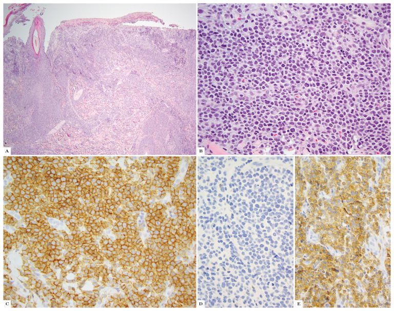
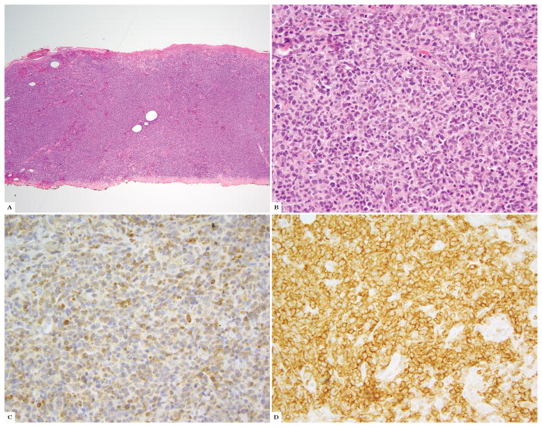
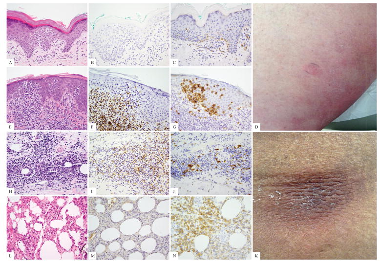
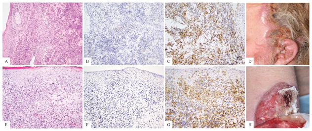
Methods and results: Twelve cases (10 patients) with TCRγ+ CTCL and 132 additional CLPD cases (127 patients) were examined, including mycosis fungoides (MF) (n = 60), cutaneous GDTCL (n = 15), subcutaneous panniculitis-like T-cell lymphoma (SPTCL) (n = 11), and CD30+ lymphoproliferative disorder (LPD) (n = 24). Clone H-41 against TCRδ was used on a Leica Bond-3 automated stainer to label FFPE slides. H-41 immunostaining was graded as percentage infiltrate: high (50-100%), moderate (10-49%), and low (0-9%). In TCRγ+ tumours, 12 of 12 (100%) patients showed TCRδ expression comparable to TCRγ expression. No (0%) TCRγ+ cases were negative for TCRδ. In all CLPDs, TCRδ expression was as follows: GDTCL, 16 of 20 cases (14 of 15 patients) high, two moderate, and two low; MF, 0 of 60 cases high, nine moderate, and 51 low; CD30+ LPD, one of 24 cases high, two moderate, and 21 low; and SPTCL, 0 of 11 cases (0 of 9 patients) high, two moderate, and two low. Three MF-like cases and one SPTCL-like case showed high expression; the remainder showed low expression.
Conclusions: mAb H-41 against TCRδ matches TCRγ in immunostaining FFPE tissues from GDTCL, supporting H-41 as a replacement for mAb γ3.20. TCRδ expression in our study suggests that the true occurrence of γδ+ non-GDTCL CTCL/CLPD may be lower than suggested by the recent literature.
Keywords: T-cell lymphoma; classification; immunohistochemistry; immunological techniques; lymphoproliferative disorders.
© 2018 John Wiley & Sons Ltd.
Conflict of interest statement
A conflict of interest statement:
SH has consulting relationships with Celgene Millenium/Takeda Kyowa-Hakka-Kirin Seattle Genetics Forty-Seven Mundipharma Verastem, and research relationship with Celgene Millenium/Takeda Kyowa-Hakka-Kirin Seattle Genetics Forty-Seven Infinity/Verastem Spectrum Pharmaceuticals, ADCT Therapeutics and Aileron Therapeutics. AM consulting relationships with Seattle Genetics and BMS. MP, SG, DF, PM, MK, AC, AD and AJ have no conflicts of interest to declare.
Figures
References
-
- Taghon T, Rothenberg EV. Molecular mechanisms that control mouse and human tcr-alphabeta and tcr-gammadelta t cell development. Semin Immunopathol. 2008;30:383–398. - PubMed
-
- Tripodo C, Iannitto E, Florena AM, et al. Gamma-delta t-cell lymphomas. Nat Rev Clin Oncol. 2009;6:707–717. - PubMed
-
- Guitart J, Weisenburger DD, Subtil A, et al. Cutaneous gammadelta t-cell lymphomas: A spectrum of presentations with overlap with other cytotoxic lymphomas. Am J Surg Pathol. 2012;36:1656–1665. - PubMed
-
- Toro JR, Liewehr DJ, Pabby N, et al. Gamma-delta t-cell phenotype is associated with significantly decreased survival in cutaneous t-cell lymphoma. Blood. 2003;101:3407–3412. - PubMed
-
- Willemze R, Jansen PM, Cerroni L, et al. Subcutaneous panniculitis-like t-cell lymphoma: Definition, classification, and prognostic factors: An eortc cutaneous lymphoma group study of 83 cases. Blood. 2008;111:838–845. - PubMed
MeSH terms
Substances
Grants and funding
LinkOut - more resources
Full Text Sources
Other Literature Sources
Medical
Research Materials
