. 2018 Sep 15;27(18):3165-3176.
doi: 10.1093/hmg/ddy219.

Ulk2 controls cortical excitatory-inhibitory balance via autophagic regulation of p62 and GABAA receptor trafficking in pyramidal neurons

Affiliations

Ulk2 controls cortical excitatory-inhibitory balance via autophagic regulation of p62 and GABAA receptor trafficking in pyramidal neurons

Akiko Sumitomo et al. Hum Mol Genet. .

Abstract

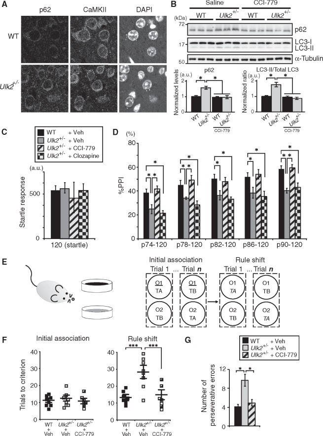
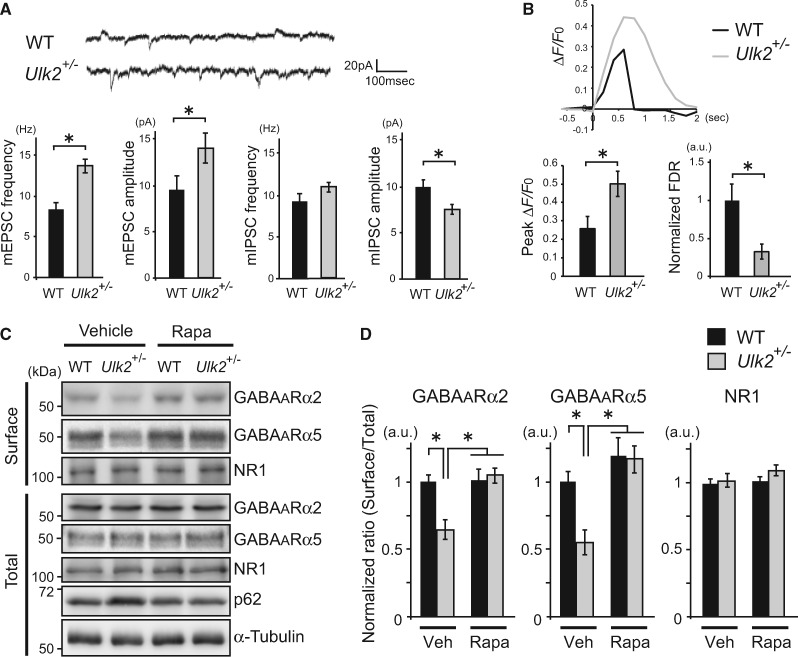
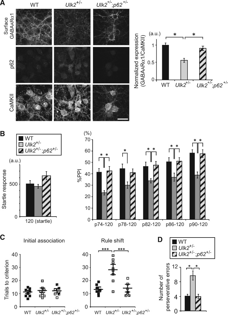
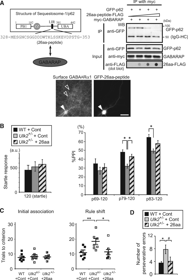
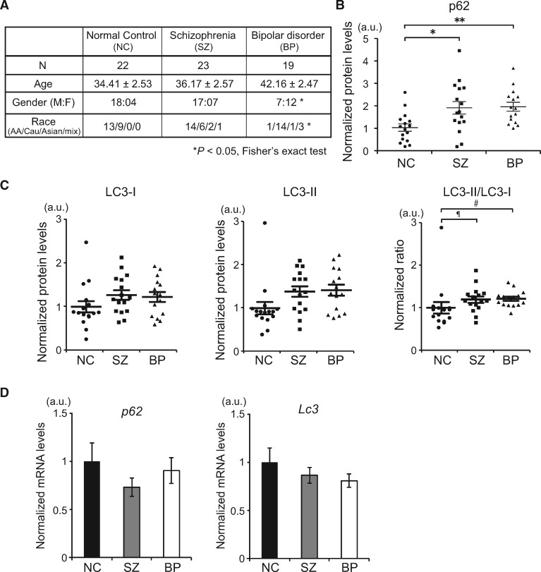
Autophagy plays an essential role in intracellular degradation and maintenance of cellular homeostasis in all cells, including neurons. Although a recent study reported a copy number variation of Ulk2, a gene essential for initiating autophagy, associated with a case of schizophrenia (SZ), it remains to be studied whether Ulk2 dysfunction could underlie the pathophysiology of the disease. Here we show that Ulk2 heterozygous (Ulk2+/-) mice have upregulated expression of sequestosome-1/p62, an autophagy-associated stress response protein, predominantly in pyramidal neurons of the prefrontal cortex (PFC), and exhibit behavioral deficits associated with the PFC functions, including attenuated sensorimotor gating and impaired cognition. Ulk2+/- neurons showed imbalanced excitatory-inhibitory neurotransmission, due in part to selective down-modulation of gamma-aminobutyric acid (GABA)A receptor surface expression in pyramidal neurons. Genetically reducing p62 gene dosage or suppressing p62 protein levels with an autophagy-inducing agent restored the GABAA receptor surface expression and rescued the behavioral deficits in Ulk2+/- mice. Moreover, expressing a short peptide that specifically interferes with the interaction of p62 and GABAA receptor-associated protein, a protein that regulates endocytic trafficking of GABAA receptors, also restored the GABAA receptor surface expression and rescued the behavioral deficits in Ulk2+/- mice. Thus, the current study reveals a novel mechanism linking deregulated autophagy to functional disturbances of the nervous system relevant to SZ, through regulation of GABAA receptor surface presentation in pyramidal neurons.


Figures

Figure 1.
Figure 1.
Upregulated p62 protein levels in pyramidal neurons of the PFC and PFC-associated behavioral deficits in Ulk2+/− mice. (A) Selective upregulation of p62 protein in CaMKII+ neurons in the PFC of Ulk2+/− mice. Scale bar, 10 μm. (B) Western blot of p62 and LC3 in the frontal cortex of Ulk2+/− mice chronically treated with saline or a rapamycin analog CCI-779. The expression levels were measured by densitometric analysis using ImageJ. Error bars indicate SEM. *<0.05. (C, D) The amplitude of startle response (C) and the percentage of PPI (D) were evaluated for WT+saline (n=10), Ulk2+/−+saline (n=10), Ulk2+/−+CCI-779 (n=10) and Ulk2+/−+clozapine (n=10). No significant difference in startle response [F(3, 36)=0.9683, P=0.4183] (one-way ANOVA). Statistical significance for %PPI: F(3, 36)=11.28, <0.0001 (two-way ANOVA with repeated measures); *<0.05 (Bonferroni post hoc test). (E) Schematic diagram of the rule shift assay. Upon rule shifting, the stimulus associated with food reward is changed from odor cue 1 (underlined) to texture cue A (italicized) in this diagram. (F) Numbers of trials to criterion during the initial association phase, as well as the rule shift phase of the assays, were scored for WT (n=8), Ulk2+/− (n=8) and Ulk2+/−+CCI-779 (n=7). No significant difference during the initial association phase [F(2, 20)=1.54, P=0.832]. Statistical significance during the rule-shift phase: F(2, 20)=7.67, P<0.0001 (one-way ANOVA); ***<0.001 (Bonferroni post hoc test). (G) Numbers of perseverative errors during the rule shift phase were scored. F(2, 20)=4.83, P=0.024 (one-way ANOVA); *<0.05 (Bonferroni post hoc test).
Figure 2.
Figure 2.
Reduced surface expression of GABAA receptors and imbalanced excitatory−inhibitory neurotransmission in Ulk2+/− pyramidal neurons. (A) Patch-clamp recording of miniature synaptic currents from WT and Ulk2+/− primary neurons (=20 each) (16–25 DIV). Averages of mEPSC and mIPSC frequencies and amplitudes are plotted in the graph. *P<0.05 (Mann–Whitney test, two-tailed). (B) WT and Ulk2+/− primary cortical neurons were transfected with pCAG-GCaMP6, and [Ca2+]i in each transfected neuron was measured following depolarization. Representative [Ca2+]i changes (ΔF/F0) are plotted, and the averaged peak value and the FDR are shown in the graph (=10 each). *P<0.05 (Mann–Whitney test, two-tailed). (C, D) Surface-biotinylated cell lysates from primary neurons cultured with or without rapamycin (100 nM, 30 min) were analyzed by western blot using anti-GABAA receptor α2 and α5 subunit and NR1 antibodies. Experiments were done in triplicate and densitometry analyses were done to evaluate the surface levels of each receptor by normalizing them against the total levels of respective receptor, and plotted in the graph. *P<0.05 (Kruskal–Wallis test).
Figure 3.
Figure 3.
Reducing p62 gene dosage rescues GABAA receptor surface presentation and behavioral deficits in Ulk2+/− mice. (A) Primary cortical neurons were prepared from mice with the indicated genotypes, and the cell surface staining was done at 16 DIV with anti-GABAA receptor α1 subunit antibody, followed by permeabilization and further staining with anti-CaMKII and p62 antibodies. Scale bar, 20 μm. Fluorescence intensities per soma were measured using ImageJ. *P< 0.05 (Kruskal–Wallis test). Lower magnification images are shown in Supplementary Material, Fig. S3A. (B) The amplitude of startle response (left) and %PPI (right) were evaluated for WT (n=9), Ulk2+/− (n=9) and Ulk2+/−; p62+/− (n=12). Statistical significance in startle response [F(2, 28)=5.548, P=0.0093] (one-way ANOVA) and in %PPI [F(2, 28)=8.761, P=0.0011] (two-way ANOVA with repeated measures); *<0.05, **<0.01 (Bonferroni post hoc test). (C) Numbers of trials to criterion during the initial association phase, as well as the rule shift phase of the assays, were scored for WT (n=8), Ulk2+/− (n=8) and Ulk2+/−; p62+/− (n=7). No significant difference during the initial association phase [F(2, 20)=1.24, P=0.968]. Statistical significance during the rule-shift phase: F(2, 20)=8.42, P<0.0001 (one-way ANOVA); ***<0.001 (Bonferroni post hoc test). (D) Numbers of perseverative errors during the rule shift phase were scored. F(2, 20)=5.27, P=0.038 (one-way ANOVA); *<0.05 (Bonferroni post hoc test).
Figure 4.
Figure 4.
Interfering with p62–GABARAP interaction rescues GABAA receptor surface presentation and behavioral deficits in Ulk2+/− mice. (A) Schematic illustration of sequestosome-1/p62 protein structure and designing strategy for the 26-amino acid peptide that interferes with p62–GABARAP interaction. The LIR (LC3-interacting region) contains a DDDWxxL motif that was shown to interact with GABARAP (23). Primary cortical neurons prepared from Ulk2+/− mice were transfected at 16DIV with an expression plasmid encoding GFP-26aa-peptide, and the cell surface GABAA receptor (α1 subunit) was immuno-stained without cell permeabilization. White arrow indicates a cell transfected with GFP-26aa-peptide, and open arrows indicate neighboring non-transfected cells. Scale bar, 20 μm. (B) The amplitude of startle response (left) and %PPI (right) were evaluated for WT injected with control AAV (n=7), Ulk2+/− with control AAV (n=7), and Ulk2+/− with 26aa-peptide AAV (n=6). No significant difference in startle response [F(2, 17)=1.045, P=0.833] (one-way ANOVA). Statistical significance for %PPI: F(2, 17)=8.53, =0.0126 (two-way ANOVA with repeated measures); *<0.05 (Bonferroni post hoc test). (C) Numbers of trials to criterion during the initial association phase, as well as the rule shift phase of the assays, were scored for WT injected with control AAV (n=7), Ulk2+/− with control AAV (=7), and Ulk2+/−; p62+/− with 26aa-peptide AAV (n=6). *<0.05, **<0.01. No significant difference during the initial association phase [F(2, 17)=1.02, =0.827]. Statistical significance during the rule-shift phase: F(2, 17)=7.74, P=0.018 (one-way ANOVA); *<0.05, **<0.01 (Bonferroni post hoc test). (D) Numbers of perseverative errors during the rule shift phase were scored. F(2, 17)=4.58, =0.046 (one-way ANOVA); *<0.05, #P=0.057 (Bonferroni post hoc test).
Figure 5.
Figure 5.
Attenuated autophagy in olfactory neuronal cells in sporadic cases of psychiatric disorders. (A) Demographics of human subjects in this study. *<0.05 (Fisher’s exact test). (B) The relative protein expression levels of p62 for each subject of control (circle), SZ (square) and BP (triangle) evaluated by western blot. *P<0.05, **P<0.01 (one-way ANOVA). See also Supplementary Material, Fig. S4A and B. (C) The relative protein expression levels of LC3-I, LC3-II and their ratio (LC3-II/LC3-I) for each subject of control (circle), SZ (square) and BP (triangle) evaluated by western blot. P=0.067175, #P=0.054516. (D) qPCR analysis of p62 and Lc3 mRNA expression in control or patients-derived olfactory neuronal cells. No statistical significance (one-way ANOVA).

Similar articles

Cited by

References

    1. Mizushima N., Komatsu M. (2011) Autophagy: renovation of cells and tissues. Cell, 147, 728–741. - PubMed
    1. Hara T., Nakamura K., Matsui M., Yamamoto A., Nakahara Y., Suzuki-Migishima R., Yokoyama M., Mishima K., Saito I., Okano H., Mizushima N. (2006) Suppression of basal autophagy in neural cells causes neurodegenerative disease in mice. Nature, 441, 885–889. - PubMed
    1. Komatsu M., Waguri S., Chiba T., Murata S., Iwata J., Tanida I., Ueno T., Koike M., Uchiyama Y., Kominami E., Tanaka K. (2006) Loss of autophagy in the central nervous system causes neurodegeneration in mice. Nature, 441, 880–884. - PubMed
    1. Komatsu M., Wang Q.J., Holstein G.R., Friedrich V.L. Jr, Iwata J., Kominami E., Chait B.T., Tanaka K., Yue Z. (2007) Essential role for autophagy protein Atg7 in the maintenance of axonal homeostasis and the prevention of axonal degeneration. Proc. Natl. Acad. Sci. U.S.A., 104, 14489–14494. - PMC - PubMed
    1. Toda H., Mochizuki H., Flores III R., Josowitz R., Krasieva T.B., LaMorte V.J., Suzuki E., Gindhart J.G., Furukubo-Tokunaga K., Tomoda T. (2008) UNC-51/ATG1 kinase regulates axonal transport by mediating motor-cargo assembly. Genes Dev., 22, 3292–3307. - PMC - PubMed

Publication types