. 2018 Jul 27;46(13):6773-6784.
doi: 10.1093/nar/gky470.

Gyrase containing a single C-terminal domain catalyzes negative supercoiling of DNA by decreasing the linking number in steps of two

Affiliations

Gyrase containing a single C-terminal domain catalyzes negative supercoiling of DNA by decreasing the linking number in steps of two

Jampa Tsedön Stelljes et al. Nucleic Acids Res. .

Abstract

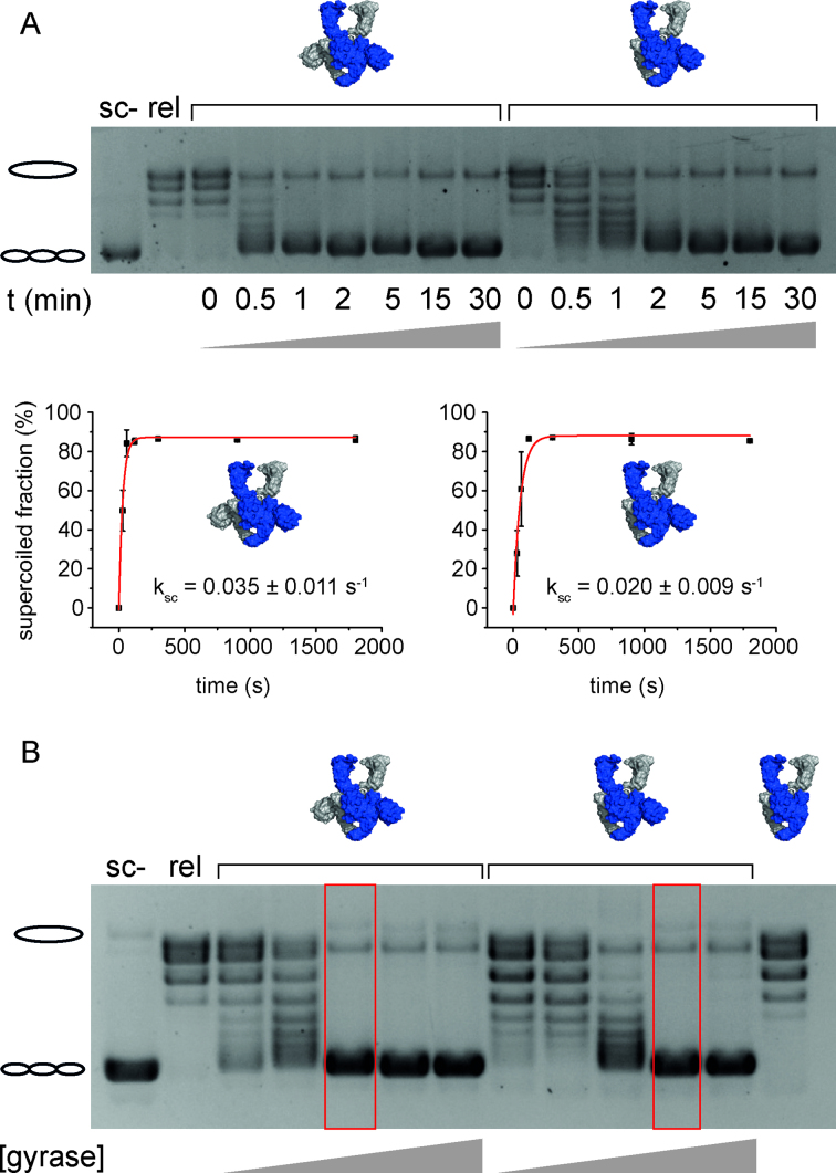
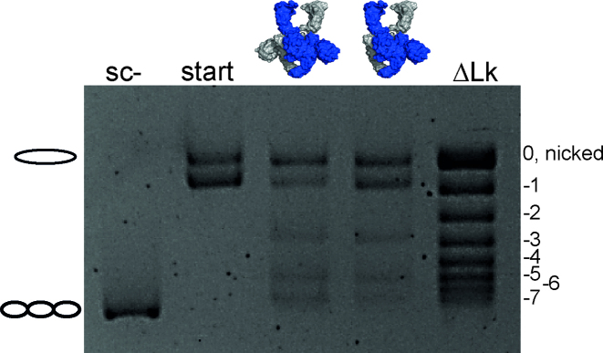
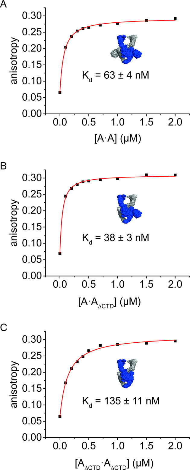
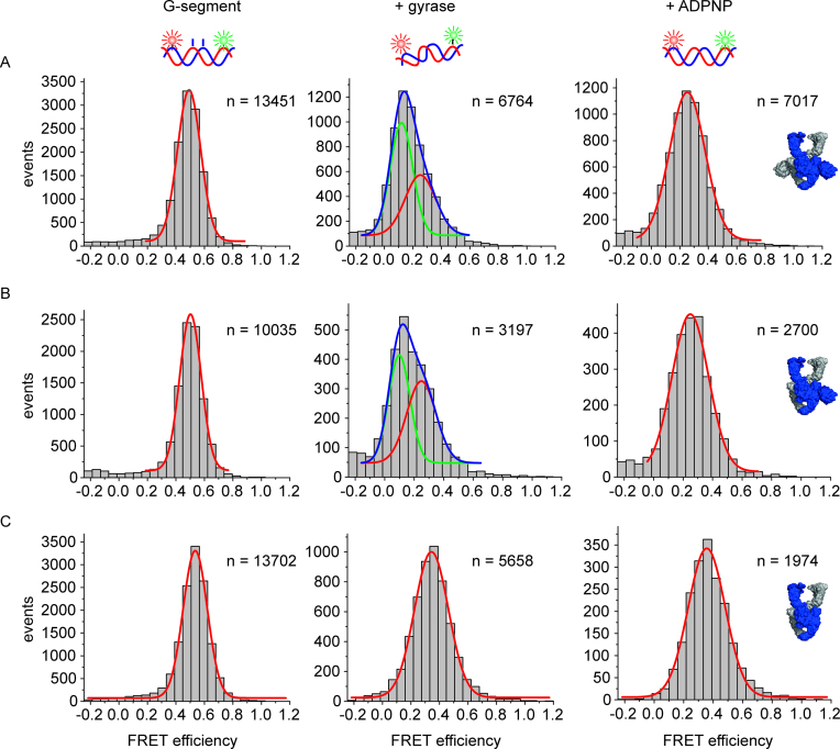
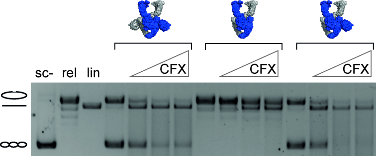
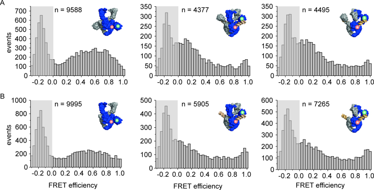
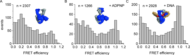
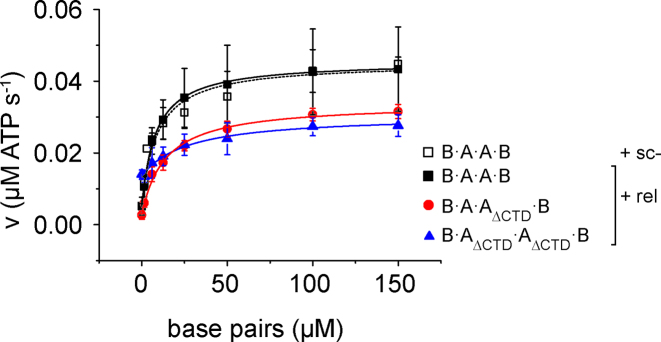
The topological state of DNA in vivo is regulated by topoisomerases. Gyrase is a bacterial topoisomerase that introduces negative supercoils into DNA at the expense of ATP hydrolysis. According to the strand-passage mechanism, a double-strand of the DNA substrate is cleaved, and a second double-stranded segment is passed through the gap, converting a positive DNA node into a negative node. The correct orientation of these DNA segments for strand passage is achieved by wrapping of the DNA around gyrase, which involves the C-terminal domains (CTDs) of both GyrA subunits in the A2B2 heterotetramer. Gyrase lacking both CTDs cannot introduce negative supercoils into DNA. Here, we analyze the requirements for the two CTDs in individual steps in the supercoiling reaction. Gyrase that contains a single CTD binds, distorts, and cleaves DNA similarly to wildtype gyrase. It also shows wildtype-like DNA-dependent ATPase activity, and undergoes DNA-induced movement of the CTD as well as N-gate narrowing. Most importantly, the enzyme still introduces negative supercoils into DNA in an ATP-dependent reaction, with a velocity similar to wildtype gyrase, and decreases the linking number of the DNA in steps of two. One CTD is thus sufficient to support DNA supercoiling.


Figures

Figure 1.
Figure 1.
DNA supercoiling by gyrase containing two or one CTD(s). (A) Time trace of ATP-dependent DNA supercoiling by gyrase with two CTDs and gyrase with one CTD (see cartoons of structures). 20 nM relaxed pUC18 was supercoiled by 40 nM GyrA and 160 nM GyrB in presence of 1.5 mM ATP at 37°C, and the reaction was stopped at indicated time points. Error bars depict the standard deviation from three independent experiments. Quantification of the fraction of supercoiled DNA as a function of time gives rate constants of supercoiling of ksc = 0.035 ± 0.011 s−1 for gyrase with both CTDs, and ksc = 0.020 ± 0.009 s−1 for gyrase with a single CTD. Errors are standard deviation from three independent experiments. Gyrase with one CTD thus catalyzes negative supercoiling of DNA only slightly more slowly than gyrase with two CTDs. (B) Concentration dependence of DNA supercoiling for gyrase with two, one, and no CTD(s). 20 nM relaxed pUC18 were incubated with 10, 20, 40, 80, or 160 nM GyrA and 20, 40, 80, 160, or 320 nM GyrB in the presence of 1.5 mM ATP, and reactions were stopped after 5 min. Compared to gyrase with two CTDs, about twice the concentration of gyrase with one CTD is needed to achieve complete supercoiling (red boxes). Gyrase lacking the CTDs (160 nM GyrAΔCTD, 320 nM GyrB) does not supercoil DNA. sc-: negatively supercoiled DNA, rel: relaxed DNA.
Figure 2.
Figure 2.
DNA supercoiling of a single topoisomer by gyrase containing two or one CTD(s). Supercoiling reactions were performed with 6 nM single topoisomer (start), 10 nM GyrA, and 20 nM GyrB in the presence of 1.5 mM ATP at 37°C, and reactions were stopped after 5 min. Gyrase with one and two CTDs decreases the linking number of the DNA in steps of two. ΔLk denotes the topoisomer ladder. Gyrase with one and two CTD(s) catalyzes negative DNA supercoiling by decreasing the linking number in steps of two.
Figure 3.
Figure 3.
DNA binding to gyrase with two, one, or no CTD(s). Fluorescence anisotropy titrations of an Alexa488/Alexa546-labeled 60 bp DNA with 0.2–2 μM GyrA (monomer concentration) containing two CTDs (A·A, panel A), one CTD (A·AΔCTD, panel B), or no CTDs (AΔCTD·AΔCTD, panel C), in presence of 8 μM GyrB at 37°C. Binding was followed using the fluorescence anisotropy of Alexa546 as a probe. The curves depicted are representatives of duplicate measurements; errors (±) denote the error of the mean. Deletion of one CTD does not reduce the affinity of gyrase for the 60 bp DNA, but deletion of both CTDs leads to a two-fold decrease in DNA affinity.
Figure 4.
Figure 4.
Conformation of the G-segment bound to gyrase with two, one, or no CTDs. Single-molecule FRET histograms of an Alexa488/Alexa546-labeled 60 bp DNA (G-segment) containing a phosphorothioate modification at the cleavage site, bound to gyrase with two CTDs (A), with one CTD (B), and without CTDs (C). DNA on its own (left panels) shows a unimodal FRET histogram characteristic of regular B-form DNA. DNA bound to gyrase (center panels) with two or one CTDs exists in a low-FRET state (EFRET = 0.12, green Gaussian; severely distorted from B-form geometry (18)) and a medium-FRET state (EFRET = 0.25, red Gaussian, slightly distorted from B-form geometry (18)); the sum of the two distributions is shown in blue. Addition of ADPNP to the gyrase/DNA complex to induce N-gate closure leads to disappearance of the low-FRET state for both enzymes. Gyrase lacking the CTDs does not distort the G-segment. Addition of ADPNP has no effect on the FRET distribution. The DNA concentration was 50 pM, the concentrations of GyrA and GyrB were 2 and 5 μM, respectively. n denotes the number of events summarized in each histogram.
Figure 5.
Figure 5.
DNA cleavage by gyrase with two, one, or no CTD(s) in the presence of CFX. DNA cleavage reactions were performed at 37°C with 20 nM plasmid DNA, 200 nM GyrA, 800 nM GyrB, 1.5 mM ATP and increasing concentrations of CFX (0, 10, 100 or 250 μM) for 5 min. All enzymes show similar levels of DNA cleavage at all CFX concentrations tested, irrespective of the number of CTDs present.
Figure 6.
Figure 6.
DNA-induced CTD movement monitored by single-molecule FRET. Single-molecule FRET histograms for gyrase labeled with donor (green) and acceptor (red) in the NTD and CTD of one GyrA subunit. The area at EFRET < 0 contains the peak representing molecules that carry only the donor fluorophore, and is shaded in gray. n denotes the number of events accumulated in each histogram. (A) Gyrase with both CTDs (B·AT140C/K594C·A·B), labeled with donor and acceptor fluorophores in the NTD and CTD of one GyrA subunit, shows a broad FRET histogram (left), in agreement with flexible attachment of the CTDs to the GyrA NTD. Addition of DNA (center) leads to a decrease in FRET efficiencies, indicative of an upward movement of the CTDs. Addition of ADPNP (right) does not further affect the FRET efficiency histogram and thus the position of the CTDs. (B) Gyrase with one CTD (B·AT140C/K594C·AΔCTD·B) shows a similar FRET histogram as gyrase with two CTDs. Addition of DNA (center) also leads to a decrease in FRET efficiency. Addition of ADPNP does not induce any further changes in FRET efficiency and CTD position. Concentrations were 150 pM GyrA (donor concentration), 8 μM GyrB, 20 nM relaxed pUC18, and 2 mM ADPNP.
Figure 7.
Figure 7.
Gyrase with a single CTD undergoes DNA-induced N-gate narrowing. Single-molecule FRET histograms for gyrase (BE17CA·BE17CAΔCTD) labeled with donor and acceptor fluorophores at the N-gate in the absence (A) and presence of 2 mM ADPNP (B) or 20 nM plasmid DNA (C). In the absence of DNA or ADPNP, the FRET efficiency is low (EFRET = 0.2), consistent with an open N-gate. ADPNP addition leads to N-gate closure, evidenced by a shift of the FRET efficiency to EFRET = 0.95–1. In the presence of DNA, the FRET efficiency is also high (EFRET = 0.95–1), which is consistent with DNA-induced narrowing of the N-gate. n denotes the number of events summarized in each histogram.
Figure 8.
Figure 8.
Effect of the CTDs on DNA-stimulated ATPase activity of gyrase. Steady-state ATPase activity of gyrase containing two, one, or no CTD(s) at 37°C with 0.1 μM GyrB, 0.5 μM GyrA and 1.5 mM ATP as a function of the concentration of negatively supercoiled DNA (sc-; B·A·A·B; black, open squares) or relaxed DNA (rel; B·A·A·B: black squares, B·A·AΔCTD·B: red circles; B·AΔCTD·AΔCTD·B: blue triangles). Error bars denote standard deviations from three to five independent experiments. The kcat and Kapp,DNA values obtained from analyses with the Michaelis-Menten equation are summarized in Table 1.

References

    1. Brown P.O., Cozzarelli N.R.. A sign inversion mechanism for enzymatic supercoiling of DNA. Science. 1979; 206:1081–1083. - PubMed
    1. Goto T., Wang J.C.. Yeast DNA topoisomerase II. An ATP-dependent type II topoisomerase that catalyzes the catenation, decatenation, unknotting, and relaxation of double-stranded DNA rings. J. Biol. Chem. 1982; 257:5866–5872. - PubMed
    1. Gellert M., Mizuuchi K., O’Dea M.H., Nash H.A.. DNA gyrase: an enzyme that introduces superhelical turns into DNA. Proc. Natl. Acad. Sci. U.S.A. 1976; 73:3872–3876. - PMC - PubMed
    1. Peng H., Marians K.J.. Decatenation activity of topoisomerase IV during oriC and pBR322 DNA replication in vitro. Proc. Natl. Acad. Sci. U.S.A. 1993; 90:8571–8575. - PMC - PubMed
    1. Wang J.C. Moving one DNA double helix through another by a type II DNA topoisomerase: the story of a simple molecular machine. Q. Rev. Biophys. 1998; 31:107–144. - PubMed

Publication types