. 2018 Aug 1;128(8):3583-3594.
doi: 10.1172/JCI120972. Epub 2018 Jul 23.

Purinergic P2X4 receptors and mitochondrial ATP production regulate T cell migration

Affiliations

Purinergic P2X4 receptors and mitochondrial ATP production regulate T cell migration

Carola Ledderose et al. J Clin Invest. .

Abstract

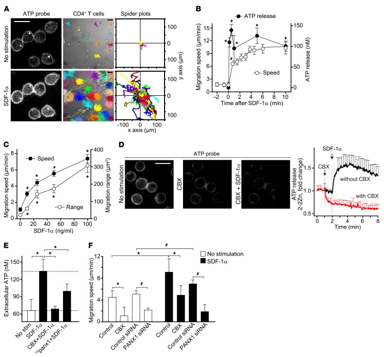
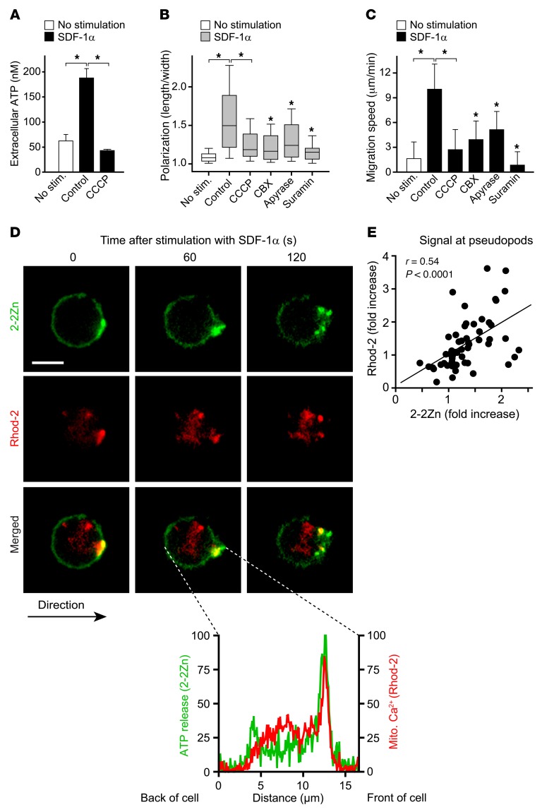
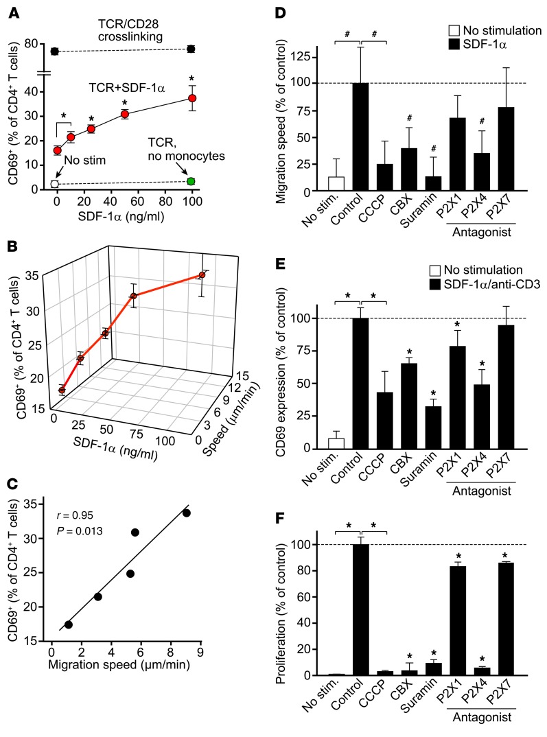
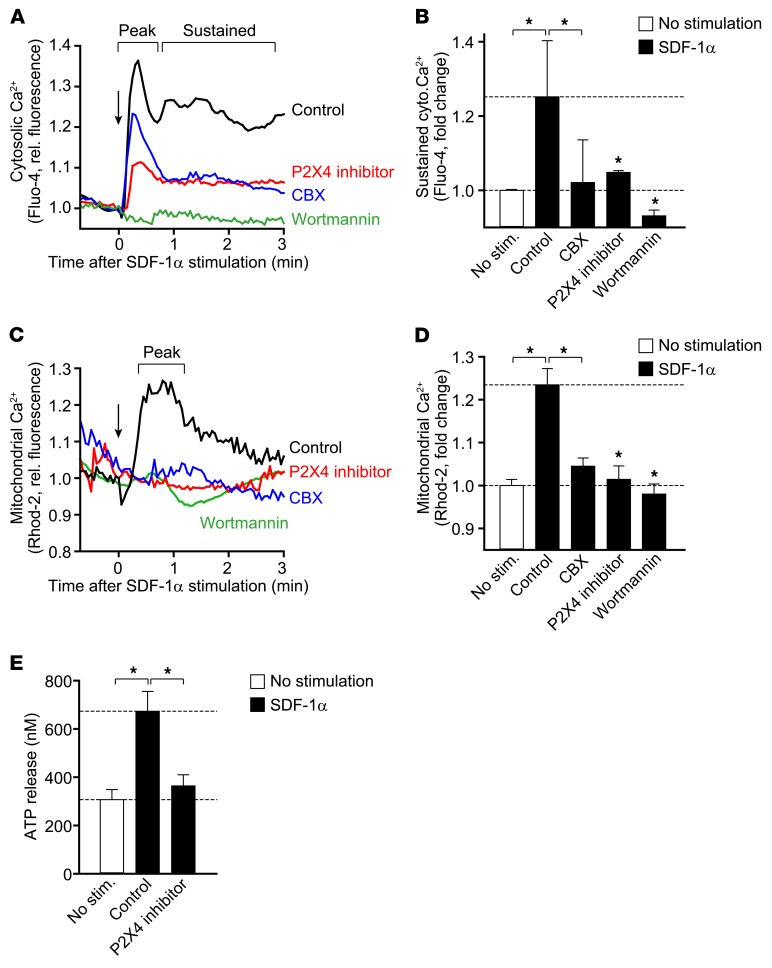
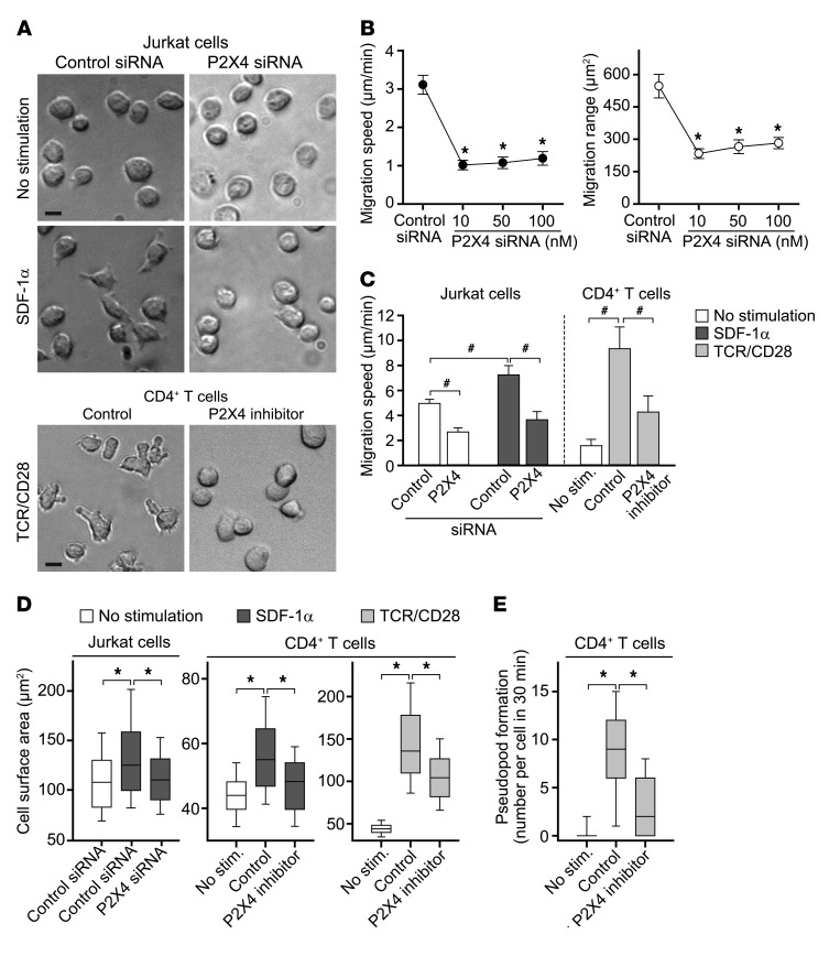
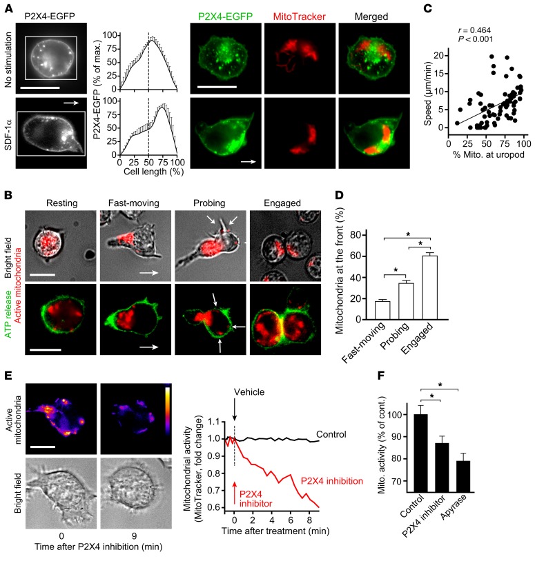
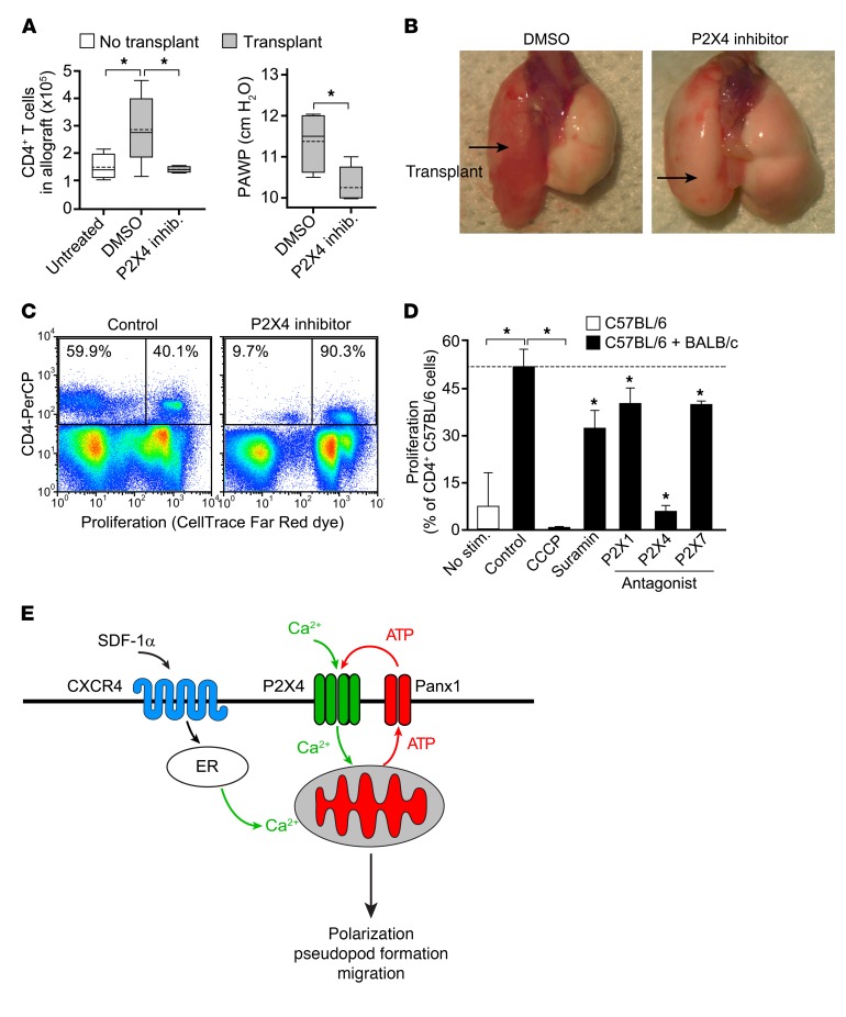
T cells must migrate in order to encounter antigen-presenting cells (APCs) and to execute their varied functions in immune defense and inflammation. ATP release and autocrine signaling through purinergic receptors contribute to T cell activation at the immune synapse that T cells form with APCs. Here, we show that T cells also require ATP release and purinergic signaling for their migration to APCs. We found that the chemokine stromal-derived factor-1α (SDF-1α) triggered mitochondrial ATP production, rapid bursts of ATP release, and increased migration of primary human CD4+ T cells. This process depended on pannexin-1 ATP release channels and autocrine stimulation of P2X4 receptors. SDF-1α stimulation caused localized accumulation of mitochondria with P2X4 receptors near the front of cells, resulting in a feed-forward signaling mechanism that promotes cellular Ca2+ influx and sustains mitochondrial ATP synthesis at levels needed for pseudopod protrusion, T cell polarization, and cell migration. Inhibition of P2X4 receptors blocked the activation and migration of T cells in vitro. In a mouse lung transplant model, P2X4 receptor antagonist treatment prevented the recruitment of T cells into allograft tissue and the rejection of lung transplants. Our findings suggest that P2X4 receptors are therapeutic targets for immunomodulation in transplantation and inflammatory diseases.

Keywords: Cell Biology; Mitochondria; T cells; Transplantation.


Conflict of interest statement

Conflict of interest: The authors have declared that no conflict of interest exists.

Figures

Figure 1
Figure 1. ATP release through pannexin-1 channels is required for T cell migration.
(A) ATP release at the cell surface from human CD4+ T cells before and 1 minute after stimulation with SDF-1α was visualized with the ATP probe 2-2Zn (left column; ×100 objective; scale bar, 10 μm). Cell migration was tracked for 30 minutes in the presence or absence of SDF-1α. Paths of individual cells superimposed (center column; ×63 objective; scale bar, 10 μm) or aligned with their origins at x = y = 0 (right column) are shown. Data are representative of 5 experiments. (See also Supplemental Video 1). (B) Migration speed and ATP release of CD4+ T cells in response to SDF-1α. (C) Speed and migration range of CD4+ T cells treated with increasing concentrations of SDF-1α (30-minute observation). (D) CD4+ T cells were stained with 2-2Zn and the response to CBX (100 μM) or SDF-1α was analyzed with fluorescence microscopy. Representative images before, 0.5 minutes after addition of CBX, and 1 minute after addition of SDF-1α (left) and traces of cells derived from 2 separate experiments (control, n = 31; CBX, n = 39) are shown. Data are mean ± SD; ×100 objective; scale bar, 10 μm. (See also Supplemental Video 2.) (E) ATP concentrations in the supernatants of CD4+ T cells treated with CBX (50 μM) or 10panx1 (100 μM) and stimulated with SDF-1α for 5 minutes. (F) Spontaneous or SDF-1α–induced migration speed of Jurkat cells after silencing of PANX1 or treatment with CBX (100 μM; see also Supplemental Video 3; *P < 0.05, #P < 0.05 by Kruskal-Wallis test). Data in B (migration speed), C, and F represent mean ± SD of 60 cells analyzed in 3 independent experiments. Data in B (ATP release) and E represent mean ± SD of 3 independent experiments; *P < 0.05 vs. control (1-way ANOVA); #P < 0.05 (unpaired 2-tailed t test).
Figure 2
Figure 2. Mitochondria produce the ATP that is released from migrating T cells.
(A) ATP concentrations were measured in the supernatants of CD4+ T cells treated with CCCP (5 μM) for 10 minutes and stimulated with SDF-1α for 5 minutes (mean ± SD, n = 3; *P < 0.05 vs. control; 1-way ANOVA). (B and C) CD4+ T cells were treated with CCCP (5 μM), CBX (50 μM), apyrase (20 U/ml), suramin (100 μM), or cell culture medium (control) for 10 minutes, and polarization (B) and migration speed (C) in response to SDF-1α were analyzed. Data represent mean ± SD of 86 (no stimulation), 237 (control), 133 (CCCP), 110 (CBX), 87 (apyrase), and 49 (suramin) cells analyzed in 6 (control) or 3 separate experiments. *P < 0.05 vs. control (Kruskal-Wallis test). (D and E) CD4+ T cells were stained with the mitochondrial Ca2+ indicator Rhod-2 and the ATP probe 2-2Zn, stimulated with SDF-1α, and ATP release and mitochondrial Ca2+ influx were analyzed with fluorescence microscopy (see also Supplemental Video 4). Representative images of 6 individual experiments comprising a total of 55 cells are shown in D (scale bar, 5 μm; ×100 objective). The histogram shows the distribution of the 2-2Zn and Rhod-2 signal across the cell axis as indicated. (E) 2-2Zn and Rhod-2 fluorescence intensities were measured at the front of polarizing CD4+ T cells and normalized to the fluorescence intensities at the back of the same cell. Data are derived from 55 cells analyzed in 6 individual experiments.
Figure 3
Figure 3. T cell migration and activation depends on P2X4 receptors.
(A) CD69 expression in CD4+ T cells stimulated for 5 hours with SDF-1α and anti-CD3 antibodies in a PBMC culture was measured by flow cytometry. Positive controls (stimulation with anti-CD3/anti-CD28 coated beads) and negative controls (no stimulation or stimulation with anti-CD3 in monocyte-depleted cultures) were included as indicated. Data represent mean ± SD of 3 individual experiments. *P < 0.05 vs. 0 ng/ml SDF-1α (1-way ANOVA); TCR, T cell receptor. (B) PBMCs were placed into fibronectin-coated glass-bottom chamber slides, stained with APC-labeled anti-CD4 antibodies, stimulated with SDF-1α, and migration speed of CD4+ T cells was analyzed by time-lapse microscopy. Data are mean ± SD of 50 cells analyzed in 3 separate experiments. CD69 expression following stimulation with SDF-1α and anti-CD3 antibodies for 5 hours was analyzed as in A. Data represent mean ± SD of 3 separate experiments. (C) Correlation between CD69 expression and migration speed. Data are the mean values of the experiments shown in B. (D) CD4+ T cells were treated with CCCP (5 μM), CBX (100 μM), suramin (100 μM), or inhibitors of P2X1 (NF023; 10 μM), P2X4 (5-BDBD; 10 μM), or P2X7 (A438079; 10 μM) receptors, and migration speed in response to SDF-1α was analyzed. Data represent mean ± SD of 80 cells analyzed in 3 experiments; #P < 0.05 vs. control (Kruskal-Wallis test). (E) CD69 expression following TCR stimulation with anti-CD3 for 3 hours was analyzed as in A. (F) Proliferation of CD4+ T cells in a PBMC culture stimulated with anti-CD3 antibodies for 72 hours was determined by analyzing CFSE dilution. Data in E and F represent mean ± SD of 6 (E) or 3 (F) individual experiments. *P < 0.05 vs. control (1-way ANOVA).
Figure 4
Figure 4. P2X4 receptors regulate Ca2+ signaling.
Cytosolic (A and B) or mitochondrial (C and D) Ca2+ levels in CD4+ T cells stimulated in the presence of CBX (50 μM), the P2X4 receptor antagonist 5-BDBD (10 μM), or the PI3K inhibitor wortmannin (10 μM) were recorded by time-lapse fluorescence microscopy. (A) Data are mean Fluo-4 fluorescence traces of 65 (control), 42 (CBX), 54 (P2X4 inhibitor), or 58 (wortmannin) cells from 1 experiment and are representative of 3–6 experiments. (B) Averaged plateau fluorescence values ± SD of 7 (no stimulation), 6 (control), 5 (P2X4 inhibitor), or 3 (CBX, wortmannin) separate experiments each comprising averaged data from all cells in a microscopic field (range 38–71). (C) Data are mean Rhod-2 fluorescence traces of 32 (control), 31 (CBX), 35 (P2X4 inhibitor), or 30 (wortmannin) cells derived from 1 experiment and are representative of 3–5 experiments. (D) Averaged peak fluorescence values ± SD of 5 (no stimulation, control) or 3 (CBX, P2X4 inhibitor, wortmannin) separate experiments each comprising averaged data from all cells in a microscopic field (range 16–39). (E) Jurkat T cells were treated with 5-BDBD (P2X4 inhibitor; 20 μM) or vehicle control for 10 minutes. ATP release in response to stimulation with SDF-1α or vehicle control was measured after 5 minutes (mean ± SD, n = 3); *P < 0.05 vs. control (1-way ANOVA).
Figure 5
Figure 5. P2X4 receptors regulate T cell polarization, pseudopod formation, and migration.
(AC) Cell migration after silencing of P2X4 receptors in Jurkat cells or pharmacological P2X4 inhibition (5-BDBD, 10 μM) in CD4+ T lymphoblasts in the presence or absence of SDF-1α. (A) Representative images of 4 experiments; ×20 objective; scale bar, 10 μm (see also Supplemental Video 6). (B) Jurkat cells were treated with control or P2X4-targeting siRNA at the indicated concentrations, and migration speed and range (in 30 minutes) were analyzed after 48 hours. Data represent mean ± SEM of 60 cells derived from 3 experiments. (C) Jurkat cells were transfected with control or P2X4-targeting siRNA (10 nM) and migration was analyzed after 48 hours. Data represent mean ± SD of 3 (Jurkat cells) or 4 (T cells) separate experiments each comprising 40 cells. (D) Effect of P2X4 silencing or inhibition on the cell surface area of Jurkat cells, primary CD4+ T cells, and CD4+ T lymphoblasts stimulated or not with SDF-1α. Box plots show the median and the distribution of 262, 553, and 276 Jurkat cells, 297, 290, and 127 primary CD4+ T cells, 290 control lymphoblasts, and 127 lymphoblasts treated with 5-BDBD. Cells were analyzed in 4 separate experiments. (E) Migration of primary CD4+ T cells (no stimulation) or CD4+ T lymphoblasts treated or not with 5-BDBD was monitored by time-lapse microscopy. The number of pseudopodia formed by a particular cell during the 30-minute observation period was recorded. Box plots show the median and the distribution of 212 (no stimulation), 140 (control), and 82 (P2X4 inhibitor) analyzed cells derived from 5 (no stimulation) or 3 independent experiments. *P < 0.05 vs. control (Kruskal-Wallis test). #P < 0.05 (1-way ANOVA); TCR, T cell receptor.
Figure 6
Figure 6. P2X4 receptors promote mitochondrial activation and localized ATP release from migrating T cells.
(A) Distribution of EGFP-tagged P2X4 receptors and mitochondria in unstimulated and SDF-1α–stimulated Jurkat cells. Histograms show the distribution of P2X4 receptor fluorescence along the cell axis as indicated (rectangle) and represent mean ± SD of 7 independent experiments. Arrow indicates direction of migration (see also Supplemental Video 7); ×100 objective. Scale bar, 10 μm. (B) Mitochondria are in the back of fast-moving cells and translocate to the front of cells probing their surroundings or engaging with other cells. CD4+ T lymphoblasts stained with MitoTracker Red CM-H2Xros (top row; ×63 objective) or with MitoTracker and 2-2Zn (bottom row; ×100 objective) are shown. Images are representative of 30 (top) or 15 (bottom row) experiments. Arrows in cells probing their environment indicate spots of increased mitochondrial activity (see also Supplemental Video 8). Scale bar, 10 μm. (C) Migration speed and mitochondrial localization were analyzed in 30-second increments in cells derived from 5 different experiments. The results shown comprise 73 single experiments. (D) Localization of mitochondria in the front half of fast-moving, probing, or interacting cells. Data represent mean ± SD of 30 cells, derived from 7 separate experiments; *P < 0.05 (Kruskal-Wallis test). (E) Representative images (left) and fluorescence intensity traces (right) of mitochondrial activity (MitoTracker Red CM-H2Xros) in CD4+ T lymphoblasts before and after P2X4 receptor inhibition (5-BDBD, 10 μM). Color coding was applied to demonstrate differences in mitochondrial activity. Right panel: Change in mitochondrial activity over time following treatment with 5-BDBD or culture medium (control). Data are representative of 20 cells; ×100 objective (see also Supplemental Video 9). Scale bar, 10 μm. (F) Averaged mitochondrial activity (mean ± SEM) of 22 (control), 20 (P2X4 inhibitor), or 11 (apyrase; 10 U/ml) cells analyzed in 2 (apyrase) or 3 individual experiments; *P < 0.05 (1-way ANOVA).
Figure 7
Figure 7. Inhibition of purinergic signaling prevents T cell recruitment in vivo.
(A and B) Recipient C57BL/6 mice were treated with the P2X4 inhibitor 5-BDBD or vehicle (DMSO) 24 hours before and immediately after transplantation of BALB/c lung allografts. (A) The number of CD4+ T cells infiltrating the lung allograft (left) and peak airway pressures (PAWP) in the allograft (right) were measured 24 hours after transplantation. For nontransplanted control and P2X4 inhibitor-treated group, n = 4; for DMSO-treated group, n = 6. Box plots: solid line indicates median, dotted line indicates mean; *P < 0.05, 1-way ANOVA (left) or unpaired 2-tailed Student’s t test (right). (B) Representative images of lung allografts 24 hours after transplantation. (C and D) In vitro proliferation of C57BL/6 CD4+ T cells (responders) cocultured in a mixed lymphocyte reaction with BALB/c splenocytes (stimulators) in the presence or absence of CCCP (1 μM), suramin (100 μM), NF279 (P2X1 antagonist; 20 μM), 5-BDBD (P2X4 antagonist; 20 μM), or A438079 (P2X7 antagonist; 20 μM) for 4 days. Representative dot plots (C) and averaged (mean ± SD) results (D) of 3 separate experiments are shown; *P < 0.05 vs. control (1-way ANOVA). (E) Purinergic regulation of T cell migration by P2X4 receptors. Chemokine receptors (e.g., CXCR4) trigger the production of ATP by mitochondria, ATP release through PANX1 channels, and autocrine stimulation of P2X4 receptors that facilitate Ca2+ influx, sustain mitochondrial ATP production, and promote pseudopod protrusion at the front of cells.

Similar articles

Cited by

References

    1. Gérard A, et al. Detection of rare antigen-presenting cells through T cell-intrinsic meandering motility, mediated by Myo1g. Cell. 2014;158(3):492–505. doi: 10.1016/j.cell.2014.05.044. - DOI - PMC - PubMed
    1. Germain RN, Robey EA, Cahalan MD. A decade of imaging cellular motility and interaction dynamics in the immune system. Science. 2012;336(6089):1676–1681. doi: 10.1126/science.1221063. - DOI - PMC - PubMed
    1. Masopust D, Schenkel JM. The integration of T cell migration, differentiation and function. Nat Rev Immunol. 2013;13(5):309–320. doi: 10.1038/nri3442. - DOI - PubMed
    1. Krummel MF, Bartumeus F, Gérard A. T cell migration, search strategies and mechanisms. Nat Rev Immunol. 2016;16(3):193–201. doi: 10.1038/nri.2015.16. - DOI - PMC - PubMed
    1. Moreau HD, Bousso P. Visualizing how T cells collect activation signals in vivo. Curr Opin Immunol. 2014;26:56–62. doi: 10.1016/j.coi.2013.10.013. - DOI - PubMed

Publication types

Substances