Review
. 2018 Jul;47(6):675-689.
doi: 10.1097/MPA.0000000000001075.

Advances in Diagnostic and Intraoperative Molecular Imaging of Pancreatic Cancer

Affiliations
Review

Advances in Diagnostic and Intraoperative Molecular Imaging of Pancreatic Cancer

Willemieke S Tummers et al. Pancreas. 2018 Jul.

Abstract

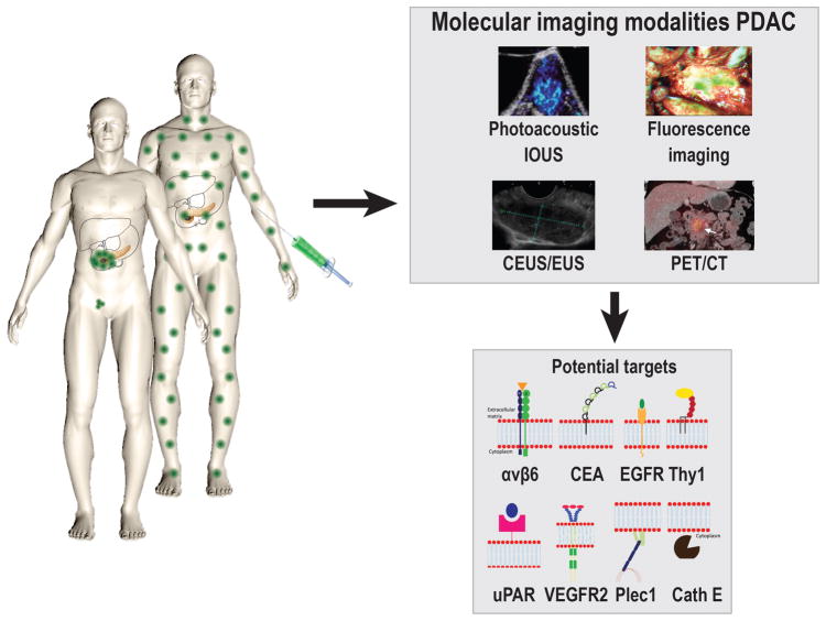
Pancreatic ductal adenocarcinoma (PDAC) has a dismal prognosis. To improve outcomes, there is a critical need for improved tools for detection, accurate staging, and resectability assessment. This could improve patient stratification for the most optimal primary treatment modality. Molecular imaging, used in combination with tumor-specific imaging agents, can improve established imaging methods for PDAC. These novel, tumor-specific imaging agents developed to target specific biomarkers have the potential to specifically differentiate between malignant and benign diseases, such as pancreatitis. When these agents are coupled to various types of labels, this type of molecular imaging can provide integrated diagnostic, noninvasive imaging of PDAC as well as image-guided pancreatic surgery. This review provides a detailed overview of the current clinical imaging applications, upcoming molecular imaging strategies for PDAC, and potential targets for imaging, with an emphasis on intraoperative imaging applications.


Conflict of interest statement

Conflict of Interest Disclosure: The authors declare no conflict of interest to the submitted work.

Figures

FIGURE 1
FIGURE 1
Schematic illustration of the key imaging modalities used for the diagnostics and potential intraoperative modalities for pancreatic cancer. Ultrasound, (endoscopic) ultrasound; CT, computed tomography; MRI, magnetic resonance imaging; PET, positron emission tomography. Representative images are shown of pancreatic cancer with the displayed modalities, expect for photoacoustic and raman optical imaging.
FIGURE 2
FIGURE 2
A, Schematic overview of the principle of ultrasound molecular imaging. A molecularly-targeted contrast agent (microbubble) is administered intravenously into the subject (in this case a mouse). Sound waves are transmitted into the subject by the transducer, the sound wave reflections are recorded and converted into images. Because of the size of microbubbles of several micrometers, the contrast agent remains intravascular and attaches to the target of choice (for example VEGFR2). Examples of in vivo molecular ultrasound images with microbubbles in (B) transgenic mouse model of PDAC, showing a strong signal when targeting VEGFR2 in focus of PDAC compared to normal pancreatic tissue, even in small PDAC lesions [From Pysz et al, 2015], and (C) in human with breast cancer using microbubbles targeting kinase insert domain receptor (MBKDR). Left panel: the anatomical image for reference, right panel: MBKDR accumulation in breast cancer lesion [Willmann et al, 2017]
FIGURE 3
FIGURE 3
A, Schematic overview of the principle of tumor-targeted PET imaging, a suitable tracer will be administered into the subject (in this case a mouse). Depending on the size of the tracer, the tracer can target the cancer at multiple locations; e.g. intravascular, receptors on the cell membrane, or intracellular. B, Small-animal PET imaging. BxPC-3 (integrin αvβ6 pos) and 293 (integrin αvβ6 negative) cells were xenografted in nude mice. PET images were acquired in tumor-bearing mice using a αvβ6-targeted cysteine knot (18F-fluorobenzoate-R01) [From Hackel et al, 2013].
FIGURE 4
FIGURE 4
A, Schematic overview of the principle of fluorescent imaging, a suitable targeted imaging agent with a fluorescent dye will be administered into the subject (in this case a mouse). The agent is visualized using a fluorescence imaging system, with an adequate excitation laser and camera able to detect the emitted light. The targeted agents migrate to the cellular targets to visualize the tumor in a target-specific manner, the imaging agent can target the cancer at multiple locations depending on the size; e.g. intravascular, receptors on the cell membrane, or intracellular. B, Top: schematic overview showing the principle of fluorescent imaging. Bottom: Intraoperative image showing the use of tumor-targeted fluorescent guided imaging during pancreatic cancer surgery.
FIGURE 5
FIGURE 5
A, Schematic overview of photoacoustic imaging principle; after injection of a tumor-targeting agent the imaging agent will target tumor cells and produces an enhanced photoacoustic signal, after excitation with a laser. The agent can target the cancer at multiple locations depending on the size; e.g. intravascular, receptors on the cell membrane, or intracellular. B, Top: Schematic overview showing the principle of photoacoustic imaging; the thermo-elastic expansion caused by heating of the tissue due to the laser will lead to acoustic waves that can be converted into both ultrasound and molecular images. Bottom: Tumor-targeted photoacoustic imaging. Mice bearing FTC133 tumors were photoacoustically imaged using 680 and 750 nm light before and after the injection of a MMP-targeting probe [From Levi et al, 2013].
FIGURE 6
FIGURE 6
A schematic overview of the principle of tumor-targeted imaging in pancreatic cancer, showing the most promising imaging modalities for early diagnosis and improved surgical treatment, and most promising targets for this purpose. Avβ6; Integrin αvβ6, CEA; Carcinoembryonic Antigen, EGFR; Epidermal growth factor receptor, Thy1; Thy-1 cell surface antigen, uPAR; Urokinase receptor, VEGFR2; Vascular endothelial growth factor receptor 2, Plec1; Plectin 1, Cath E; Cathepsin E.

Similar articles

Cited by

References

    1. Vincent A, Herman J, Schulick R, et al. Pancreatic cancer. Lancet. 2011;378:607–620. - PMC - PubMed
    1. Jang JY, Kang MJ, Heo JS, et al. A prospective randomized controlled study comparing outcomes of standard resection and extended resection, including dissection of the nerve plexus and various lymph nodes, in patients with pancreatic head cancer. Ann Surg. 2014;259:656–664. - PubMed
    1. Tummala P, Junaidi O, Agarwal B. Imaging of pancreatic cancer: An overview. J Gastrointest Oncol. 2011;2:168–174. - PMC - PubMed
    1. Agarwal B, Correa AM, Ho L. Survival in pancreatic carcinoma based on tumor size. Pancreas. 2008;36:e15–20. - PubMed
    1. Stathis A, Moore MJ. Advanced pancreatic carcinoma: current treatment and future challenges. Nat Rev Clin Oncol. 2010;7:163–172. - PubMed

Publication types

MeSH terms