. 2018 Jun 12;32(12):fj201701151RRR.
doi: 10.1096/fj.201701151RRR. Online ahead of print.

Galectin-3 and N-acetylglucosamine promote myogenesis and improve skeletal muscle function in the mdx model of Duchenne muscular dystrophy

Affiliations

Galectin-3 and N-acetylglucosamine promote myogenesis and improve skeletal muscle function in the mdx model of Duchenne muscular dystrophy

Ann Rancourt et al. FASEB J. .

Abstract

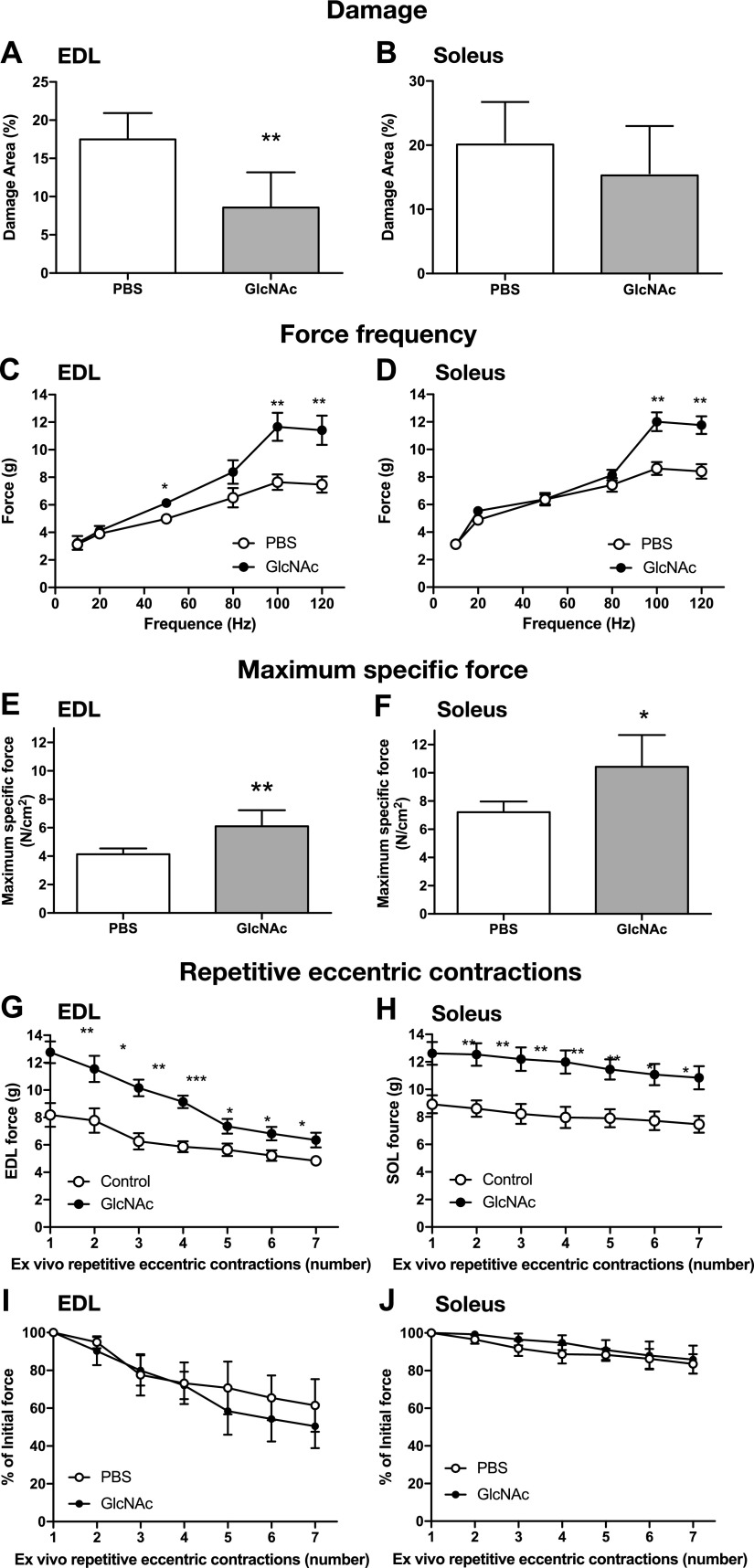
The muscle membrane, sarcolemma, must be firmly attached to the basal lamina. The failure of proper attachment results in muscle injury, which is the underlying cause of Duchenne muscular dystrophy (DMD), in which mutations in the dystrophin gene disrupts the firm adhesion. In patients with DMD, even moderate contraction causes damage, leading to progressive muscle degeneration. The damaged muscles are repaired through myogenesis. Consequently, myogenesis is highly active in patients with DMD, and the repeated activation of myogenesis leads to the exhaustion of the myogenic stem cells. Therefore, approaches to reducing the risk of the exhaustion are to develop a treatment that strengthens the interaction between the sarcolemma and the basal lamina and increases the efficiency of the myogenesis. Galectin-3 is an oligosaccharide-binding protein and is known to be involved in cell-cell interactions and cell-matrix interactions. Galectin-3 is expressed in myoblasts and skeletal muscle, although its function in muscle remains elusive. In this study, we found evidence that galectin-3 and the monosaccharide N-acetylglucosamine, which increases the synthesis of binding partners (oligosaccharides) of galectin-3, promote myogenesis in vitro. Moreover, in the mdx mouse model of DMD, treatment with N-acetylglucosamine increased muscle-force production. The results suggest that treatment with N-acetylglucosamine might mitigate the burden of DMD.-Rancourt, A., Dufresne, S. S., St-Pierre, G., Lévesque, J.-C., Nakamura, H., Kikuchi, Y., Satoh, M. S., Frenette, J., Sato, S. Galectin-3 and N-acetylglucosamine promote myogenesis and improve skeletal muscle function in the mdx model of Duchenne muscular dystrophy.

Keywords: glycobiology; lectins; monosaccharide.


Conflict of interest statement

The authors acknowledge the Bioimaging Platform at the Centre de Recherche Centre Hospitalier Universitaire (CHU) de Quebec. The authors thank the Consortium for Functional Glycomics for galectin-3 knockout mice. The mAb directed against MHC was obtained from the Developmental Studies Hybridoma Bank, created by the U.S. National Institutes of Health, Institute of Child Health and Human Development, and maintained at the Department of Biology, University of Iowa (Iowa City, IA, USA). The authors declare no conflicts of interest.

Figures

Figure 1
Figure 1
Expression of galectin-3 in differentiating myoblasts, myotubes, and muscles. A, B). Differentiation of murine myoblast C2C12 cells was induced by changing the medium from GM to DM. The levels of galectin-3 in the cells (A) and medium (B) were detected with Western blotting using an anti–galectin-3 antibody. Representative images from 3 independent experiments and results of densitometry analyses (means ± sd) are shown. Significance compared to 0 h of differentiation by 1 way-ANOVA is shown. *P < 0.05, **P < 0.01, ***P < 0.001. When ANOVA–on ranks was used, the detected significance was as follows: for cells, 48 h and 72 h; and for medium, 24 h and 48 h. C) Galectin-3 in myoblasts and myotubes after 55 h (FOV 1 and 2) or 72 h (FOV 3, 4, and 5) of differentiation was visualized with immunostaining using an anti–galectin-3 antibody (red). Extended-focus images of the fluorescence (red) and an optical plane image of stained nuclei (DAPI, blue) were merged (a, b, d, e, g, h, j, k, m, n), as were a focal-plane DIC images and DAPI images are merged and shown (c, f, i, l, o). Myotubes are marked with thin, white lines, and myoblasts that attached onto the myotubes are marked with yellow lines in FOVs 3, 4, and 5. A z-optical image close to the bottom of the cover glass (p) and a z-optical image close to the top of a myotube of FOV 5 (images mo) show a lack of galectin-3 expression in the myotubes and concentrations of galectin-3 on the extended myoblasts attached to the myotubes. Representative images from 2 independent experiments are shown. D) Galectin-3 expression in muscle derived from wild type (C57BL/6) and galectin-3 null mice.
Figure 2
Figure 2
Galectin-3 promotes myogenesis. A) Myogenesis was induced in the presence or absence of different concentrations of lactose, an antagonist of galectin-3, for 72 h. Cells were fixed and stained with anti-MHC antibody and DAPI. The total areas of MHC+ multinucleated myotubes were calculated using in-house software, and the fold increase in myogenesis was calculated with cells treated with PBS(−) as 1. Each data point represents the mean of the fold increase obtained by 3 independent experiments with error bars indicating sd. Significance compared with control (0 mM) by 1-way ANOVA is shown. *P < 0.05, **P < 0.01. When ANOVA–on ranks was used, the detected significance was as follows: lactose 5 mM. B, C) Myogenesis was induced in the presence or absence of galectin-3 for 62 h (B) and 72 h (C). The number of nuclei in MHC+ and multinuclear myotubes were counted and the fold increase in myogenesis was calculated taking cells treated with PBS(−) as 1. Each data point represents the mean of the fold increase obtained from 3 independent experiments with error bars indicating sd. Significance compared to control (0 µM) by 1 way -ANOVA is shown. *P < 0.05, **P < 0.01. When ANOVA–on ranks was used, the detected significance was the following: galectin-3, 0.2 µM and 2 µM. C) The number of nuclei in each MHC+ multinucleated myotube found in the stitched FOVs was counted and categorized into 3 groups based on the number of nuclei in each myotube. The representative results obtained from 2 independent experiments are shown. D, E) Stitched images (7 × 5 FOVs) of extended-focus images of myotubes (MHC+ cells are in white) formed in the absence (D) and presence (E) of galectin-3 after differentiation for 72 h are shown together with the size of 1 FOV. Representative images of 3 independent experiments are shown.
Figure 3
Figure 3
GlcNAc promotes myogenesis. A) Regulation of the synthesis of Asn-linked glycans by GlcNAc in muscle cells. Glucosamine is taken up by a glucose transporter. Most glucosamine is used for glycolysis and glycogen synthesis, and only a small percentage is transformed into UDP-GlcNAc. In contrast, extracellular GlcNAc is rapidly endocytosed to synthesize UDP-GlcNAc. UDP-GlcNAc is then incorporated into Asn-linked glycans, which are attached to proteins in the Golgi apparatus. The glycoproteins are then transported to the cell surface. Galectin-3 binds to the glycoproteins. B) Myogenesis was induced for 62 h in the presence or absence of GlcNAc. The number of nuclei in MHC+ multinucleated myotube was counted, and the fold increase in myogenesis was calculated taking cells treated with PBS(−) as 1. Each data point represents the mean of the fold increase obtained in 3 independent experiments, with error bars indicating sd. Significance compared with control (0 mM) by 1-way ANOVA is shown. **P < 0.01. When ANOVA–on ranks was used, the detected significance was as follows: GlcNAc, 0.2 mM and 1 mM, where P < 0.05. C) Myogenesis was induced in the presence or absence of saccharides (Gal and mannose) at a concentration of 10 mM. The total areas of MHC+ multinucleated myotubes were calculated, and the fold increase in the myogenesis was calculated taking cells treated with PBS(−) as 1. Each data point represents the mean of the fold increase obtained in 3 independent experiments with error bars indicating sd. Significance compared with control by 1-way ANOVA is shown. **P < 0.01. When ANOVA–on ranks was used, the detected significance was the following: Gal, where P < 0.05. D, E) Stitched, extended-focus images (5 × 5 FOVs) of myotubes (MHC+ cells are white) formed in the absence (D) or presence (E) of GlcNAc after differentiation for 72 h. Representative images from 3 independent experiments are shown. F) Myogenesis was induced for 72 h, and the number of nuclei in each MHC+ multinucleated myotube found in the stitched FOVs was counted and categorized into 3 groups based on the number of nuclei in each myotube. The representative results obtained from 2 independent experiments are shown. G) Myogenesis was induced in the presence of GlcNAc (1 mM) together with different concentrations of the galectin-3 antagonist, lactose. The total areas of MHC+ multinucleated myotubes were calculated and the fold increase in the myogenesis was calculated taking cells treated with GlcNAc as 1. Each data point represents the mean of the fold increase obtained by 3 independent experiments with error bars indicating sd. Significance compared to control by 1 way -ANOVA is shown. **P < 0.01. When ANOVA–on ranks was used, the detected significance was the following: lactose 10 mM, where P < 0.05.
Figure 4
Figure 4
Lack of galectin-3 impairs the force-production capacity of soleus muscles. Soleus muscles were isolated from galectin-3 knockout mice, and the maximum specific force was measured. Data represent the mean of n = 6, with error bars representing sd. Significance compared with wild-type by 1-way ANOVA is shown. **P < 0.01.
Figure 5
Figure 5
GlcNAc treatment improves the function of dystrophic muscles in mdx mice. DMD mdx mice were treated intraperitoneally with GlcNAc (250 mg/kg bodyweight/d) for 10 d, starting 25 d after birth. Data represents the mean of n = 6, with error bars representing sd. A, B) Soleus and EDL muscles were collected on d 35 and stained with hematoxylin and eosin. The areas of muscle damage (pixels) and muscle tissues (pixels) were estimated with Photoshop software (Adobe Systems, San Jose, CA, USA), and the percentage of damage was calculated. CH) Soleus and EDL muscles were collected on d 35, and the force-frequency curves (C, D), maximum specific force (sP0) (E, F), and resistance to repetitive eccentric contractions [force (G, H) and percentage of initial force (I, J)] were measured. Significance compared with PBS-treated mice by 1-way ANOVA is shown. *P < 0.05, **P < 0.01.

Similar articles

Cited by

References

    1. Cummings R. D., Liu F.-T., Vasta G. R. (2017) Galectins. In: Essentials of Glycobiology [Internet], 3rd ed. (Varki, A., Esko, J. D., Stanley, P., Hart, G. W., Aebi, M., Darvill, A.G ., Kinoshita, T., Packer, N. H., Prestegard, J. H., Schnaar, R. L., Seeberger, P. H., Cummings, R. D., Liu, F. T., Vasta, G. R., eds.), Chapter 36, Cold Spring Harbor Laboratory Press, Cold Spring Harbor, NY, USA
    1. Sato S., St-Pierre C., Bhaumik P., Nieminen J. (2009) Galectins in innate immunity: dual functions of host soluble β-galactoside-binding lectins as damage-associated molecular patterns (DAMPs) and as receptors for pathogen-associated molecular patterns (PAMPs). Immunol. Rev. 230, 172–187 - PubMed
    1. Rabinovich G. A., Toscano M. A. (2009) Turning ‘sweet’ on immunity: galectin–glycan interactions in immune tolerance and inflammation. Nat. Rev. Immunol. 9, 338–352 - PubMed
    1. Hirabayashi J., Kasai K. (1993) The family of metazoan metal-independent β-galactoside-binding lectins: structure, function and molecular evolution. Glycobiology 3, 297–304 - PubMed
    1. Sato S., Hughes R. C. (1994) Regulation of secretion and surface expression of Mac-2, a galactoside-binding protein of macrophages. J. Biol. Chem. 269, 4424–4430 - PubMed

LinkOut - more resources