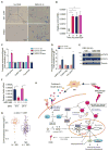
. 2018 Jun 11;33(6):1061-1077.e6.
doi: 10.1016/j.ccell.2018.05.003.

Liver Cancer Initiation Requires p53 Inhibition by CD44-Enhanced Growth Factor Signaling

Affiliations

Liver Cancer Initiation Requires p53 Inhibition by CD44-Enhanced Growth Factor Signaling

Debanjan Dhar et al. Cancer Cell. .

Abstract

How fully differentiated cells that experience carcinogenic insults become proliferative cancer progenitors that acquire multiple initiating mutations is not clear. This question is of particular relevance to hepatocellular carcinoma (HCC), which arises from differentiated hepatocytes. Here we show that one solution to this problem is provided by CD44, a hyaluronic acid receptor whose expression is rapidly induced in carcinogen-exposed hepatocytes in a STAT3-dependent manner. Once expressed, CD44 potentiates AKT activation to induce the phosphorylation and nuclear translocation of Mdm2, which terminates the p53 genomic surveillance response. This allows DNA-damaged hepatocytes to escape p53-induced death and senescence and respond to proliferative signals that promote fixation of mutations and their transmission to daughter cells that go on to become HCC progenitors.

Keywords: CD44; DNA damage response; EGFR; HCC; MDM2 nuclear translocation; cancer initiation; hepatocellular carcinoma; liver cancer; p53; p53 termination.


Conflict of interest statement

DECLARATION OF INTERESTS

The authors declare no competing interests.

Figures
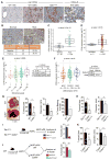
Figure 1
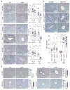
Figure 1. CD44 is upregulated in HCC and is needed for its development
(A) Pan CD44 and CD44v6 IHC of vehicle (Veh) or DEN-challenged 5-month-old WT and 9-month-old Tak1ΔHep livers. (B) CD44 IHC of human normal liver and HCC. (C–D) CD44 mRNA expression in human normal liver and HCC specimens analyzed using Affymetrix Genome U133A 2.0 array (C) or Q-RT-PCR (D). (E–F) Edmonson tumor grading (E) and tumor differentiation (F) were categorized based on CD44 expression using patient samples shown in (D). For panels C–F, the results are expressed as Tukey’s boxplots where box indicates the 1st and 3rd quartiles, bar indicates median, whiskers indicate 1.5 interquartile range (IQR) and data beyond the end of the whiskers represent outliers. Mann-Whitney test was used to test the difference between two groups and Kruskal Wallis test for more than two groups. (G) Gross morphology of 9-month-old DEN-challenged WT and Cd44−/− livers. Tumor multiplicity, tumor size, and tumor incidence were determined. (H) Tumor multiplicity and tumor size in 9-month-old Tak1ΔHep and Cd44−/−;Tak1ΔHep livers. (I) 104 HcPC from 2-month-old Tak1ΔHep and Cd44−/−;Tak1ΔHep mice were transplanted into MUP-uPA mice. Tumor multiplicity was assessed 6 months later (n ≥ 6 mice/group). (J) 104 HcPC from 5-month-old DEN-treated WT mice were transplanted into either MUP-uPA or MUP-uPA;Cd44−/− hosts and tumor multiplicity was assessed 6 months later (n ≥ 3 mice/group). (K) Cd44F/F and Cd44ΔHep males were DEN-challenged and tumor multiplicity and size were determined 9 months later. All bar graphs in panels G–K represent the mean ± SEM. See also Figure S1.
Figure 2
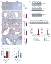
Figure 2. Proliferative pericentral hepatocytes exit the cell cycle in response to DNA damage in the absence of CD44
(A–C) Fifteen-day-old males of indicated genotypes were treated with ± DEN (25 mg/kg), and 9 months later liver sections were stained with either Ki67 (A and C) or p53 (B) antibodies. The numbers of stained zone 3 cells per high magnification field (HMF) were determined (n ≥ 3 mice/group) (“C” = pericentral area; ND = not detectable). Student’s t-test was used to test the difference between two groups and one-way ANOVA with Tukey’s multiple comparison test was used for more than two groups. All bar graphs represent the mean ± SEM. See also Figure S2.
Figure 3
Figure 3. CD44 inhibits killing of DEN-exposed adult pericentral hepatocytes
(A–F) 8–12-week-old WT and Cd44−/− males were DEN-challenged (100 mg/kg). Livers and serum were collected when indicated and analyzed as shown. (A) Liver lysates were IB-analyzed with the indicated antibodies. (B) Serum ALT was measured (n=3). (C) IHC of livers for cleaved-caspase-3 (CC3) (n ≥ 8 different fields from 3 different mice for each time point). (D–E) Ki67 IHC (D) and quantification of Ki67+ hepatocytes (E). (F) Compensatory proliferation index was calculated by dividing the average number of Ki67+ hepatocytes at days 3 and 6 from (E) by the number of CC3+ hepatocytes from (C) at 48 hr post-DEN (n ≥ 6 different fields from 3 different mice for each time point). All bar graphs represent the mean ± SEM. “C” = pericentral area; ND = not detectable. See also Figure S3.
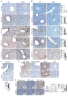
Figure 4
Figure 4. Impaired termination of the p53 response in the absence of CD44
(A–D) 8–12-week-old male mice of indicated genotypes were DEN-challenged (100 mg/kg). Livers and sera were collected when indicated and analyzed: (A) p53 IHC. Bar graphs on the right show number of hepatocytes with nuclear p53 per field (n ≥ 7 different fields from 3 different mice for each time point; “C” = pericentral area). (B–C) IB analysis of liver lysates probing phospho-S15 p53 (B) and p21 (C). (D) Serum ALT (n = 3 mice for each genotype per time point). (E) Fifteen-day-old male mice of indicated genotypes were DEN-challenged (25 mg/kg) and tumor multiplicity was assessed 9 months later. All bar graphs represent the mean ± SEM. See also Figure S4.
Figure 5
Figure 5. CD44 is required for optimal Akt activation and Mdm2 nuclear translocation
(A–C) WT and Cd44−/− males (8–12-week-old) were DEN-challenged (100 mg/kg), their livers were collected when indicated and IHC-analyzed for Mdm2 (A), phospho-S473 Akt (B) and phospho-S166 Mdm2 (C) (n ≥ 6 different fields from 3 different mice for each time point; mean ± SEM). (D) WT mice were treated with Veh or MK2206 (100 mg/kg/day) starting one day prior to DEN challenge (100 mg/kg). Livers were collected 48 hr later and IHC analyzed with the indicated antibodies (n ≥ 3 mice/group). (E) Human HCC tissue array was IHC-analyzed for CD44 and phospho-S166 MDM2. (C = central vein). See also Figure S5.
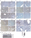
Figure 6
Figure 6. CD44-dependent EGFR activation in pericentral hepatocytes
(A–E) 8–12-week-old male mice of indicated genotypes were DEN-challenged (100 mg/kg) and their livers were collected when indicated. (A) Stained for phospho-Y1068 EGFR. (B) Cd44F/F and Cd44ΔHep livers were IHC-analyzed with indicated antibodies at 3 hr post-DEN. (C) WT and Cd44−/− livers were stained for tEGFR. (D) Hepatocytes were isolated 3 hr post DEN, and IB-analyzed with indicated antibodies. (E) Livers were collected 48 hr after DEN treatment and IHC analyzed for Mdm2 and p53. (n ≥ 15 different fields from ≥ 3 different mice/group; mean ± SEM). “C” = pericentral and “P” = periportal areas. See also Figure S6.
Figure 7
Figure 7. STAT3 controls CD44 expression in pericentral hepatocytes
(A–B) WT males were DEN-challenged (100 mg/kg) and the Cd44 mRNA was analyzed by ISH in their livers (A) or by Q-RT-PCR in isolated hepatocytes (B). (n ≥ 3 mice/group; ND = not detected). (C–D) ChIP assays probing STAT3 recruitment to the Cd44 promoter in DihXY cells with or without serum starvation (C), and with or without IL-6 stimulation (30 min) after serum starvation (D). (E–F) WT mice were treated with Veh or AZD1480 (30 mg/kg/day) starting one day prior to DEN injection (100 mg/kg). Isolated hepatocytes were IB-analyzed as indicated (E), whereas Cd44 mRNA was quantitated by Q-RT-PCR (F) (n ≥ 3 mice/group). All bar graphs in panels B–F represent the mean ± SEM. (G) Human liver adenomas were grouped based on IL6ST mutation status (M=mutated, NM=Not mutated) and CD44 mRNA expression was quantitated. Results are expressed as Tukey’s boxplots where box indicates the 1st and 3rd quartiles, bar indicates median, whiskers indicate 1.5 IQR and data beyond the whiskers represent outliers. (H) Schematic representation of the CD44-Mdm2-p53 circuit that controls HCC initiation. DEN-exposed pericentral hepatocytes undergo DNA damage and mutagenesis. Extensively damaged cells die and release damage associated molecular patterns (DAMPs) that activate macrophages to produce cytokines (IL-6) and growth factors, including EGFR ligands. Hepatocytes with moderate DNA damage mount a DNA damage response that leads to p53 activation and induction of p21Waf1, Noxa, and Puma which mediate cell-cycle arrest or apoptosis. p53 also leads to Mdm2 induction. In pericentral cells, growth factors and IL-6 lead to induction of CD44 which potentiates EGFR and Akt activation, resulting in Mdm2 phosphorylation and nuclear translocation. Nuclear Mdm2 inhibits p53 activation and accumulation. Termination of the p53 response allows carcinogen-exposed pericentral hepatocytes to survive, proliferate and transmit potentially oncogenic mutations to their progeny. See also Figure S7.

Comment in

References

    1. Aubrey BJ, Strasser A, Kelly GL. Tumor-Suppressor Functions of the TP53 Pathway. Cold Spring Harb Perspect Med. 2016;6 - PMC - PubMed
    1. Bahar Halpern K, Shenhav R, Matcovitch-Natan O, Toth B, Lemze D, Golan M, Massasa EE, Baydatch S, Landen S, Moor AE, et al. Single-cell spatial reconstruction reveals global division of labour in the mammalian liver. Nature. 2017;542:352–356. - PMC - PubMed
    1. Bergmann J, Muller M, Baumann N, Reichert M, Heneweer C, Bolik J, Lucke K, Gruber S, Carambia A, Boretius S, et al. IL-6 trans-signaling is essential for the development of hepatocellular carcinoma in mice. Hepatology. 2017;65:89–103. - PubMed
    1. Bettermann K, Vucur M, Haybaeck J, Koppe C, Janssen J, Heymann F, Weber A, Weiskirchen R, Liedtke C, Gassler N, et al. TAK1 suppresses a NEMO-dependent but NF-kappaB-independent pathway to liver cancer. Cancer Cell. 2010;17:481–496. - PubMed
    1. Bourguignon LY. Hyaluronan-mediated CD44 activation of RhoGTPase signaling and cytoskeleton function promotes tumor progression. Semin Cancer Biol. 2008;18:251–259. - PMC - PubMed

Publication types

MeSH terms

Substances