

Seleno-short-chain chitosan induces apoptosis in human breast cancer cells through mitochondrial apoptosis pathway in vitro
- PMID: 29895197
- PMCID: PMC6133326
- DOI: 10.1080/15384101.2018.1464845
Seleno-short-chain chitosan induces apoptosis in human breast cancer cells through mitochondrial apoptosis pathway in vitro
Abstract
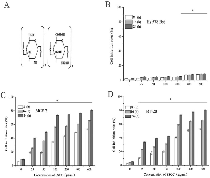
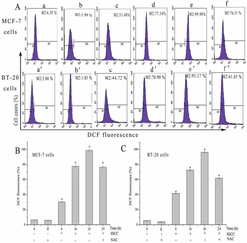
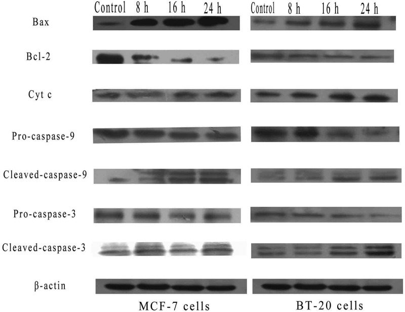
Seleno-short-chain chitosan (SSCC) was a synthesized chitosan derivative with the molecular weight of 4826.986 Da. The study is aimed to investigate cytotoxicity of SSCC on human breast cancer MCF-7 and BT-20 cells and explore apoptosis-related mechanism in vitro. The MTT (3- [4,5-Dimethylthiazol-2-yl]-2, 5-diphenylterazolium bromide) assay showed that SSCC exhibited significantly cytotoxic effects on MCF-7 and BT-20 cells in a dose- and time-dependent manner, and the effective inhibitory concentration was 100 μg/ml and 200 μg/ml, respectively. Apoptosis assay of these two kinds of cells was determined by Hoechst 33,342/PI and Annexin V-FITC/PI double staining. The cell cycle assay showed that SSCC triggered S and G2/M phase cell cycle arrest in MCF-7 cells and S phase cell cycle arrest in BT-20 cells in a time-dependent manner. Further studies demonstrated that SSCC led to the generation of reactive oxygen species (ROS) and the disruption of mitochondrial membrane potential (MMP) in these two kinds of cells. N- acetyl-L cysteine (NAC), as a radical scavenger, significantly inhibited the generation of ROS and decreased the apoptosis of MCF-7 and BT-20 cells. Moreover, the expression of mitochondrial apoptosis-related proteins was detected by western blot assay. SSCC up-regulated the expression of Bax, down-regulated the expression of Bcl-2, subsequently increased the release of cytochrome c from mitochondria to cytoplasm, and activated the cleavage of caspase-9 and -3, which finally induced apoptosis in MCF-7 and BT-20 cells in vitro. Consequently, these data indicated that SSCC could induce apoptosis of MCF-7and BT-20 cells in vitro by mitochondrial pathway.
Keywords: NAC; Seleno-short-chain chitosan; breast cancer BT-20 cells; breast cancer MCF-7 cells; mitochondrial apoptosis pathway.
Figures


Similar articles
-
Seleno-short-chain chitosan induces apoptosis in human non-small-cell lung cancer A549 cells through ROS-mediated mitochondrial pathway.Cytotechnology. 2017 Dec;69(6):851-863. doi: 10.1007/s10616-017-0098-z. Epub 2017 Apr 18. Cytotechnology. 2017. PMID: 28421411 Free PMC article.
-
Antitumor effects of seleno-short-chain chitosan (SSCC) against human gastric cancer BGC-823 cells.Cytotechnology. 2019 Dec;71(6):1095-1108. doi: 10.1007/s10616-019-00347-w. Epub 2019 Oct 9. Cytotechnology. 2019. PMID: 31598888 Free PMC article.
-
Combination treatment of ligustrazine piperazine derivate DLJ14 and adriamycin inhibits progression of resistant breast cancer through inhibition of the EGFR/PI3K/Akt survival pathway and induction of apoptosis.Drug Discov Ther. 2014 Feb;8(1):33-41. doi: 10.5582/ddt.8.33. Drug Discov Ther. 2014. PMID: 24647156
-
Chimaphilin induces apoptosis in human breast cancer MCF-7 cells through a ROS-mediated mitochondrial pathway.Food Chem Toxicol. 2014 Aug;70:1-8. doi: 10.1016/j.fct.2014.04.014. Epub 2014 May 1. Food Chem Toxicol. 2014. PMID: 24793375
-
Antitumor effects of seleno-β-lactoglobulin (Se-β-Lg) against human gastric cancer MGC-803 cells.Eur J Pharmacol. 2018 Aug 15;833:109-115. doi: 10.1016/j.ejphar.2018.05.042. Epub 2018 May 30. Eur J Pharmacol. 2018. PMID: 29859147
Cited by
-
Designing and the anticancer activity of chitosan and chitosan oligosaccharide lactate nanobeads loaded with Biginelli hybrid.RSC Adv. 2024 Oct 4;14(43):31526-31534. doi: 10.1039/d4ra05783j. eCollection 2024 Oct 1. RSC Adv. 2024. PMID: 39372042 Free PMC article.
-
Chitosan gold nanoparticles induce different ROS-dependent cell death modalities in leukemic cells.Int J Nanomedicine. 2019 Sep 4;14:7173-7190. doi: 10.2147/IJN.S221021. eCollection 2019. Int J Nanomedicine. 2019. PMID: 31564872 Free PMC article.
-
BRD4 inhibitor nitroxoline enhances the sensitivity of multiple myeloma cells to bortezomib in vitro and in vivo by promoting mitochondrial pathway-mediated cell apoptosis.Ther Adv Hematol. 2020 Jun 8;11:2040620720932686. doi: 10.1177/2040620720932686. eCollection 2020. Ther Adv Hematol. 2020. PMID: 32551032 Free PMC article.
-
Chitosan-Based Nano-Smart Drug Delivery System in Breast Cancer Therapy.Pharmaceutics. 2023 Mar 8;15(3):879. doi: 10.3390/pharmaceutics15030879. Pharmaceutics. 2023. PMID: 36986740 Free PMC article. Review.
-
Marine Compounds, Mitochondria, and Malignancy: A Therapeutic Nexus.Mar Drugs. 2022 Sep 30;20(10):625. doi: 10.3390/md20100625. Mar Drugs. 2022. PMID: 36286449 Free PMC article. Review.
References
-
- McNamara KM, Kannai A. Possible roles for glucocorticoid signalling in breast cancer. Mol Cell Endocrinol. 2018;5:38–50. - PubMed
-
- Vishal M, Swetha R, Thejaswini G, et al. Role of Runx2 in breast cancer-mediated bone metastasis. Int J Biol Macromol. 2017;99:608–614. - PubMed
-
- Wang S, Li W, Fang W, et al. 36 cases adenoid cystic carcinoma of the breast in China: comparison with matched grade one invasive ductal carcinoma-not otherwise specified. Pathol Res Pract. 2017;213(4):310–315. - PubMed
-
- Wakeam E, Acuna SA, Leighl NB, et al. Surgery versus chemotherapy and radiotherapy for early and locally advanced small cell lung cancer: a propensity-matched analysis of survival. Lung Cancer. 2017;109:78–88. - PubMed
-
- Saltz LB, Clarke S, Diazrubio E, et al. Bevacizumab in combination with oxaliplatin-based chemotherapy as first-line therapy in metastatic colorectal cancer: a randomized phase III study. J Clin Oncol. 2008;26(12):2013–2019. - PubMed
MeSH terms
Substances
LinkOut - more resources
Full Text Sources
Other Literature Sources
Medical
Research Materials
Miscellaneous