. 2018 Jun 13;9(1):156.
doi: 10.1186/s13287-018-0907-0.

Use of bioreactors for culturing human retinal organoids improves photoreceptor yields

Affiliations

Use of bioreactors for culturing human retinal organoids improves photoreceptor yields

Patrick Ovando-Roche et al. Stem Cell Res Ther. .

Abstract

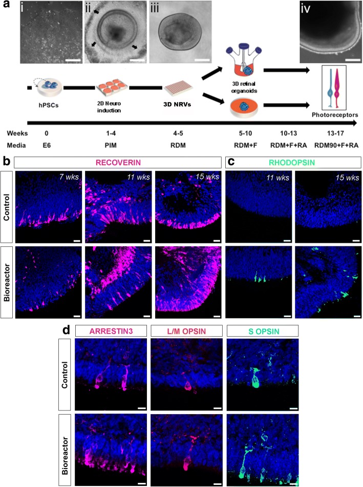
Background: The use of human pluripotent stem cell-derived retinal cells for cell therapy strategies and disease modelling relies on the ability to obtain healthy and organised retinal tissue in sufficient quantities. Generating such tissue is a lengthy process, often taking over 6 months of cell culture, and current approaches do not always generate large quantities of the major retinal cell types required.

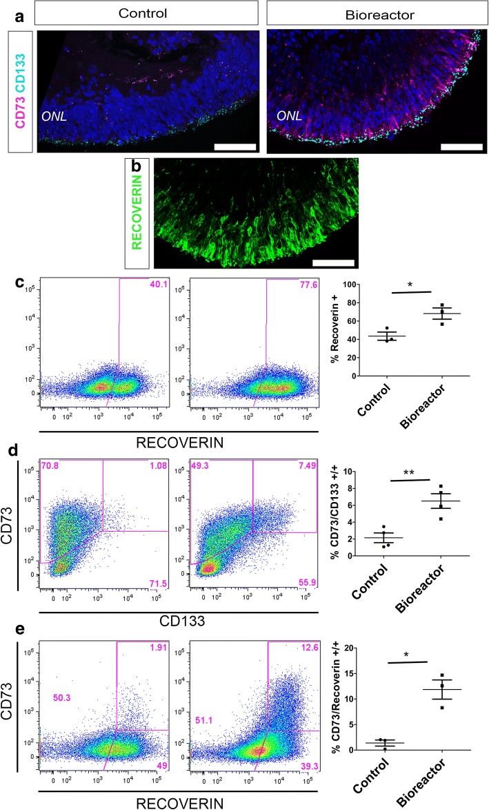
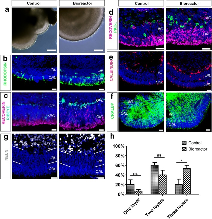
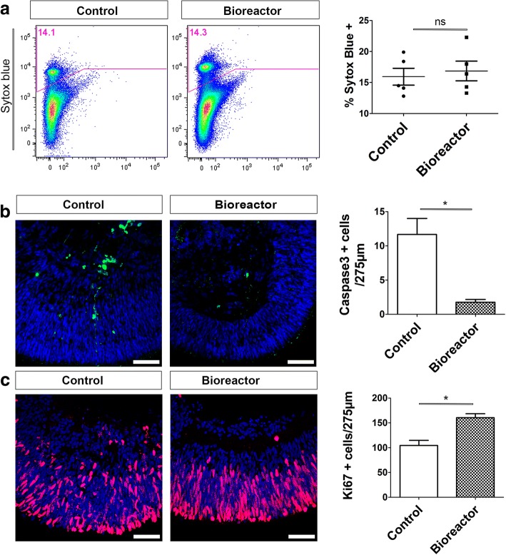
Methods: We adapted our previously described differentiation protocol to investigate the use of stirred-tank bioreactors. We used immunohistochemistry, flow cytometry and electron microscopy to characterise retinal organoids grown in standard and bioreactor culture conditions.

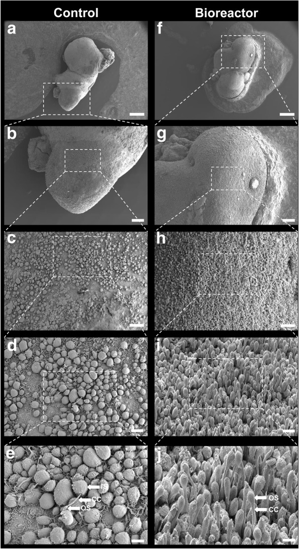
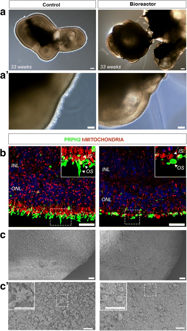
Results: Our analysis revealed that the use of bioreactors results in improved laminar stratification as well as an increase in the yield of photoreceptor cells bearing cilia and nascent outer-segment-like structures.

Conclusions: Bioreactors represent a promising platform for scaling up the manufacture of retinal cells for use in disease modelling, drug screening and cell transplantation studies.

Keywords: Bioreactors; Photoreceptors; Pluripotent stem cells; Retinal organoids.


Conflict of interest statement

Ethics approval and consent to participate

Not applicable.

Competing interests

The authors declare that they have no competing interests.

Publisher’s Note

Springer Nature remains neutral with regard to jurisdictional claims in published maps and institutional affiliations.

Figures

Fig. 1
Fig. 1
Photoreceptor maturation following bioreactor differentiation. a Schematic showing different stages of differentiation protocol. Representative phase-contrast images: (i) hPSCs at day 0 of differentiation; (ii) developing neuroepithelia surrounded by RPE cells (black arrows) at week 4; (iii) isolated retinal cup at week 5; (iv) mature retinal organoid at week 17. Following retinal cup formation, samples cultured in 100-mm cell culture plates or 100-ml bioreactors to form retinal organoids. Mature photoreceptors observed between days 90 and 120. b, c RECOVERIN and RHODOPSIN-positive photoreceptor cells in both control and bioreactor conditions at indicated number of weeks of differentiation. d Immunohistochemistry of L/M-OPSIN, Cone Arrestin (ARRESTIN3) and S-OPSIN cone markers at week 16 of differentiation in control and bioreactor conditions. Scale bars: 20 μm (a), 25 μM (b, c), 10 μM (d). hPSC human pluripotent stem cell, NRV neuroretinal vesicle, RDM + F RDM supplemented with 10% foetal bovine serum + 2% Glutamax + 100 μM taurine, RA retinoic acid
Fig. 2
Fig. 2
Photoreceptor quantification following bioreactor differentiation. a, b Cryosections from week 16 bioreactor cultures showing both CD73 and CD133 cell surface markers localised to ONL-like region (a) where RECOVERIN-positive photoreceptors are also located (b). c–e Representative FC analysis of week 16 bioreactor cultures vs control cultures: RECOVERIN-positive photoreceptor cells (68.17% ± 6.15 vs 43.53% ± 4.47) (c); CD133/CD73 double-positive developing rods (6.50% ± 0.86 vs 2.14% ± 0.57) (d); RECOVERIN/CD73 mature photoreceptors (11.87% ± 1.88 vs 1.38% ± 0.59) (e). Error bars, mean ± SEM; n = 50 retinal organoids per individual FC experiment, N = 3–4 independent differentiation experiments; *P < 0.05, **P < 0.01, two-tailed unpaired t test with Welch’s correction. Scale bars: 50 μM (a, b). ONL outer nuclear layer
Fig. 3
Fig. 3
Characterisation of neuroepithelia lamination. a–f Phase-contrast images (a) and immunohistochemical analysis (b–f) of week 16 hPSC-derived retinal organoids grown in bioreactors and in control conditions. RHODOPSIN-positive (b) and RECOVERIN-positive photoreceptors in ONL and RIBEYE-positive ribbon synapses in OPL (c). PKCα-positive rod bipolar cells (d) and CALBINDIN-positive amacrine and horizontal interneurons in INL (e). CRALBP-positive Müller glia cells also present in both culture conditions (f). g Presence of NEUN-positive RGCs and subset of amacrine cells. h Quantification of number of retinal layers in control and bioreactor retinal organoids following DAPI staining. n = 10 random regions of retinal neuroepithelia per independent experiment, N = 3 independent experiments. Error bars, mean ± SEM; ns, P > 0.05; *P < 0.05, two-way ANOVA. Scale bars: 25 μM (a, b, d–g), 10 μM (c). GCL ganglion cell layer, INL inner nuclear layer, ns not significant, ONL outer nuclear layer, OPL outer plexiform layer
Fig. 4
Fig. 4
Bioreactor culture of retinal organoids increases cell proliferation and reduces cell death. a FC analysis of week 16 control vs bioreactor hPSC-derived retinal organoids stained with SYTOX Blue (error bars, mean ± SEM; n = 50 retinal organoids, N = 5 independent experiments; ns, P > 0.05, two-tail unpaired t test with Welch’s correction). b Immunohistochemistry analysis and quantification of cleaved caspase-3 retinal organoids showing reduction in number of caspase-3-positive apoptotic cells (error bars, mean ± SEM; n = 10, N = 3; *P < 0.05, two-tail unpaired t test with Welch’s correction). c Immunohistochemistry and quantification of Ki67 proliferative cells revealed significant increase in number of proliferating cells present in neuroepithelia of bioreactor cultures (error bars, mean ± SEM; n = 10 images, N = 3 independent experiments; *P < 0.05, two-tail unpaired t test with Welch’s correction). ns not significant
Fig. 5
Fig. 5
Ultrastructural and topographical analysis of human hPSC-derived neuroepithelia and photoreceptor cells at early stages of development. a Diagrammatic representation of key structures of mature photoreceptor. b Immunohistochemical staining of cone photoreceptor (L/M-OPSIN, magenta) and its ribbon synapse (RIBEYE, green, arrow). c TEM image illustrating morphology of hPSC-derived photoreceptor OLM (arrows), IS, CC and OS-like structures. d SEM image from etched resin section 2 μm thick showing IS, CC and OS. e TEM images illustrating internal organisation of CC (high magnification box) and OS. f Backscatter EM images from different levels of 3view dataset showing photoreceptor ribbon synapse. g 3view sequence of backscatter EM images of hPSC-derived photoreceptor OS (yellow), CC (green) and IS (red). See Additional file 2: Movie 1. h 3view 3D reconstruction of 150 sections with thickness of 100 nm each. Representative photoreceptor OS, CC and IS observed in yellow, green and red respectively. CC connecting cilium, IS inner segment, OLM outer limiting membrane, OS outer segment
Fig. 6
Fig. 6
SEM images showing topography of whole retinal organoids. Topographic features of neuroepithelia showing photoreceptor cell density and morphology from control (a–e) vs bioreactor (f–j) at ascending magnifications. Scale bars: 400 μM (a, f), 100 μM (b, g), 30 μM (c, h), 10 μM (d, i), 5 μM (e, j). CC connecting cilium, IS inner segment, OS outer segment
Fig. 7
Fig. 7
Characterisation of human hPSC-derived neuroepithelia and photoreceptor cells at late stages of development. a Representative bright-field images of retinal organoids cultured in standard plates and bioreactors. a′ High-magnification images of neuroepithelia region showing CC/OS brush border in control but not in bioreactor samples. b Immunohistochemistry analysis for hMITOCHONDRIA and PRPH2 to delineate IS and OS, respectively, showed elongated OS-like structures in control samples only. c, c′ SEM images showing the topography of whole retinal organoids. Photoreceptor cell density and morphology from control vs bioreactor at ascending magnifications. Scale bars: 200 μM (a), 100 μM (a′), 25 μM (b), 20 μM (c), 10 μM (c′ and high magnification box). INL inner nuclear layer, IS inner segment, ONL outer nuclear layer, OS outer segment

References

    1. Aghaizu ND, Kruczek K, Gonzalez-Cordero A, et al. Pluripotent stem cells and their utility in treating photoreceptor degenerations. Prog Brain Res. 2017;231:191–223. doi: 10.1016/bs.pbr.2017.01.001. - DOI - PubMed
    1. Di Foggia V, Makwana P, Ali RR, et al. Induced pluripotent stem cell therapies for degenerative disease of the outer retina: disease modeling and cell replacement. J Ocul Pharmacol Ther. 2016;32(5):240–252. doi: 10.1089/jop.2015.0143. - DOI - PMC - PubMed
    1. Ovando-Roche P, Georgiadis A, Smith AJ, et al. Harnessing the potential of human pluripotent stem cells and gene editing for the treatment of retinal degeneration. Curr Stem Cell Rep. 2017;3(2):112–123. doi: 10.1007/s40778-017-0078-4. - DOI - PMC - PubMed
    1. Duong TT, Vasireddy V, Mills JA, et al. Retinas in a dish peek into inherited retinal degeneration. Cell Stem Cell. 2016;18(6):688–689. doi: 10.1016/j.stem.2016.05.021. - DOI - PubMed
    1. Khurana V, Tardiff DF, Chung CY, et al. Toward stem cell-based phenotypic screens for neurodegenerative diseases. Nat Rev Neurol. 2015;11(6):339–350. doi: 10.1038/nrneurol.2015.79. - DOI - PubMed

Publication types