

Conditioned medium from bone marrow-derived mesenchymal stem cells inhibits vascular calcification through blockade of the BMP2-Smad1/5/8 signaling pathway
- PMID: 29895327
- PMCID: PMC5998505
- DOI: 10.1186/s13287-018-0894-1
Conditioned medium from bone marrow-derived mesenchymal stem cells inhibits vascular calcification through blockade of the BMP2-Smad1/5/8 signaling pathway
Abstract
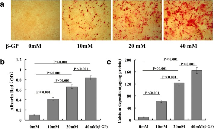
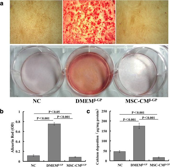
Background: Arterial calcification is associated with cardiovascular disease as a complication of advanced atherosclerosis and is a significant contributor to cardiovascular morbidity and mortality. Osteoblastic differentiation of vascular smooth muscle cells (VSMCs) plays an important role in arterial calcification and is characterized by cellular necrosis, inflammation, and lipoprotein and phospholipid complexes, especially in atherosclerotic calcification. The conditioned medium from bone marrow-derived mesenchymal stem cells (MSC-CM) is well known as a rich source of autologous cytokines and is universally used for tissue regeneration in current clinical medicine. Here, we demonstrate that MSC-CM inhibits beta-glycerophosphate (β-GP)-induced vascular calcification through blockade of the bone morphogenetic protein-2 (BMP2)-Smad1/5/8 signaling pathway.
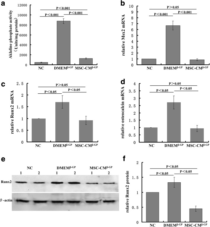
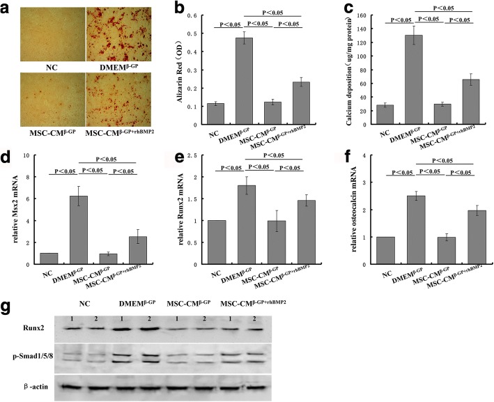
Methods: VSMC calcification was induced by β-GP followed by treatment with MSC-CM. Mineral deposition was assessed by Alizarin Red S staining. Intracellular calcium content was determined colorimetrically by the o-cresolphthalein complexone method and alkaline phosphatase (ALP) activity was measured by the para-nitrophenyl phosphate method. Expression of BMP2, BMPR1A, BMPR1B, BMPR2, msh homeobox 2 (Msx2), Runt-related transcription factor 2 (Runx2), and osteocalcin (OC), representative osteoblastic markers, was assessed using real-time polymerase chain reaction analysis while the protein expression of BMP2, Runx2, and phosphorylated Smad1/5/8 was detected by western blot analysis.
Results: Our data demonstrated that MSC-CM inhibits osteoblastic differentiation and mineralization of VSMCs as evidenced by decreased calcium content, ALP activity, and decreased expression of BMP-2, Runx2, Msx2, and OC. MSC-CM suppressed the expression of phosphorylated Smad1/5/8 and the β-GP-induced translocation from the cytoplasm to the nucleus. Further study demonstrated that human recombinant BMP-2 overcame the suppression of VSMC calcification by MSC-CM.
Conclusion: MSC-CM may act as a novel therapy for VSMC calcification by mediating the BMP2-Smad1/5/8 signaling pathway.
Keywords: Atherosclerosis; BMP2–Smad1/5/8 signaling; Conditioned medium from bone marrow-derived mesenchymal stem cells; Vascular calcification.
Conflict of interest statement
Ethics approval and consent to participate
Not applicable.
Consent for publication
All authors have agreed to the publication of this manuscript.
Competing interests
The authors declare that they have no competing interests.
Publisher’s Note
Springer Nature remains neutral with regard to jurisdictional claims in published maps and institutional affiliations.
Figures


Similar articles
-
The Bioactive Substance Secreted by MSC Retards Mouse Aortic Vascular Smooth Muscle Cells Calcification.Biomed Res Int. 2018 Jun 3;2018:6053567. doi: 10.1155/2018/6053567. eCollection 2018. Biomed Res Int. 2018. PMID: 29967775 Free PMC article.
-
Ginkgo Biloba Extract EGB761 Alleviates Warfarin-induced Aortic Valve Calcification Through the BMP2/Smad1/5/Runx2 Signaling Pathway.J Cardiovasc Pharmacol. 2021 Sep 1;78(3):411-421. doi: 10.1097/FJC.0000000000001082. J Cardiovasc Pharmacol. 2021. PMID: 34132687 Free PMC article.
-
Noggin suppression decreases BMP-2-induced osteogenesis of human bone marrow-derived mesenchymal stem cells in vitro.J Cell Biochem. 2012 Dec;113(12):3672-80. doi: 10.1002/jcb.24240. J Cell Biochem. 2012. PMID: 22740073
-
The use of cell conditioned medium for musculoskeletal tissue regeneration.J Cell Physiol. 2018 Jun;233(6):4423-4442. doi: 10.1002/jcp.26291. Epub 2017 Dec 29. J Cell Physiol. 2018. PMID: 29159853 Review.
-
The Role of Vitamin D in Modulating Mesenchymal Stem Cells and Endothelial Progenitor Cells for Vascular Calcification.Int J Mol Sci. 2020 Apr 2;21(7):2466. doi: 10.3390/ijms21072466. Int J Mol Sci. 2020. PMID: 32252330 Free PMC article. Review.
Cited by
-
The Role of SIRT3 in the Osteoporosis.Front Endocrinol (Lausanne). 2022 May 25;13:893678. doi: 10.3389/fendo.2022.893678. eCollection 2022. Front Endocrinol (Lausanne). 2022. PMID: 35692409 Free PMC article. Review.
-
Supercritical Carbon Dioxide Decellularized Bone Matrix Seeded with Adipose-Derived Mesenchymal Stem Cells Accelerated Bone Regeneration.Biomedicines. 2021 Dec 3;9(12):1825. doi: 10.3390/biomedicines9121825. Biomedicines. 2021. PMID: 34944642 Free PMC article.
-
Development of stem cell therapy for atherosclerosis.Mol Cell Biochem. 2024 Apr;479(4):779-791. doi: 10.1007/s11010-023-04762-8. Epub 2023 May 13. Mol Cell Biochem. 2024. PMID: 37178375 Review.
-
Stem Cell Based Approaches to Modulate the Matrix Milieu in Vascular Disorders.Front Cardiovasc Med. 2022 Jun 15;9:879977. doi: 10.3389/fcvm.2022.879977. eCollection 2022. Front Cardiovasc Med. 2022. PMID: 35783852 Free PMC article. Review.
-
The Cell Origin and Role of Osteoclastogenesis and Osteoblastogenesis in Vascular Calcification.Front Cardiovasc Med. 2021 Apr 23;8:639740. doi: 10.3389/fcvm.2021.639740. eCollection 2021. Front Cardiovasc Med. 2021. PMID: 33969008 Free PMC article. Review.
References
Publication types
MeSH terms
Substances
LinkOut - more resources
Full Text Sources
Other Literature Sources
Research Materials
Miscellaneous
