. 2018 Jun 12;19(6):1737.
doi: 10.3390/ijms19061737.

Generation of Human Breg-Like Phenotype with Regulatory Function In Vitro with Bacteria-Derived Oligodeoxynucleotides

Affiliations

Generation of Human Breg-Like Phenotype with Regulatory Function In Vitro with Bacteria-Derived Oligodeoxynucleotides

Jorge Gallego-Valle et al. Int J Mol Sci. .

Abstract

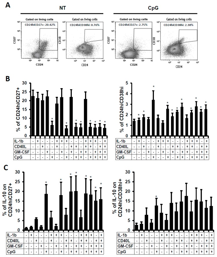
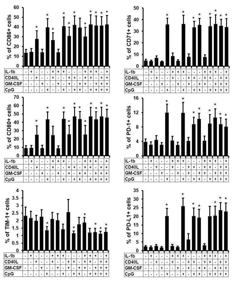
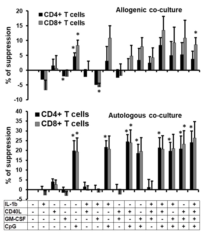
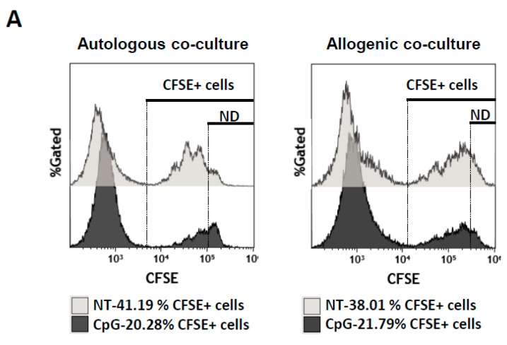
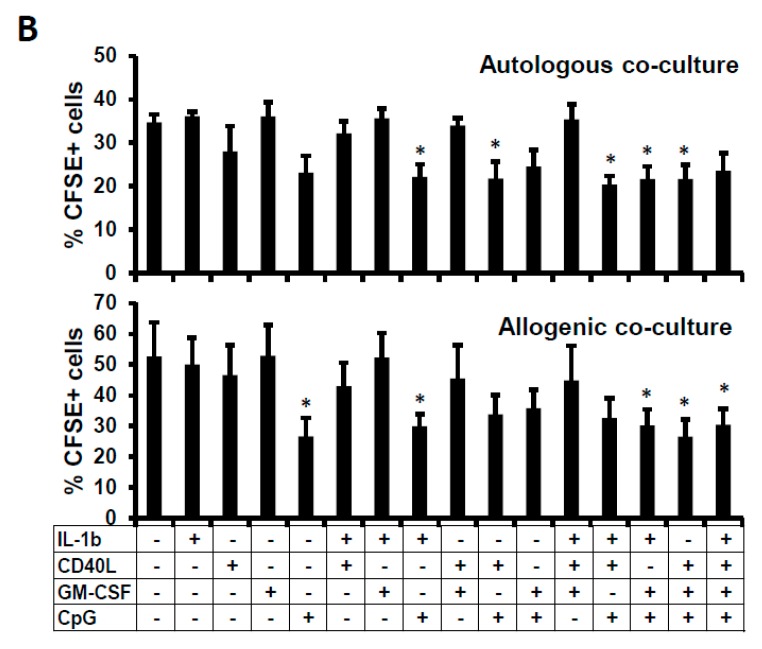
Regulatory B cells (Bregs) participate in auto-tolerance maintenance and immune homeostasis. Despite their impact on many diseases and due to the difficulty to define them, knowledge about their origin and their physiological inducers is still unclear. The incomplete understanding about the generation of Bregs and their limited numbers in periphery make it difficult to develop Breg-based therapy. Therefore, identifying factors that promote their development would allow their ex-vivo production in order to create new immunotherapy. This project aims to test the capacity of several cytokines (Interleukin 1-beta (IL-1β), Granulocyte Macrophage Colony-Stimulating Factor (GM-CSF), and Cluster of differentiation 40 ligand (CD40L)) and bacteria-derived oligodeoxynucleotides (CpG-ODN), alone or in combination, to generate B cells with regulatory phenotype and function. We have demonstrated that the Breg-associated phenotypes were heterogeneous between one and other stimulation conditions. However, the expression of other markers related to Bregs such as IL-10, CD80, CD86, CD71, Programmed cell death-1 (PD-1), and Programmed death-ligand 1 (PD-L1) was increased when cells were stimulated with CpG alone or in combination. Moreover, stimulated B cells presented a suppressive function on autologous activated peripheral blood mononuclear cells (PBMC) proliferation. Therefore, this work is the first step to demonstrate the feasibility to induce functional Breg-like cells in vitro and will then facilitate the way to produce Breg-like cells as a potential future cellular therapy.

Keywords: Breg-like B cells; IL-10-producing B cells; suppressive function.


Conflict of interest statement

The authors declare no conflict of interest.

Figures

Figure 1
Figure 1
IL-10-producing B cells after B-cell stimulation. B cells were treated for 48 h and analysed by flow cytometry. (A) Total IL-10-producing cells were analysed within viable cells using intracellular labelling in non-treated (NT) and CpG-treated B cells (dot plots). Dot plot of one representative experiment out of five is shown. Numbers represent the percentages of living cells and total IL-10-producing B cells. (B) Average percentages of IL-10-producing viable non-stimulated or stimulated B cells was analysed. Cultures were performed in the presence of IL-1β, CD40L, GM-CSF, or CpG. Average + SEM of five experiments for each condition are shown. * p < 0.05 when comparing non-treated condition versus treated condition.
Figure 2
Figure 2
Frequency of two Breg subsets after B-cell stimulation. B cells were non-treated (NT) or treated with IL-1β, CD40L, GM-CSF, or CpG alone, or in combination. 48 h post-stimulation, percentages of (A) CD24hiCD27+ and CD24hiCD38hi viable cells were followed. Dot plot of one representative experiment out of five is shown. Numbers represent the percentages of the two Breg subsets. (B) Average percentages of CD24hiCD27+ and CD24hiCD38hi non stimulated or stimulated viable B cells. (C) Percentages of IL-10 positive cells were detected within CD24hiCD27+ and CD24hiCD38hi viable B cells. Average + SEM of five experiments. * p < 0.05 when comparing non-treated condition versus treated condition.
Figure 3
Figure 3
Frequency of Breg-related surface marker subsets. B cells were non-treated (NT) or treated with IL-1β, CD40L, GM-CSF, or CpG alone, or in combination. 48 h post-stimulation, surface markers such as CD86, CD80, TIM-1, CD71, PD-1 and PD-L1 were followed by flow cytometry. Average percentages of these markers are presented. Average + SEM of five experiments. * p < 0.05 when comparing non-treated condition versus treated condition.
Figure 4
Figure 4
Anti-proliferative function of the stimulated B cells. Percentages of suppression of proliferation of allogenic (upper panel) or autologous PBMCs (lower panel) treated with CFSE and co-cultured with non-treated or treated-B cells (ratio B cells:PBMCs of 2:1) at day 3 post-co-culture. B cells were non-treated or treated with IL-1β, CD40L, GM-CSF, or CpG alone, or in combination for 2 days before co-culture with CFSE+-PBMCs. Average + SEM of the percentages of suppression of CD4+ and CD8+ T cell proliferation from four different experiments are represented. * p < 0.05 when comparing non-treated condition versus treated condition.
Figure 5
Figure 5
Relation between suppression of proliferation and cell death. (A) Histograms of CFSE+ cells in the co-culture experiment between non-treated (NT) or CpG-stimulated B cells and CFSE+-PBMCs (ratio B cells: CFSE+-PBMCs of 2:1) in autologous and allogenic conditions. Numbers represent the percentages of the CFSE+ cells in the co-culture. ND for non-divided cells (B) Average percentages of CFSE+ cells after 3 days of co-culture with B cells either non-treated or treated with IL-1β, CD40L, GM-CSF, or CpG alone, or in combination for 2 days. Average + SEM of four experiments. Loss of CFSE+ cells might be due to a lack of cellular proliferation or due to CFSE+ cell death. Percentage of suppression of proliferation of CD4+ or CD8+ T cells was calculated as follows: (frequency of dividing cells = 100 − (proliferation of CFSE+-labeled cells in co-culture with stimulated B cells × 100/proliferation of CFSE-labeled T cells in co-culture with non-treated B cells)) [20]. * p < 0.05 when comparing non-treated condition versus treated condition.
Figure 5
Figure 5
Relation between suppression of proliferation and cell death. (A) Histograms of CFSE+ cells in the co-culture experiment between non-treated (NT) or CpG-stimulated B cells and CFSE+-PBMCs (ratio B cells: CFSE+-PBMCs of 2:1) in autologous and allogenic conditions. Numbers represent the percentages of the CFSE+ cells in the co-culture. ND for non-divided cells (B) Average percentages of CFSE+ cells after 3 days of co-culture with B cells either non-treated or treated with IL-1β, CD40L, GM-CSF, or CpG alone, or in combination for 2 days. Average + SEM of four experiments. Loss of CFSE+ cells might be due to a lack of cellular proliferation or due to CFSE+ cell death. Percentage of suppression of proliferation of CD4+ or CD8+ T cells was calculated as follows: (frequency of dividing cells = 100 − (proliferation of CFSE+-labeled cells in co-culture with stimulated B cells × 100/proliferation of CFSE-labeled T cells in co-culture with non-treated B cells)) [20]. * p < 0.05 when comparing non-treated condition versus treated condition.

References

    1. Medzhitov R. Inflammation 2010: New adventures of an old flame. Cell. 2010;140:771–776. doi: 10.1016/j.cell.2010.03.006. - DOI - PubMed
    1. Chaudhry A., Rudensky A.Y. Control of inflammation by integration of environmental cues by regulatory T cells. J. Clin. Investig. 2013;123:939–944. doi: 10.1172/JCI57175. - DOI - PMC - PubMed
    1. Ohkura N., Kitagawa Y., Sakaguchi S. Development and maintenance of regulatory T cells. Immunity. 2013;38:414–423. doi: 10.1016/j.immuni.2013.03.002. - DOI - PubMed
    1. Balkwill F., Montfort A., Capasso M. B regulatory cells in cancer. Trends Immunol. 2013;34:169–173. doi: 10.1016/j.it.2012.10.007. - DOI - PubMed
    1. Berthelot J.M., Jamin C., Amrouche K., le Goff B., Maugars Y., Youinou P. Regulatory B cells play a key role in immune system balance. Joint Bone Spine. 2013;80:18–22. doi: 10.1016/j.jbspin.2012.04.010. - DOI - PubMed

MeSH terms

LinkOut - more resources