. 2018 Jun 12;8(1):8973.
doi: 10.1038/s41598-018-27190-x.

A System of Cytokines Encapsulated in ExtraCellular Vesicles

Affiliations

A System of Cytokines Encapsulated in ExtraCellular Vesicles

Wendy Fitzgerald et al. Sci Rep. .

Erratum in

Abstract

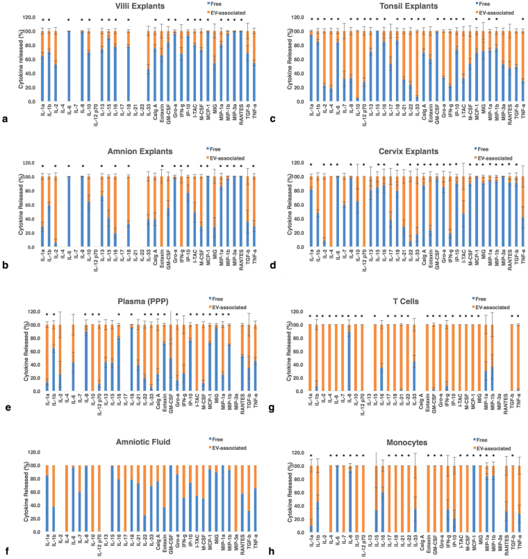
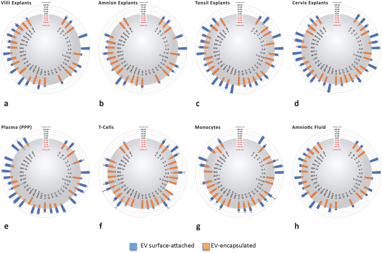
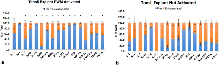
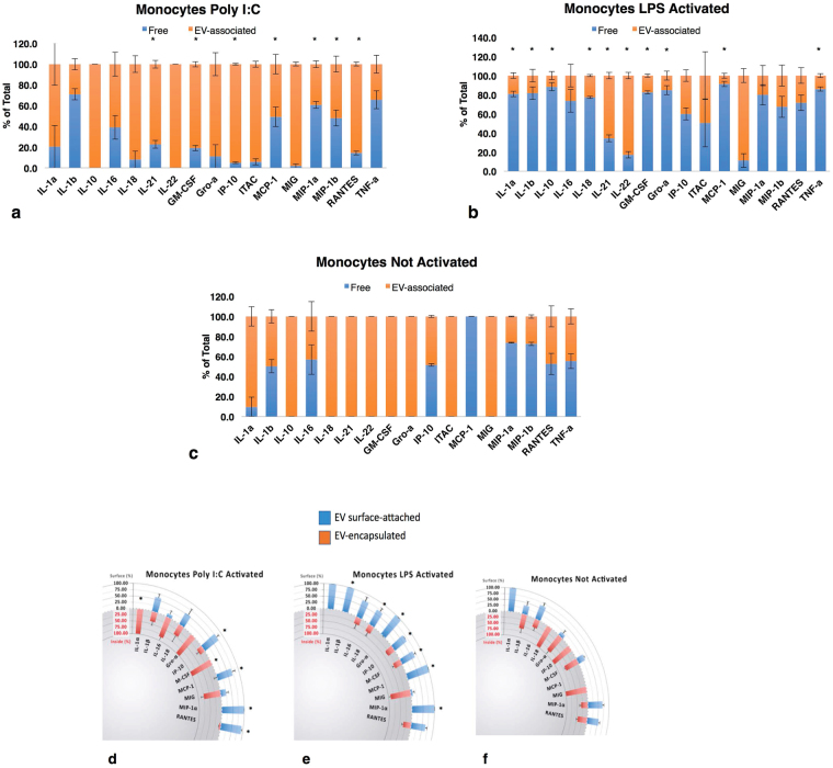
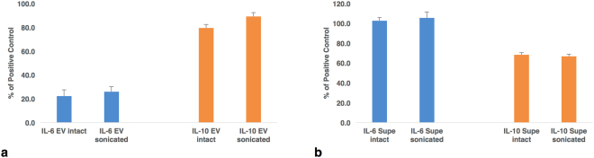
Cytokines are soluble factors that mediate cell-cell communications in multicellular organisms. Recently, another system of cell-cell communication was discovered, which is mediated by extracellular vesicles (EVs). Here, we demonstrate that these two systems are not strictly separated, as many cytokines in vitro, ex vivo, and in vivo are released in EV-encapsulated forms and are capable of eliciting biological effects upon contact with sensitive cells. Association with EVs is not necessarily a property of a particular cytokine but rather of a biological system and can be changed upon system activation. EV-encapsulated cytokines were not detected by standard cytokine assays. Deciphering the regulatory mechanisms of EV-encapsulation will lead to a better understanding of cell-cell communications in health and disease.


Conflict of interest statement

The authors declare no competing interests.

Figures

Figure 1
Figure 1
Free and EV-associated cytokines released by tissue explants. Cytokines in free form and EV-associated are expressed as percent of total cytokine released ± SEM. Blue bars: cytokine in “free” form, orange: EV-associated. Note that the same cytokines can be predominantly in free form or in EV-associated form depending on the biological system. (a) Placental villous explants: cumulative amounts of products released over 7 days, n = 10; (b) Amnion explants: amounts released over 7 days, n = 10; (c) Tonsillar explants: amounts released over 9 days, n = 5; (d) Cervical explants: amounts released over 9 days, n = 6; (e) PPP from healthy donors, n = 52; (f) Amniotic fluid from 3 donors; (g) T cells: amounts released over one day, n = 6; (h) Monocytes: amounts released over one day, n = 6. *Indicates significant difference p < 0.05 between free and EV- associated cytokines. Cumulative amounts are the sum of measurements made at day 3, 6, and 9 for tonsil and cervix, and day 1, 4, and 7 for villi and amnion explants.
Figure 2
Figure 2
Distribution of cytokines between the surface and the inner volume of EVs. Fractions of total EV-associated cytokines ± SEM. Samples collected as in Fig. 1. (a) Placental villous explants n = 10; (b) Amnion explants, n = 10; (c) Tonsillar explants n = 5; (d) Cervical explants n = 6; (e) PPP from healthy donors n = 52; (f) Amniotic fluid from 3 donors (g) T cells n = 6; (h) Monocytes n = 6. *Indicates significant difference p < 0.05 between surface and encapsulated cytokines.
Figure 3
Figure 3
Cytokine distributions between EV-associated and free forms in stimulated tonsils. Tonsil explants with or without stimulation by pokeweed mitogen for 3 days. Supernatants free of EV and EVs lysed with 1% Triton-X. Free and EV-associated cytokines are expressed as percent of total ± SEM. Blue bars: “free” cytokines, orange: EV-associated, n = 4. Cytokines released by tonsil explants: (a) stimulated with pokeweed mitogen; (b) without stimulation. *Indicates significant difference p < 0.05 in cytokine distribution between PWM-activated and controls.
Figure 4
Figure 4
Cytokine distributions between EV-associated and free forms in stimulated monocytes. Monocytes were cultured without stimuli (c) or stimulated by poly I:C (a) or LPS (b) Free and EV-associated cytokines are expressed as percent of total ± SEM. Blue bars: “free” cytokines, orange: EV-associated. Distribution between encapsulated and surface attached selected cytokines which changed with stimulation a and b. Stimulation with poly I:C (d), LPS (e) and control (f) n = 3. *Indicates significant difference p < 0.05 in cytokine distribution between activated and control monocytes.
Figure 5
Figure 5
Biological activity of EV-encapsulated cytokines. The average levels of metabolic activity ± SEM compared to that of positive control cells in optimal growth medium as measured by MTT. IL-6 activity was measured in TF-1 cells; IL-10 activity in MC/9 cells. (a) Intact or sonicated EVs from PPP assayed for IL-6 activity (n = 3) and from activated monocytes for IL-10 activity (n = 3). (b) Indicator cells incubated with intact or sonicated supernatants from EVs from tonsil explants assayed for IL-6 (n = 5) and IL-10 (n = 4).

References

    1. Raposo G, Stoorvogel W. Extracellular vesicles: exosomes, microvesicles, and friends. The Journal of cell biology. 2013;200:373–383. doi: 10.1083/jcb.201211138. - DOI - PMC - PubMed
    1. Datta A, et al. Manumycin A suppresses exosome biogenesis and secretion via targeted inhibition of Ras/Raf/ERK1/2 signaling and hnRNP H1 in castration-resistant prostate cancer cells. Cancer Lett. 2017;408:73–81. doi: 10.1016/j.canlet.2017.08.020. - DOI - PMC - PubMed
    1. Khan FM, et al. Inhibition of exosome release by ketotifen enhances sensitivity of cancer cells to doxorubicin. Cancer Biol Ther. 2018;19:25–33. doi: 10.1080/15384047.2017.1394544. - DOI - PMC - PubMed
    1. Bianco F, et al. Acid sphingomyelinase activity triggers microparticle release from glial cells. The EMBO journal. 2009;28:1043–1054. doi: 10.1038/emboj.2009.45. - DOI - PMC - PubMed
    1. Mire-Sluis AR, Thorpe R. Laboratory protocols for the quantitation of cytokines by bioassay using cytokine responsive cell lines. Journal of immunological methods. 1998;211:199–210. doi: 10.1016/S0022-1759(97)00123-3. - DOI - PubMed

Publication types

MeSH terms