Review
. 2018 Sep;18(9):575-589.
doi: 10.1038/s41577-018-0025-3.

Immunological mechanisms of human resistance to persistent Mycobacterium tuberculosis infection

Affiliations
Review

Immunological mechanisms of human resistance to persistent Mycobacterium tuberculosis infection

Jason D Simmons et al. Nat Rev Immunol. 2018 Sep.

Abstract

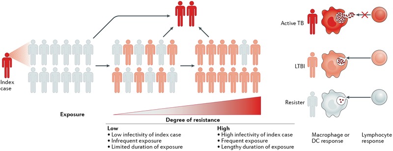
Mycobacterium tuberculosis is a leading cause of mortality worldwide and establishes a long-lived latent infection in a substantial proportion of the human population. Multiple lines of evidence suggest that some individuals are resistant to latent M. tuberculosis infection despite long-term and intense exposure, and we term these individuals 'resisters'. In this Review, we discuss the epidemiological and genetic data that support the existence of resisters and propose criteria to optimally define and characterize the resister phenotype. We review recent insights into the immune mechanisms of M. tuberculosis clearance, including responses mediated by macrophages, T cells and B cells. Understanding the cellular mechanisms that underlie resistance to M. tuberculosis infection may reveal immune correlates of protection that could be utilized for improved diagnostics, vaccine development and novel host-directed therapeutic strategies.


Conflict of interest statement

Competing interests

The authors declare no competing interests.

Figures

Fig. 1 |
Fig. 1 |. The spectrum of human resistance to infection by Mycobacterium tuberculosis.
The extent of resistance to Mycobacterium tuberculosis infection is proportional to the duration and the intensity of exposure to M. tuberculosis. Individuals who resist infection despite heavy exposure to M. tuberculosis are more likely to have immunogenetic mechanisms of resistance than those who resist infection after a lower extent of exposure. Individuals with resistance to M. tuberculosis infection after intense exposure are termed ‘resisters’ (white), whereas individuals who test positive in the purified protein derivative (PPD) skin reactivity test and/or the IFNγ release assay (IGRA) can either be asymptomatic (latent M. tuberculosis infection (LTBI); pink) or be symptomatic with tuberculosis (TB; red). DC, dendritic cell.
Fig. 2 |
Fig. 2 |. Macrophage-mediated resistance to Mycobacterium tuberculosis.
Macrophages can provide resistance to Mycobacterium tuberculosis infection at multiple points in the infection process, including the initial uptake of bacilli into phagosomes and through any of the possible fates of these phagosomes. These fates include effective (versus delayed) phagosome maturation (resulting in the generation of microbicidal products, such as nitric oxide, reactive oxygen species and cathelicidin), fusion with the lysosome (resulting in degradation of the phagosomal contents), recruitment of autophagy factors (resulting ultimately in autophagosome-lysosome fusion) or phagosomal escape of bacteria into the cytosol. Pro-inflammatory cytokine production may result from Toll-like receptor (TLR) ligation or the recognition of signals following phagosomal rupture, including inflammasome-triggered maturation of pro-IL-1β or pro-IL-18 and recognition of cytosolic M. tuberculosis DNA by cyclic GMP-AMP synthase (cGAS) and the resultant stimulator of interferon genes (STING)-dependent interferon regulatory factor 3 (IRF3)-driven transcriptional response. Vitamin D3 receptor (VDR)-dependent transcription, which requires prior activation of vitamin D to 1α,25(OH)2D3 through TLR or IFNγ signalling events, results in cathelicidin expression. Genetic and cellular evidence exists for a role for the negative TLR regulator Toll-interacting protein (TOLLIP), the autophagy factor Unc51-like kinase 1 (ULK1) and histone deacetylases (HDACs; which are master transcriptional regulators) in resistance. AIM2, absent in melanoma 2; cGAMP, cyclic GMP-AMP; ESAT6, 6 kDa early secretory antigenic target; mTOR, mechanistic target of rapamycin; MYD88, myeloid differentiation primary response protein MYD88; NF-κB, nuclear factor-κB; NLRP3, NOD-, LRR- and pyrin domain-containing 3; TBK1, TANK-binding kinase 1; TFs, transcription factors; TNF, tumour necrosis factor.
Fig. 3 |
Fig. 3 |. T cell-mediated resistance to Mycobacterium tuberculosis.
After phagocytosis of Mycobacterium tuberculosis by macrophages and dendritic cells (not shown), various T cell responses are stimulated, including conventional MHC-restricted responses (peptide-specific T cells) and MHC-independent responses (γδ T cells, which detect phosphorylated prenyl metabolites (also known as phosphoantigens)), CD1-restricted (lipid-specific) T cells and MHC class I-related gene protein (MR1)-restricted T cells (also known as mucosal-associated invariant T (MAIT) cells); MR1 presents small molecules, such as vitamin B2 derivatives. Production of IFNγ by CD4+T cells is a hallmark of the T cell response in individuals with latent M. tuberculosis infection (LTBI), whereas by definition, ‘resisters’ lack this IFNγ response. Qualitative differences in T cell responses between resisters and individuals with LTBI lead to the production of different macrophage-activating cytokines and chemokines, resulting in either the elimination of M. tuberculosis by resisters or persistence of M. tuberculosis in individuals with LTBI. BTN3A1, butyrophilin subfamily 3 member A1; NTM, non-tuberculous mycobacteria; TCR, T cell receptor; TLR, Toll-like receptor.
Fig. 4 |
Fig. 4 |. Antibody-mediated resistance to Mycobacterium tuberculosis.
Beyond their role in clearing pathogens, antibodies also direct the rapid destruction of infected cells via the recruitment of innate immune cells (such as phagocytes) that express crystallizable fragment (Fc) receptors. Two modifications to the Fc domain of an antibody control its affinity for Fc receptors, namely, changes in the antibody subclass or isotype and its glycosylation. Fab, antigen-binding fragment; NK cell, natural killer cell; ROS, reactive oxygen species.

References

    1. Dye C, Scheele S, Dolin P, Pathania V & Raviglione MC Consensus statement. Global burden of tuberculosis: estimated incidence, prevalence, and mortality by country. WHO global surveillance and monitoring project. JAMA 282, 677–686 (1999). - PubMed
    1. Houben RM & Dodd PJ The global burden of latent tuberculosis infection: a re-estimation using mathematical modelling. PLoS Med. 13, e1002152 (2016). - PMC - PubMed
    1. World Health Organization. Global Tuberculosis Report 2017. (WHO, 2017).
    1. Thompson EG et al. Host blood RNA signatures predict the outcome of tuberculosis treatment. Tuberculosis 107, 48–58 (2017). - PMC - PubMed
    1. Zak DE et al. A blood RNA signature for tuberculosis disease risk: a prospective cohort study. Lancet 387, 2312–2322 (2016). - PMC - PubMed

Publication types

LinkOut - more resources