. 2018 Jun 12;8(1):8945.
doi: 10.1038/s41598-018-26879-3.

Europe-wide reassessment of Dictyocoela (Microsporidia) infecting native and invasive amphipods (Crustacea): molecular versus ultrastructural traits

Affiliations

Europe-wide reassessment of Dictyocoela (Microsporidia) infecting native and invasive amphipods (Crustacea): molecular versus ultrastructural traits

Karolina Bacela-Spychalska et al. Sci Rep. .

Abstract

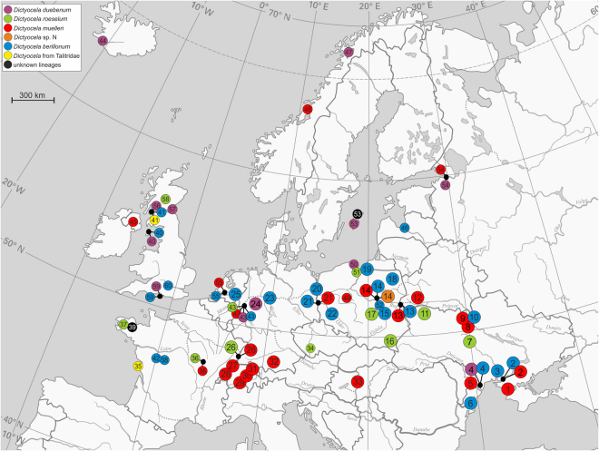
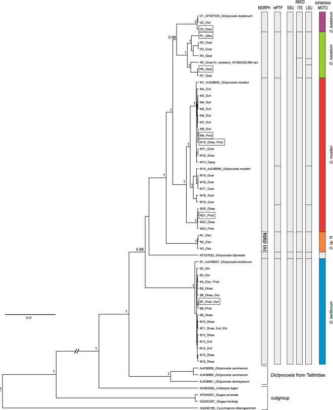
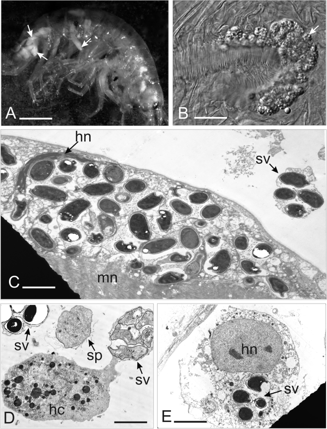
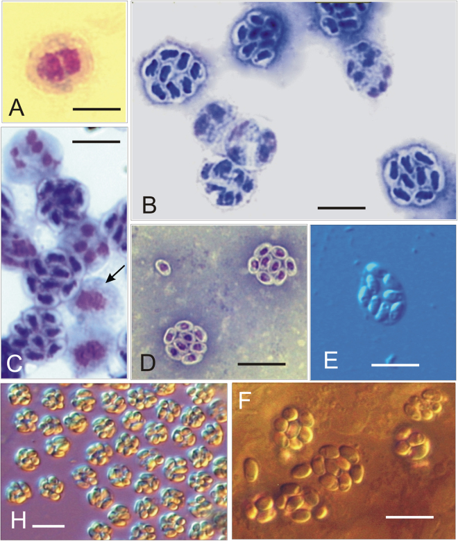
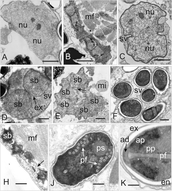
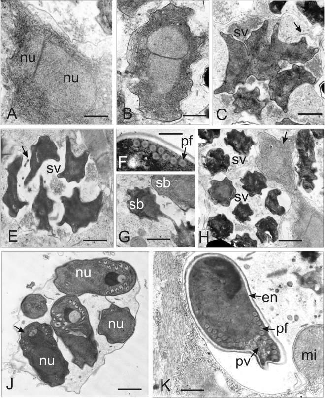
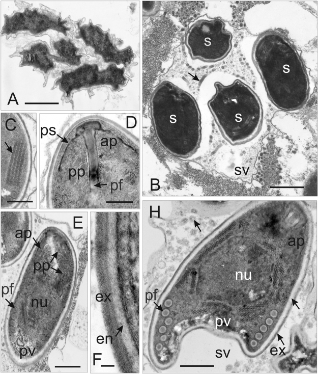
Microsporidia are common parasites infecting animals and protists. They are specifically common pathogens of amphipods (Crustacea, Malacostraca), with Dictyocoela spp. being particularly frequent and highly prevalent, exhibiting a range of phenotypic and ecological effects. Until now, seven species of Dictyocoela were defined, predominantly based on the genetic distance. However, neither the taxonomic status of this provisionally erected genus (based on eight novel sequences and one micrograph of the spore), nor its internal phylogenetic relationships have been clearly revealed. The formal description of the genus and of most of the putative species are still lacking. Here we aimed to fill this gap and performed both ultrastructural and molecular studies (based on SSU, ITS and partial LSU) using different species delimitation methods. As a consensus of these results and following conservative data interpretation, we propose to distinguish five species infecting gammarid hosts, and to keep the names introduced by the authors of the type sequences: Dictyocoela duebenum, D. muelleri, D. berillonum and D. roeselum. We provide full descriptions of these species. Moreover, thanks to our extensive sampling, we extend the known host and geographic range of these Microsporidia.


Conflict of interest statement

The authors declare no competing interests.

Figures

Figure 1
Figure 1
Records of Dictyocoela in Europe: big circles – from this study (site numbers 1–33), small circles – GenBank data (site numbers 34–62). Numbers correspond to locations as in Supplementary Tables S1 and S2. Different clades of Dictyocoela are indicated by colours as in Figs 1 and 2, Supplementary Fig. S1.
Figure 2
Figure 2
Bayesian phylogenetic reconstruction based on a 1833 bp long alignment of combined SSU, ITS and partial LSU rDNA. Above the branches the Bayesian posterior probabilities for major clades higher than 0.75 are given. Bars annotated on the right represents results of the species delimitation based on morphology and molecular (mPTP and ABGD) methods as well as consensus species delimitation. In frames, the individuals for which the ultrastructural analysis was done are indicated.
Figure 3
Figure 3
Pathogenicity of Dictyocoela spp.: (A) Gammarus varsoviensis infected with Dictyocoela muelleri. Pale-colored musculature indicates microsporidian infection (arrow). (B) Infected abdominal muscles (arrow) of the same specimens (Nomarski contrast). (C) Ultrathin section of muscle cell of Pontogammarus robustoides infected with D. berillonum. Sporophorous vesicles are located in the sarcoplasm neighboring myofibrils (mn). Deformation of host nucleus (hn) is observed. Single sporophorous vesicle (sv) is located outside the muscle cell in haemolymph. (D) Diplokaryotic sporont (sp) and sporophorous vesicles (sv) of D. muelleri, located in hemolymph of Dikerogammarus haemobaphes. Sporophorous vesicles (sv) adhering to haemocyte (hc) are visible. (E). Sporophorous vesicle of Dictyocoela berillonum (arrow) inside the cytoplasm of uninucleate haemocyte of P. robustoides. Single nucleus of haemocyte (hn) contains two nucleoli. Scale bars: 2.0 mm (A), 0.2 mm (B); 3.5 µm (C); 2.0 µm (D); 4.5 µm (E).
Figure 4
Figure 4
Light micrographs of Giemsa-stained (AD) and fresh (E,H,F) smears. (A) Diplokaryotic merozoite of Dictyocoela muelleri from Pontogammarus robustoides (B) Sporogonial plasmodia and mature octospores of Dictyocoela roeselum from Gammarus balcanicus. (C) Merogonial plasmodium with two diplokaryotic nuclei (arrow), sporonts with four and eight nuclei (beginning of second sporontal division) and mature octospores of D. berillonum from P. robustoides. (D) Mature spores of D. muelleri from D. haemobaphes. (E,F) Live spores of D. roeselum from G. balcanicus (E), D. muelleri from P. robustoides (H), and D. duebenum from Echinogammarus berilloni (F). Scale bars: 4.9 µm (D); 5.2 µm (B,C); 5.8 µm (E,F); 6.5 µm (H).
Figure 5
Figure 5
Ultrastructure of the developmental stages of Dictyocoela muelleri from Pontogammarus robustoides (A–E,H,J) and Dikerogammarus haemobaphes (F,K). (A) Diplokaryotic merozoite. The cytoplasm is homogenously granular. A few cisternae of rough cytoplasmic reticulum are visible. (B) Merogonial plasmodium neighboring myofibrils (mf). Dividing presporont with two diplokarya. (C) Beginning of meiotical division of the sporont market by detaching the two parts of the diplokaryon from each other. The separation of the sporophorous vesicle membrane (sv) is visible. (D) Part of the sporophorous vesicle (SV) with uninucleate sporoblasts (sb). The sporoblast mother cells surrounded by granular secretory material. Episporontal cover (arrow) mark the start of future exospore (ex) development. (E) Ultrathin section of sporophorous vesicle containing young uninucleate sporoblasts (sb). Granular material, forming tubular episporontal inclusions is arrowed. (F) Sporophorous vesicle (sv) with mature spores and tubular inclusions. (H) Lateral section of the sporophorous vesicle containing late sporoblasts (sb). The space between the sporoblasts is filled with granular and tubular inclusions (arrow). (J) Immature spore with 11–12 polar filament coils (pf) and posterosome (ps). Thin layer of electron-lucent endospore and complete exospore are clearly visible. (K) Longitudinal section of the anterior part of the mature spore showing the endospore (en), exospore (ex), manubrial part of polar filament (pf), fine lamellar anterior polaroplast, wide lamellar posterior polaroplast (pp), and anchoring disc (ad). Scale bars: 200 nm (K); 500 nm (A,C,D); 600 nm (J); 700 nm (B); 800 nm (E,F); 1.5 nm (H).
Figure 6
Figure 6
Ultrastructure of developmental stages of D. roeselum from Gammarus balcanicus (AF) and D. duebenum from Echinogammarus berilloni (G–K). (A) Oblique section of merozoite showing two attached nuclei (nu) and homogenously granular cytoplasm. (B) Diplokaryotic presporont with cytoplasm containing numerous cysternae of the endoplasmic reticulum. The cell is coated with electron dense material. (C) Sporophorous vesicle (sv) with divided sporogonal plasmodium. Episporontal space contains not numerous tubules (arrow) and granular substance. (E) Sporophorous vesicle (sv) with separated late sporoblasts, rare microtubules (arrow) and aggregated granules. (F) Part of mature spores showing eight coils of isofilar polar filament (pf). (G) Part of sporophorous vesicle with rare inclusions. Divided and separated sporoblasts (sb) are visible. (H) Early merozoite, late merogonal stages (presporont) and sporophorous vesicles (sv) containing late sporoblasts are presented. The presporont is arrowed. (J) Sporophorous vesicle with immature spores (arrow) and rare loosely packed inclusions are presented. The dense Golgi-derived substance fills the core of the polar filament and forms a large globe shaped inclusion. (K) Section through the posterior part of mature uninucleate spore. Polar filament (pf), posterior vacuole (pv) and spore wall ultrastructure with exospore (e) and endospore (en) are visible. Sporophorous vesicle contains rare tubules 70–100 nm in diameter and eletron dense filaments 40–50 nm in thickness. Scale bars: 100 nm (B); 300 nm (A,F); 800 nm (K); 1.2 µm (C,E,G,J); 1.5 µm (H).
Figure 7
Figure 7
Late sporogonial stages of Dictyocoela berillonum from Pontogammarus robustiodes. (A,B) Section of the sporophorous vesicles (sv) containing sporoblasts (A) and spores (B). The sv space is filled with thin granular material and not numerous septate tubules (arrow). (C) Part of immature spore with polyribosomes (arrow). (D) Anterior part of immature spore with anchoring apparatus, polar sac (ps), manubrial part of polar filament (pf), thin lamellar anterior polaroplast (ap), and cysterns of posterior polaroplast (pp). The spore wall is narrowed at the apical pole. (E) Anterior part of mature spore with lamellar posterior polaroplast (pp) and posterior vacuole (pv). Tubular inclusions, structure of the spore wall and coiled part of polar filament (pf) are visible. (F) Ultrastructure of the spore wall. The plasmalemma, endospore (en) and two layers of exospore (ex) are presented. (H) Oblique section of mature spore. Anterior polaroplast (ap), single nucleus (nu), 5–6 coils of the polar filament (pf), posterior vacuole (pv), and inclusions (arrow) inside sporophorous vacuole (sv) are displayed. Scale bars: 40 nm (F); 300 nm (D); 400 nm (H); 500 nm (C,E); 1.5 µm (A,B).

References

    1. Bulnheim HP. Microsporidian infections of amphipods with special reference to host parasite relationships: a review. Mar. Fish. Rev. 1975;37:39–45.
    1. Grabner DS, et al. Invaders, natives and their enemies: distribution patterns of amphipods and their microsporidian parasites in the Ruhr Metropolis, Germany. Parasit. Vectors. 2015;8:4192015. doi: 10.1186/s13071-015-1036-6. - DOI - PMC - PubMed
    1. Stentiford, G. D. & Dunn, A. M. Microsporidia in aquatic invertebrates. In Microsporidia: Pathogens of Opportunity (eds Weiss, L. M. & Becnel, J. J.) 579–604 (John Wiley & Sons, Inc., 2014).
    1. Terry RS, et al. Ultrastructural Characterisation and Molecular Taxonomic Identification of Nosema granulosis n. sp., a Transovarially Transmitted Feminising (TTF) Microsporidium. J. Eukaryot. Microbiol. 1999;46:492–499. doi: 10.1111/j.1550-7408.1999.tb06066.x. - DOI - PubMed
    1. Terry RS, et al. Widespread vertical transmission and associated host sex-ratio distortion within the eukaryotic phylum Microspora. Proc. R. Soc. Lond. Ser. B – Biol. Sci. 2004;271:1783–1789. doi: 10.1098/rspb.2004.2793. - DOI - PMC - PubMed

Publication types

LinkOut - more resources