

doi: 10.1038/s41598-018-27264-w.
A Versatile Safeguard for Chimeric Antigen Receptor T-Cell Immunotherapies
Affiliations
- PMID: 29895885
- PMCID: PMC5997667
- DOI: 10.1038/s41598-018-27264-w
Item in Clipboard
A Versatile Safeguard for Chimeric Antigen Receptor T-Cell Immunotherapies
Sci Rep.
.
Abstract
CAR T-cell therapies hold great promise for treating a range of malignancies but are however challenged by the complexity of their production and by the adverse events related to their activity. Here we report the development of the CubiCAR, a tri-functional CAR architecture that enables CAR T-cell detection, purification and on-demand depletion by the FDA-approved antibody Rituximab. This novel architecture has the potential to streamline the manufacturing of CAR T-cells, allow their tracking and improve their overall safety.
Conflict of interest statement
J.V., L.P., and P.D., V.G., A.J. and A.D. are present Cellectis employees and B.B., C.A.S, T.P. and B.J.S are present Pfizer employees.
Figures
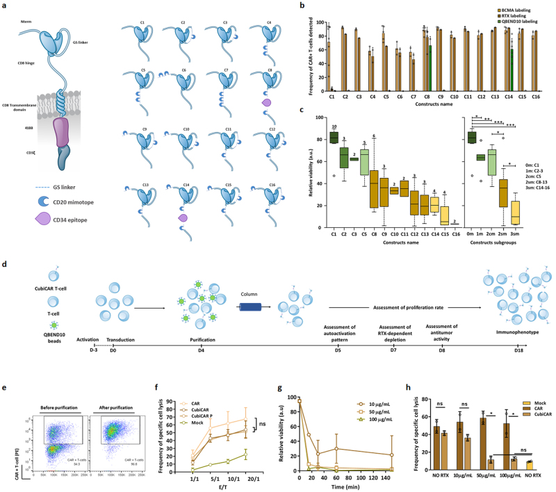
In vitro screening, identification and characterization of the CubiCAR architecture. (a, left panel) Scheme of the 2nd generation CAR construct used in this study. This construct includes an anti-BCMA ScFV, a CD8 hinge and transmembrane domain, a 4-1BB costimulatory domain and a CD3 activation domain (a, right panel) Scheme and names of the different engineered extracellular constructs tested. The location of CD20 mimotopes and CD34 epitope are indicated. (b) Flow cytometric detection of CAR constructs transiently expressed at the surface of primary T-cells using either the soluble BCMA protein, RTX or QBEND10 as surface markers. The error bars in represent the standard deviation on experimental values computed out of ≥2 biological replicates performed with ≥2 different donors (c) Box plot illustrating the median of efficiency of RTX-dependent depletion of primary T-cells transiently expressing CAR constructs. Viability of primary T-cells incubated for 150 min in the presence of 100 µg/mL RTX and complement was determined by flow cytometry and normalized to untreated control (relative viability, see Methods). Relative viability is indicated for each constructs (left panel) or for construct subgroups including those containing 2 consecutive CD20 mimotopes (2 cm) and 2 to 3 separated CD20 mimotopes (2 sm, 3 sm respectively, right panel). The number of independent biological replicates performed is indicated at the top of each box plot. The significance of the differences between subgroups was assessed using a non parametric Mann-Whitney U test (ns, non significant, *p < 0.05, **p < 0.01, ***p < 0.001). (d) Schema of the workflow used to characterize primary T-cells steadily expressing the CubiCAR (C14) construct. (e) Flow cytometry analysis of CubiCAR T-cells before and after QBEND10 coated beads purification using BCMA soluble protein as surface marker. (f) Specific cell lysis activity of unpurified and purified CubiCAR T-cells toward BCMA+ and BCMA- tumor cell lines determined at different E/T ratio. (g) Kinetic of CubiCAR T-cells depletion by complement and increasing amounts of RTX (10–100 µg/mL). (h) Effect of RTX on the specific cell lysis activities of CAR or purified CubiCAR T-cells. Activities were determined after a 30 min long incubation of cells with complement and increasing amounts of RTX. The Error bars in (f), (g) and (h) represent the standard deviation on experimental values (technical triplicate) computed out of 2 biological replicates performed with 2 different donors. The significance of the differences between subgroups in (f) and (h) was assessed using a one-way ANOVA test (ns, non significant, *p < 0.05, **p < 0.01, ***p < 0.001). Reproduced with permission from Cellectis group.
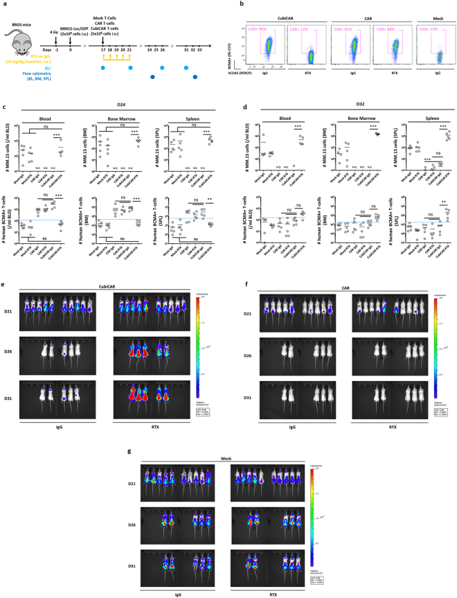
CubiCAR T-cells display anti-tumor activity and are specifically depleted by RTX in vivo. (a) Experimental workflow used to assess the anti-tumor activity of purified CubiCAR T-cells and the extent of their depletion by RTX in BRGS mice. Bioluminescence (BLI) and flow cytometry analysis of blood, bone marrow and spleen (BLD, BM and SPL respectively) are indicated by light and dark blue dots respectively. Mice were imaged at D17 for randomization and at D21, D26 and D31 to monitor tumor expansion. 5 mice/group were sacrificed at D24 and D32 for flow cytometry analysis. (b) and (c) results of flow cytometry analysis obtained at D24 and D32 respectively and expressed as number of human MM.1 S or BCMA+ T-cells (defined as T-cells, positively labeled by soluble biotinylated-BCMA protein and fluorescent streptavidin). MM.1S-Luc-GFP cells were detected as CD38+ and GFP+ among mCD45−/hCD45- cells (supplementary Fig. 16a). CAR and CubiCAR T-cells were detected by the soluble biotinylated BCMA protein staining (BCMA+) among the hCD45+/CD3+ cells. The threshold of BCMA + T-cells detection, set by the highest signal obtained from Mock transduced T-cells, is indicated by a dashed blue line. Mock transduced T-cells (Mock) were detected among hCD45+ cells as CD3+/BCMA− cells (supplementary Fig. 16a). (d) Representative flow cytometry results obtained in at D24, from the BM of mice infused by CubiCAR, CAR and Mock transduced T-cells in the presence of IgG or RTX. (e), (f) and (g) bioluminescence analysis of mice respectively infused by CubiCAR, CAR or mock transduced T-cells at D21, D26 and D31 in the presence or in the absence of RTX. All pictures illustrate the radiance (p/s/cm2/sr) of luminescence that was computed with the same dynamic range for each mouse. N.D, non detectable. Data were log-transformed before doing an ANOVA followed by a multiple comparison Tukey HSD test (ns, non-significant, *p < 0.05, **p < 0.01, ***p < 0.001). Reproduced from the Cellectis group except for the mouse model scheme that is not subject to copyright and was obtained from Pixabay web site.
References
MeSH terms
Substances
LinkOut - more resources
Full Text Sources
Other Literature Sources
