. 2018 Jun 12;9(1):2295.
doi: 10.1038/s41467-018-04592-z.

Spatio-temporal relays control layer identity of direction-selective neuron subtypes in Drosophila

Affiliations

Spatio-temporal relays control layer identity of direction-selective neuron subtypes in Drosophila

Holger Apitz et al. Nat Commun. .

Abstract

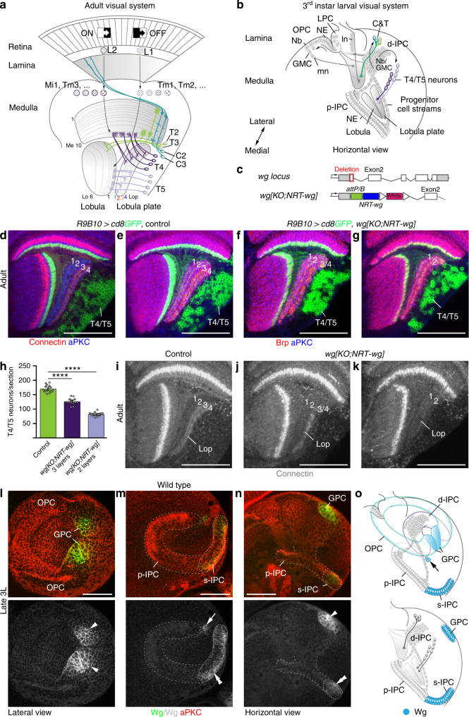
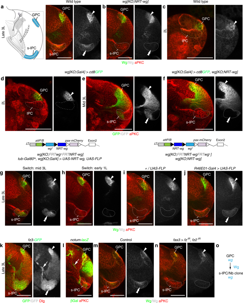
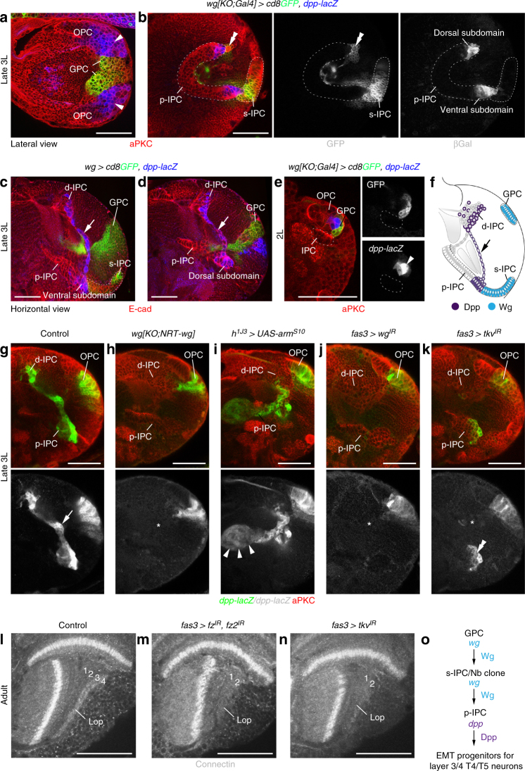
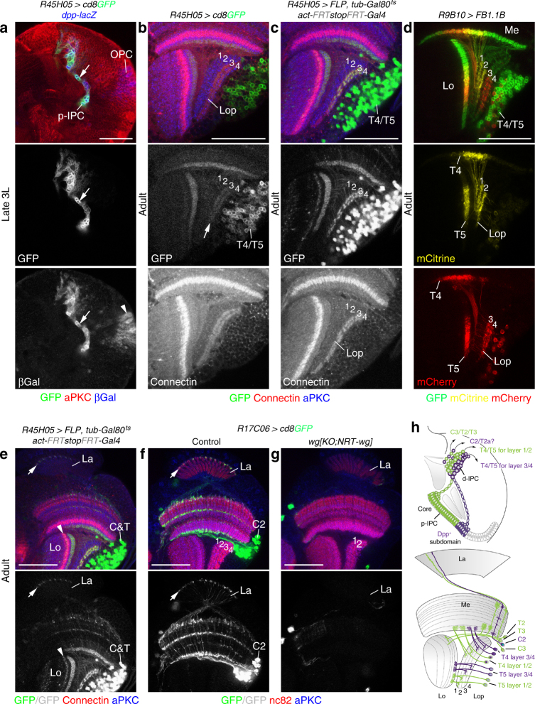
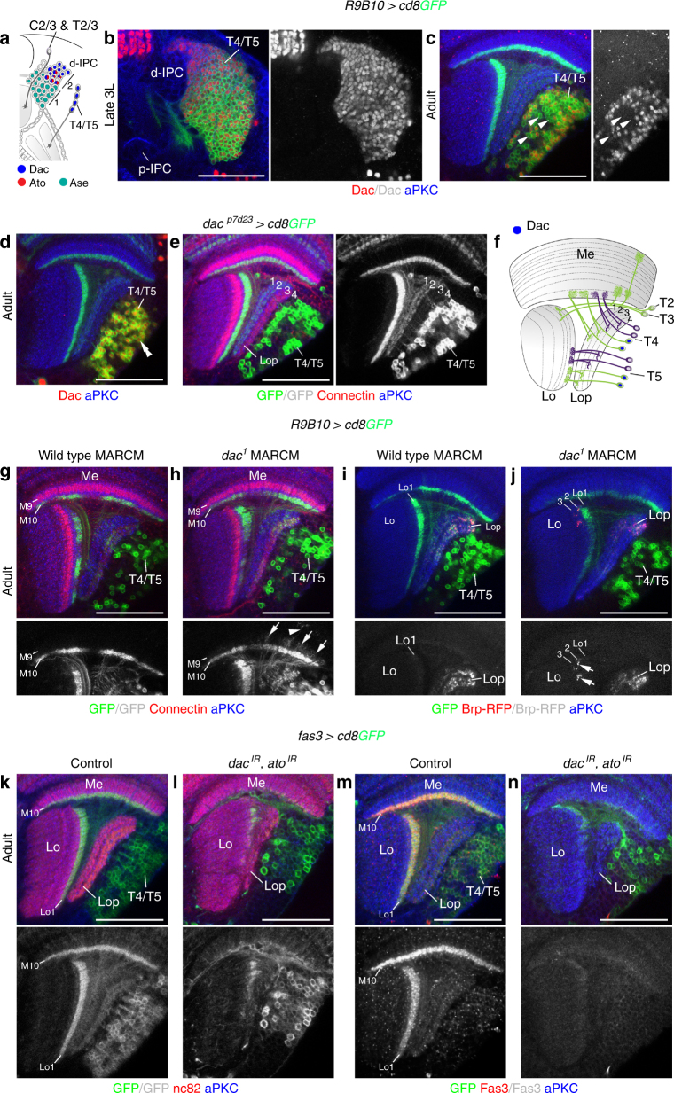
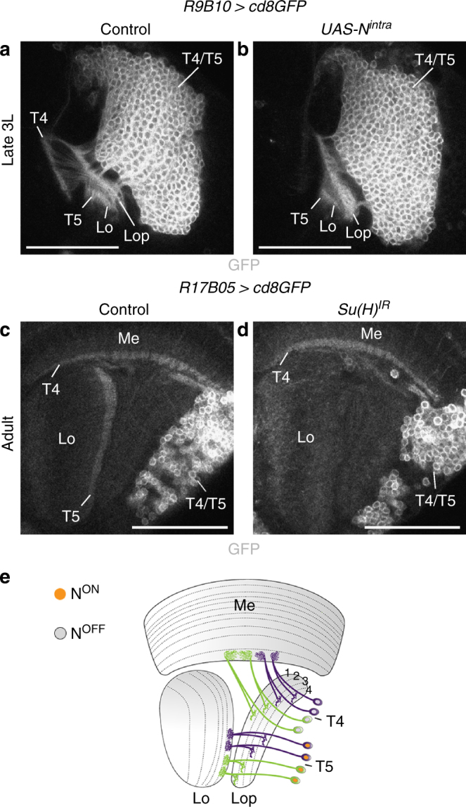
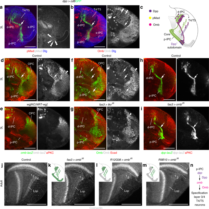
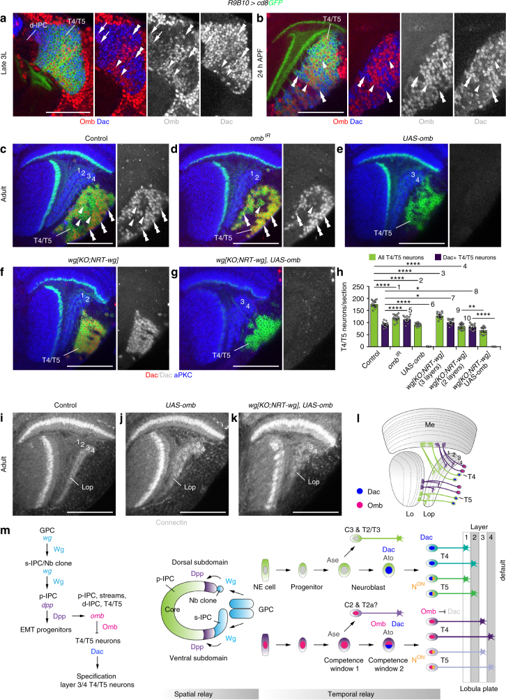
Visual motion detection in sighted animals is essential to guide behavioral actions ensuring their survival. In Drosophila, motion direction is first detected by T4/T5 neurons. Their axons innervate one of the four lobula plate layers. How T4/T5 neurons with layer-specific representation of motion-direction preferences are specified during development is unknown. We show that diffusible Wingless (Wg) between adjacent neuroepithelia induces its own expression to form secondary signaling centers. These activate Decapentaplegic (Dpp) signaling in adjacent lateral tertiary neuroepithelial domains dedicated to producing layer 3/4-specific T4/T5 neurons. T4/T5 neurons derived from the core domain devoid of Dpp signaling adopt the default layer 1/2 fate. Dpp signaling induces the expression of the T-box transcription factor Optomotor-blind (Omb), serving as a relay to postmitotic neurons. Omb-mediated repression of Dachshund transforms layer 1/2- into layer 3/4-specific neurons. Hence, spatio-temporal relay mechanisms, bridging the distances between neuroepithelial domains and their postmitotic progeny, implement T4/T5 neuron-subtype identity.


Conflict of interest statement

The authors declare no competing interests.

Figures

Fig. 1
Fig. 1
Wg release is essential for the formation of lobula plate layers 3/4. a Schematic of the adult Drosophila visual system. Neurons in the lamina (L1/L2) and medulla (e.g., Mi1,4,9, Tm1–4,9) relay ON/OFF motion cues to T4 and T5 neuron dendrites in medulla layer (Me) 10 and lobula (Lo) layer 1. T4/T5 axons innervate lobula plate (Lop) layers 1–4. C&T neurons include C2/C3 and T2/T3 subtypes. b Schematic of the 3rd instar larval optic lobe. The OPC generates lamina (ln) and medulla (mn) neurons. p-IPC NE cells give rise to migratory progenitors that mature into d-IPC Nbs. These produce C&T and T4/T5 neurons. GMC ganglion mother cells, LPC lamina precursor cells. c Structure of wild-type wg and engineered wg loci (wg{KO;NRT-wg}). Open triangles indicate loxP sites. d R9B10-Gal4 UAS-cd8GFP (green) labels T4/T5 neurons. Connectin (red) marks Lop layers 3/4. dg Neuropils were stained with nc82 (red) and aPKC (blue). Compared to controls (e), in wg{KO;NRT-wg} flies, one (f) or two (g) lobula plate layers were absent. h The decrease of layers correlates with T4/T5 neuron numbers. The scatter plot with bars shows data points and means with ±95% confidence interval error bars (n = 15; three optical sections from five samples per genotype). Unpaired, two-tailed Student’s t-test not assuming equal variance: P = 4.72 × 10−11 and P = 3.23 × 10−17. ****P < 0.0001. Unlike in controls (i), Connectin was found in one (j) or none (k) of the Lop layers in wg{KO;NRT-wg} flies. Similar to nc82 (f), Connectin labeling showed gaps in the third lobula plate layer (j), potentially consisting of both layer 3 and 4 neurons. ln In wild-type 3rd instar larvae (3L), the GPC areas (arrowheads), surface (s-)IPC (dashed line, double arrowheads), and a Nb clone (arrow) adjacent to the dorsal p-IPC subdomain (dashed line) express Wg (green). o Schematics of larval Wg expression (blue) in 3D and a horizontal section. Arrow indicates Nb lineage. For genotypes and sample numbers, see Supplementary Table 1. Scale bars, 50 μm
Fig. 2
Fig. 2
The GPC areas release Wg to induce wg in the s-IPC. a, b Schematic in a highlights the region of interest shown in subsequent panels. Unlike in controls (a), Wg immunolabeling (green) was absent in the s-IPC (dashed line, double arrowhead) in many wg{KO;NRT-wg} flies (b). The GPC areas (arrowhead) were not affected. c, d In 2nd instar larvae (2L), Wg protein (c, green) and wg{KO;Gal4} UAS-cd8GFP (d, green) were detected in GPC areas (arrowhead), but not in the adjacent IPC (dashed line). e, f s-IPC-specific wg{KO;Gal4} UAS-cd8GFP expression (green, double arrowheads) in mid 3rd instar larvae (e) was absent in wg{KO;NRT-wg} flies (f). Arrowheads indicate expression in GPC area. g, h In controls, wg{KO;Gal4} UAS-FLP mediated wg{KO;FRT wg+ FRT NRT-wg} allele switching and simultaneous UAS-NRT-wg overexpression were induced at the mid 3rd instar larval stage (g, wg+ background). Allele switching at the 1st instar larval stage (h, NRT-wg background) did not rescue s-IPC-specific Wg loss (green). i, j Unlike in controls (i), R46E01-Gal4 UAS-FLP-mediated GPC areas-specific wg{KO;FRT NRT-wg FRT wg+} allele switching (j) rescued s-IPC-specific NRT-Wg loss (green). Filled and open triangles in transgene schematics represent FRT and loxP sites, respectively (gj). k, l The Wg target gene reporter lines fz3G00357-GFP (k, green) and notumWRE-lacZ (l, green) show expression in the GPC areas, the s-IPC, and in migratory progenitors (arrow) originating from the adjacent p-IPC. m, n Unlike in controls (m), fas3NP1233-Gal4-mediated IPC-specific fz and fz2 knockdown (n) caused loss of Wg (green) in the s-IPC. o Summary of wg function in the GPC areas. For genotypes and sample numbers, see Supplementary Table 1. Scale bars, 50 μm
Fig. 3
Fig. 3
s-IPC-derived Wg is required for Dpp-dependent EMT in the p-IPC. a, b In 3rd instar larvae (3L), dpp-lacZ (blue) was detected in subdomains adjacent to wg{KO;Gal4} UAS-cd8GFP (green) regions in the dorsal and ventral OPC (a, arrowheads) and p-IPC (b). Double arrowhead indicates GFP-positive Nb clone adjacent to the dorsal p-IPC. c, d dpp-lacZ (blue) was maintained in progenitor streams (arrows) from the ventral and dorsal p-IPC subdomains. e In 2nd instar larvae (2L), dpp-lacZ (blue) was present in the OPC (arrowhead) adjacent to wg{KO;Gal4} UAS-cd8GFP-positive GPC areas (green), but was absent in the IPC (dashed line). f Schematic illustrating Wg and Dpp expression domains. Arrow indicates progenitor stream originating from the ventral p-IPC subdomain. gk Unlike in controls (g, arrow), dpp-lacZ (green) was absent from the IPC in wg{KO;NRT-wg} flies (h, asterisk). The OPC was not affected. dpp-lacZ was ectopically induced in the IPC (arrowheads) by h1J3-Gal4-mediated expression of UAS-armS10 (i). dpp-lacZ was absent following fas3NP1233-Gal4-mediated IPC-specific wg knockdown. In the Dpp-expression domain, this caused EMT defects and loss of progenitor streams (j, asterisks). Similar defects were caused by IPC-specific tkv knockdown. dpp-lacZ remains expressed in the p-IPC (k, double arrowheads). ln Compared to controls (l), fas3NP1233-Gal4-mediated IPC-specific knockdown of fz and fz2 (m) and tkv (n) caused the loss of lobula plate (Lop) layers 3/4 labeled with Connectin in adults. o Summary of wg and dpp function in the GPC areas, the s-IPC/Nb clone, and p-IPC. For genotypes and sample numbers, see Supplementary Table 1. Scale bars, 50 μm
Fig. 4
Fig. 4
The Dpp-expression domain generates C2 and T4/T5 neurons for layers 3/4. a R45H05-Gal4 UAS-cd8GFP (green) colocalizes with the dpp-lacZ (blue) expression domain in the IPC (arrow) but not the OPC (arrowhead) in 3rd instar larvae (3L). b R45H05-Gal4 UAS-cd8GFP (green) was weakly maintained in T4/T5 neurons innervating Connectin-positive lobula plate (Lop) layers 3/4 (red, arrow) in adults. c Permanent GFP-labeling (green) of the R45H05-Gal4 expression domain using act>y+>Gal4 UAS-GFP, UAS-FLP, and tub-Gal80ts was specific to T4/T5 neurons innervating lobula plate layers 3/4. d R9B10-Gal4 in conjunction with Flybow transgenes labeled T4/T5 neuron clones either innervating lobula plate (Lop) layers 1/2 (mCitrine, yellow) or 3/4 (mCherry, red). e Permanent GFP labeling (green) with R45H05-Gal4 included C2, as well as T2a and/or T3 neurons with characteristic axon terminals in the lamina (La, arrow) and lobula (Lo, arrowhead). f, g Unlike in controls (f), C2 neuron-specific R17C06-Gal4 UAS-cd8GFP expression (green) with axon terminals (arrow) in the lamina (La) was absent in wg{KO;NRT-wg} flies that had two remaining lobula plate layers (g). h Schematics illustrating the neuron subtypes derived from the Dpp-expression domain and the core p-IPC in 3rd instar larvae and adults. For genotypes and sample numbers, see Supplementary Table 1. Scale bars, 50 μm
Fig. 5
Fig. 5
dac and ato are required for T4/T5 neuron formation. a Schematic illustrating the expression of Ase (turquoise), Ato (red), and Dac (blue) in d-IPC Nbs during the first (1) and second (2) competence windows and their progeny. b, c Dac (red) was expressed in all R9B10-Gal4 UAS-cd8GFP (green) labeled T4/T5 neurons in 3rd instar larvae (b). It was downregulated (arrowheads) in approximately 50% of adult T4/T5 neurons (c). d, e dacp7d23-Gal4 UAS-cd8GFP (green) faithfully reported Dac (red, double arrowhead) expression in adults (d), specifically labeling T4/T5 neurons innervating lobula plate (Lop) layers 1 and 2 (e). f Schematic illustrating Dac expression (blue) in adult T4/T5 neurons. gj Unlike in controls (g, i), dac1 mutant T4/T5 neurons adopted T2/T3 neuron morphologies with R9B10-Gal4 UAS-cd8GFP (green), displaying neurite extensions into medulla (Me) layer M9 (arrows) and more distal layers (arrowhead) (h), and UAS-brp-RFP labeled synaptic terminals (red, arrows) in lobula (Lo) layers 2 and 3 (j). k, l Unlike in controls (k), fas3NP1233-Gal4 UAS-cd8GFP (green)-mediated IPC-specific simultaneous knockdown of dac and ato generated neurons that failed to form a four-layered lobula plate (Lop) neuropil and dendrites in medulla (Me) layer 10 and lobula (Lo) layer 1 (l). m, n Unlike in controls (m), Fas3-positive (red) T4/T5 neurons were absent following IPC-specific simultaneous knockdown of dac and ato mediated by fas3NP1233-Gal4 UAS-cd8GFP (green) (n). For genotypes and sample numbers, see Supplementary Table 1. Scale bars, 50 μm
Fig. 6
Fig. 6
Notch controls the choice between T4 and T5 neuron fate. a, b Unlike in controls (a), R9B10-Gal4 UAS-cd8GFP labeled T4 neurites were absent in 3rd instar larvae following UAS-Nintra overexpression in d-IPC Nbs and their progeny during the second competence window (b). T5 neurites in the lobula (Lo) were unaffected. c, d Unlike in controls (c), following R17B05-Gal4 UAS-cd8GFP-mediated IPC-specific Su(H) knockdown, T5 neurites were absent in the adult lobula (d). T4 neurites were present. e Schematic illustrating the Notch-dependent specification of T5 neurons. For genotypes and sample numbers, see Supplementary Table 1. Scale bars, 50 μm
Fig. 7
Fig. 7
Omb mediates Dpp-dependent specification of T4/T5 neurons for layers 3/4. a Phosphorylated Mad (pMad, red) in the dpp-Gal4 UAS-cd8GFP (green) expression domain was found in p-IPC NE cells (double arrowheads), but not in progenitors (arrow), d-IPC Nbs/GMCs, or T4/T5 neurons. b Omb (red) was maintained in the dpp-Gal4 UAS-cd8GFP (green) expression domain in progenitors (arrow), d-IPC Nbs/GMCs (arrowhead), and T4/T5 neuron subsets (double arrowheads). c Schematic illustrating pMad and Omb distribution within the Dpp expression domain. d, e Unlike in controls (d), ombP1-lacZ (green) was absent from the IPC (arrow) in wg{KO;NRT-wg} flies (e). OPC expression (arrowhead) was not affected. f, g In controls (f), Omb protein (green) was detected in p-IPC subdomains, migratory progenitors, and progeny (arrows). Following fas3NP1233-Gal4-mediated IPC-specific tkv knockdown (g), expression was severely reduced in these cells (asterisks). OPC expression (arrowhead) was not affected. h, i Indistinguishable from controls (h), fas3NP1233-Gal4-mediated IPC-specific knockdown of omb (i) did not affect EMT of dpp-lacZ-labeled progenitors (green, arrow) in the IPC. jm Unlike in controls (j), omb knockdown in the entire IPC and its progeny using fas3NP1233-Gal4 (k), in the d-IPC and postmitotic T4/T5 neurons using R12G08-Gal4 (l), and primarily postmitotic T4/T5 neurons using R9B10-Gal4 (m) resulted in the absence of Connectin-positive lobula plate layers 3/4. Schematic insets highlight cell type-specificities of Gal4 lines in green. n Schematics summarizing the role of Dpp in inducing Omb expression to specify layer 3/4 innervating T4/T5 neurons. For genotypes and sample numbers, see Supplementary Table 1. Scale bars, 50 μm
Fig. 8
Fig. 8
Omb converts layer 1/2 into layer 3/4 T4/T5 neurons by Dac downregulation. a, b In the 3rd instar larvae (a), Omb (red) and Dac (blue) were initially co-expressed (arrows) in new-born R9B10-Gal4 UAS-cd8GFP (green) labeled T4/T5 neurons close to the d-IPC. In some more mature T4/T5 neurons, that were positive for Omb, Dac expression was low (arrowheads), while in others, Dac expression was high (double arrowheads) and Omb expression was absent. At 24 h after puparium formation (APF) (b), Dac (double arrowheads) and Omb (arrowheads) show mutually exclusive expression. cg In controls (c), Dac (red, double arrowheads) was expressed in approximately 50% of T4/T5 neurons. Arrowheads indicate Dac-negative R9B10-Gal4 UAS-cd8GFP (green) labeled T4/T5 neurons. Dac was maintained in almost all T4/T5 neurons following omb knockdown (d), and downregulated following omb overexpression (e). Dac was expressed in all T4/T5 neurons in wg{KO;NRT-wg} flies with two lobula plate layers (f). Ectopic Omb was sufficient to downregulate Dac in these flies (g). h Quantification of all and Dac-positive T4/T5 neuron numbers following omb manipulations. The scatter plot with bars shows data points and means with ±95% confidence interval error bars (n = 15 corresponding to three serial optical sections, 6-μm apart, from five samples per genotype). Unpaired, two-tailed Student’s t-test not assuming equal variance: P = 5.57 × 10−12, P = 6.19 × 10−15, P = 1.26 × 10−10, P = 1.98 × 10−17, P = 3.60 × 10−5, P = 4.79 × 10−15, P = 0.020, P = 0.017, P = 0.0015, P = 2.42 × 10−13. *P < 0.05; **P< 0.01; **** P < 0.0001. ik Unlike in controls (i), R9B10-Gal4-mediated ectopic UAS-omb expression in T4/T5 neurons of wild type (j) or wg{KO;NRT-wg} (k) flies resulted in ectopic Connectin expression in the lobula plate (Lop). l Schematic illustrating Dac and Omb expression in adults. m Working model for spatial and temporal relay mechanisms regulating the formation and specification of layer-specific T4/T5 neurons. For genotypes and sample numbers, see Supplementary Table 1. Scale bars, 50 μm

References

    1. Clark DA, Demb JB. Parallel computations in insect and mammalian visual motion processing. Curr. Biol. 2016;26:R1062–R1072. doi: 10.1016/j.cub.2016.08.003. - DOI - PMC - PubMed
    1. Mauss AS, Vlasits A, Borst A, Feller M. Visual circuits for direction selectivity. Annu. Rev. Neurosci. 2017;40:211–230. doi: 10.1146/annurev-neuro-072116-031335. - DOI - PMC - PubMed
    1. Gabriel JP, Trivedi CA, Maurer CM, Ryu S, Bollmann JH. Layer-specific targeting of direction-selective neurons in the zebrafish optic tectum. Neuron. 2012;76:1147–1160. doi: 10.1016/j.neuron.2012.12.003. - DOI - PubMed
    1. Robles E, Filosa A, Baier H. Precise lamination of retinal axons generates multiple parallel input pathways in the tectum. J. Neurosci. 2013;33:5027–5039. doi: 10.1523/JNEUROSCI.4990-12.2013. - DOI - PMC - PubMed
    1. Buchner E, Buchner S, Bulthoff I. Deoxyglucose mapping of nervous activity induced in Drosophila brain by visual movement. I. Wildtype. J. Comp. Physiol. A. 1984;155:471–483. doi: 10.1007/BF00611912. - DOI

Publication types

MeSH terms

LinkOut - more resources