. 2020 Aug;25(8):1822-1834.
doi: 10.1038/s41380-018-0078-5. Epub 2018 Jun 13.

Large-scale mapping of cortical alterations in 22q11.2 deletion syndrome: Convergence with idiopathic psychosis and effects of deletion size

Affiliations

Large-scale mapping of cortical alterations in 22q11.2 deletion syndrome: Convergence with idiopathic psychosis and effects of deletion size

Daqiang Sun et al. Mol Psychiatry. 2020 Aug.

Abstract

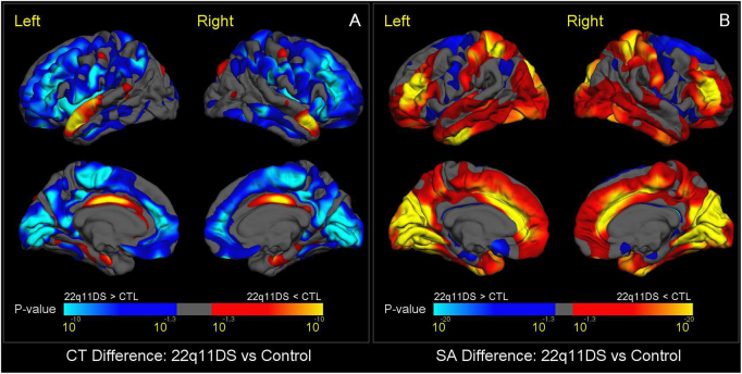
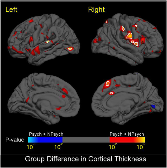
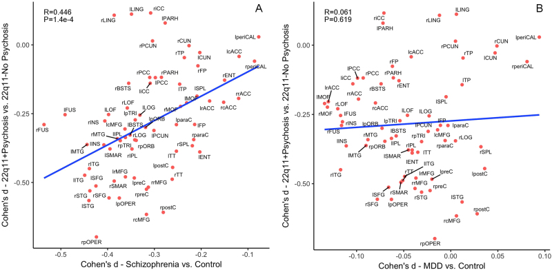
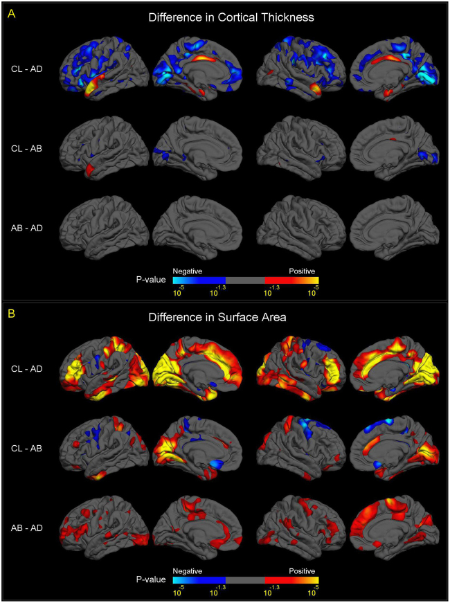
The 22q11.2 deletion (22q11DS) is a common chromosomal microdeletion and a potent risk factor for psychotic illness. Prior studies reported widespread cortical changes in 22q11DS, but were generally underpowered to characterize neuroanatomic abnormalities associated with psychosis in 22q11DS, and/or neuroanatomic effects of variability in deletion size. To address these issues, we developed the ENIGMA (Enhancing Neuro Imaging Genetics Through Meta-Analysis) 22q11.2 Working Group, representing the largest analysis of brain structural alterations in 22q11DS to date. The imaging data were collected from 10 centers worldwide, including 474 subjects with 22q11DS (age = 18.2 ± 8.6; 46.9% female) and 315 typically developing, matched controls (age = 18.0 ± 9.2; 45.9% female). Compared to controls, 22q11DS individuals showed thicker cortical gray matter overall (left/right hemispheres: Cohen's d = 0.61/0.65), but focal thickness reduction in temporal and cingulate cortex. Cortical surface area (SA), however, showed pervasive reductions in 22q11DS (left/right hemispheres: d = -1.01/-1.02). 22q11DS cases vs. controls were classified with 93.8% accuracy based on these neuroanatomic patterns. Comparison of 22q11DS-psychosis to idiopathic schizophrenia (ENIGMA-Schizophrenia Working Group) revealed significant convergence of affected brain regions, particularly in fronto-temporal cortex. Finally, cortical SA was significantly greater in 22q11DS cases with smaller 1.5 Mb deletions, relative to those with typical 3 Mb deletions. We found a robust neuroanatomic signature of 22q11DS, and the first evidence that deletion size impacts brain structure. Psychotic illness in this highly penetrant deletion was associated with similar neuroanatomic abnormalities to idiopathic schizophrenia. These consistent cross-site findings highlight the homogeneity of this single genetic etiology, and support the suitability of 22q11DS as a biological model of schizophrenia.


Conflict of interest statement

DMcD is a member of the Speaker’s Bureau for Natera. The remaining authors declare that they have no conflict of interest.

Figures

Fig. 1
Fig. 1
Vertex-Wise Mapping of Difference in CT and SA between 22q11DS and Healthy Control Subjects. a shows vertex-wise differences in CT, and b shows vertex-wise differences in SA. Colored areas show p-values for group differences after FDR correction (q = 0.05) for all vertices across both left and right cortical surfaces. Blue colors represent significant increases in 22q11DS subjects compared to healthy controls, whereas red-yellow colors represent significant reductions in 22q11DS subjects. Compared to controls, subjects with 22q11DS showed greater CT and smaller SA, most prominently in the posterior medial cortex including bilateral cuneus, precuneus, lingual gyrus, pericalcarine cortex, and bilateral medial and lateral frontal cortex. Subjects with 22q11DS showed a distinctive reduction of both CT and SA in bilateral cingulate cortex. They also had reduced  CT in the superior temporal gyrus, and greater SA in the superior parietal cortex and rostral middle frontal gyrus.
Fig. 2
Fig. 2
Mapping of CT Differences between 22q11DS + Psychosis and 22q11DS-No-Psychosis. Colored areas show p-values of group difference, and white circles include regions that pass FDR correction at q = 0.05. Blue colors represent thicker cortical gray matter in 22q11DS+ Psychosis compared to 22q11DS-No-Psychosis, and red-yellow colors represent thinner cortical gray matter in 22q11DS+ Psychosis vs. 22q11DS-No-Psychosis. Compared to those without psychosis, 22q11DS subjects with psychosis showed significantly thinner cortex in the left superior temporal gyrus and lateral occipital cortex, and right medial superior frontal, cingulate, pre- and post-central, and supramarginal gyri
Fig. 3
Fig. 3
Pattern similarity in CT deficits between 22q11DS with psychosis and idiopathic schizophrenia, in contrast to major depressive disorder (MDD). Here we correlated the effect sizes (Cohen’s d) for regional CT measures from the comparison between 22q11DS + Psychosis and 22q11DS-No-Psychosis groups with those from the ENIGMA Schizophrenia working group [47], in contrast to the ENIGMA MDD study [35]. a Correlation in the effect sizes of CT deficits between idiopathic schizophrenia and 22q11 + Psychosis; b Correlation in the effect sizes of CT deficits between MDD and 22q11 + Psychosis. The effect sizes for CT deficits from psychotic vs. non-psychotic 22q11DS comparisons were significantly correlated with those in ENIGMA idiopathic schizophrenia vs. control comparisons. In contrast, the same effect sizes were not significantly correlated with those in ENIGMA MDD vs. control comparisons. Both x- and y-axes represent effect sizes in Cohen’s d in the above-mentioned comparison for all 68 cortical regions derived from the FreeSurfer cortical parcellation. Abbreviations of the cortical regions are adopted from the brainGraph package [67] as follows: BSTS banks of superior temporal sulcus, cACC caudal anterior cingulate cortex, cMFG caudal middle frontal gyrus, CUN cuneus, ENT entorhinal cortex, FUS fusiform gyrus, IPL inferior parietal cortex, ITG inferior temporal gyrus, iCC isthmus cingulate cortex, LOG lateral occipital cortex, LOF lateral orbitofrontal cortex, LING lingual gyrus, MOF medial orbitofrontal cortex, MTG middle temporal gyrus, PARH parahippocampal gyrus, paraC paracentral, lobule, pOPER pars opercularis of inferior frontal gyrus, pORB pars orbitalis of inferior frontal gyrus, pTRI pars, triangularis of inferior frontal gyrus, periCAL pericalcarine cortex, postC post-central gyrus, PCC posterior, cingulate cortex, preC precentral gyrus, PCUN precuneus, rACC rostral anterior cingulate corte, rMFG rostral, middle frontal gyrus, SFG superior frontal gyrus, SPL superior parietal cortex, STG superior temporal gyrus, SMAR supramarginal gyrus, FP frontal pole, TP temporal pole, TT transverse temporal gyrus, INS insula. L left, R right
Fig. 4
Fig. 4
Vertex-wise mapping of differences in CT and SA, between A–B, A–D Deletion, and Control Subjects. For all figures, colored areas show p-values of group difference that remain significant after FDR correction (q = 0.05) for all vertices across both left and right cortical surfaces. The positive and negative directions in the color-bars indicate the signs of differences after subtracting one group from another labelled on the left side. a Differences in CT between A–B deletion, A–D deletion and control subjects. Compared to controls, subjects with A–B deletions showed thicker cortex (in blue colors) in bilateral pericalcarine cortex and bilateral inferior frontal gyrus, and thinner cortex (in red-yellow colors) in the left anterior superior temporal gyrus and right posterior cingulate cortex. Subjects with A–B deletions showed no significant difference in CT in any cortical region. The comparison of CT between subjects with A–D deletions and controls showed a similar pattern of group differences to the overall 22q11DS case-control analysis (Fig. 1a), although effects were diminished. b Difference in SA between A–B deletion, A–D deletion, and control subjects. Compared to controls, subjects with A–B deletions showed significantly reduced SA (in red-yellow colors), more prominent in the posterior portion of the medial and inferior cortical surface, including the bilateral cuneus, precuneus, pericalcarine, lingual, fusiform, and inferior temporal regions, and caudal anterior cingulate. Increased SA in A–B deletion cases vs. controls was observed in bilateral precentral, paracentral, and medial orbitofrontal regions (in blue colors). Compared to subjects with A–B deletions, subjects with A–D deletions showed widespread significant cortical SA reductions (in red-yellow colors), most prominently in the anterior portion of the medial cortical surface, including the paracentral lobules, cingulate, precentral, superior frontal regions, and widely distributed lateral cortical regions. Like CT, the comparisons of SA between subjects with A–D deletions and controls showed a similar pattern of group differences to the overall 22q11DS case-control analysis (Fig. 1b), although effects were diminished

References

    1. Sebat J, Levy DL, McCarthy SE. Rare structural variants in schizophrenia: one disorder, multiple mutations; one mutation, multiple disorders. Trends Genet. 2009;25:528–35. - PMC - PubMed
    1. McDonald-McGinn DM, Sullivan KE, Marino B, Philip N, Swillen A, Vorstman JA, et al. 22q11.2 deletion syndrome. Nat Rev Dis Prim. 2015;1:15071. - PMC - PubMed
    1. CNV, Schizophrenia Working Groups of the Psychiatric Genomics Consortium, Psychosis Endophenotypes International Consortium. Contribution of copy number variants to schizophrenia from a genome-wide study of 41,321 subjects. Nat Genet. 2017;49:27–35. - PMC - PubMed
    1. Bassett AS, Chow EW. Schizophrenia and 22q11.2 deletion syndrome. Curr Psychiatry Rep. 2008;10:148–57. - PMC - PubMed
    1. Green T, Gothelf D, Glaser B, Debbane M, Frisch A, Kotler M, et al. Psychiatric disorders and intellectual functioning throughout development in velocardiofacial (22q11.2 deletion) syndrome. J Am Acad Child Adolesc Psychiatry. 2009;48:1060–8. - PubMed

Publication types