Long-term follow-up of low-risk branch-duct IPMNs of the pancreas: is main pancreatic duct dilatation the most worrisome feature?
- PMID: 29895904
- PMCID: PMC5997632
- DOI: 10.1038/s41424-018-0026-3
Long-term follow-up of low-risk branch-duct IPMNs of the pancreas: is main pancreatic duct dilatation the most worrisome feature?
Erratum in
-
Correction: Long-term follow-up of low-risk branchduct IPMNs of the pancreas: is main pancreatic duct dilatation the most worrisome feature?Clin Transl Gastroenterol. 2018 Aug 1;9(7):173. doi: 10.1038/s41424-018-0036-1. Clin Transl Gastroenterol. 2018. PMID: 30068944 Free PMC article.
Abstract
Objectives: The management of branch-duct IPMN remains controversial due to the relatively low rate of malignant degeneration and the uncertain predictive role of high-risk stigmata (HRS) and worrisome features (WFs) identified by the 2012 International Consensus Guidelines. Our aim was to evaluate the evolution of originally low-risk (Fukuoka-negative) BD-IPMNs during a long follow-up period in order to determine whether the appearance of any clinical or morphological variables may be independently associated with the development of malignancy over time.
Methods: A prospectively collected database of all patients with BD-IPMN referring to our Institute between 2002 and 2016 was retrospectively analyzed. Univariate and multivariate analysis of association between changes during follow-up, including appearance of HRS/WFs, and development of malignancy (high-grade dysplasia/invasive carcinoma) was performed.
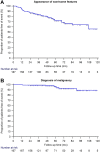
Results: A total of 167 patients were selected for analysis, and seven developed malignant disease (4.2%). During a median follow-up time of 55 months, HRS appeared in only three cases but predicted malignancy with 100% specificity. Worrisome features, on the other hand, appeared in 44 patients (26.3%). Appearance of mural nodules and MPD dilatation >5 mm showed a significant association with malignancy in multivariate analysis (p = 0.004 and p = 0.001, respectively). MPD dilatation in particular proved to be the strongest independent risk factor for development of malignancy (OR = 24.5).
Conclusions: The risk of pancreatic malignancy in this population is low but definite. The presence of major WFs, and especially MPD dilatation, should prompt a tighter follow-up with EUS and a valid cytological analysis whenever feasible.
Conflict of interest statement
Figures


Comment in
-
Long-Term Follow-Up of Low-Risk Branch Duct IPMNs of the Pancreas: Watch for Main Pancreatic Duct Dilatation, and for How Long?Clin Transl Gastroenterol. 2018 Oct 24;9(10):198. doi: 10.1038/s41424-018-0065-9. Clin Transl Gastroenterol. 2018. PMID: 30353003 Free PMC article. No abstract available.
Similar articles
-
Management of branch-duct intraductal papillary mucinous neoplasms: a large single-center study to assess predictors of malignancy and long-term outcomes.Gastrointest Endosc. 2016 Sep;84(3):436-45. doi: 10.1016/j.gie.2016.02.008. Epub 2016 Feb 18. Gastrointest Endosc. 2016. PMID: 26905937
-
Low progression of intraductal papillary mucinous neoplasms with worrisome features and high-risk stigmata undergoing non-operative management: a mid-term follow-up analysis.Gut. 2017 Mar;66(3):495-506. doi: 10.1136/gutjnl-2015-310162. Epub 2016 Jan 7. Gut. 2017. PMID: 26743012
-
Importance of main pancreatic duct dilatation in IPMN undergoing surveillance.Br J Surg. 2018 Dec;105(13):1825-1834. doi: 10.1002/bjs.10948. Epub 2018 Aug 14. Br J Surg. 2018. PMID: 30106195
-
Uptodate in the assessment and management of intraductal papillary mucinous neoplasms of the pancreas.Eur Rev Med Pharmacol Sci. 2017 Jun;21(12):2858-2874. Eur Rev Med Pharmacol Sci. 2017. PMID: 28682431 Review.
-
Intraductal papillary mucinous neoplasms of the pancreas.Eur J Surg Oncol. 2007 Aug;33(6):678-84. doi: 10.1016/j.ejso.2006.11.031. Epub 2007 Jan 4. Eur J Surg Oncol. 2007. PMID: 17207960 Review.
Cited by
-
Observation or resection of pancreatic intraductal papillary mucinous neoplasm: An ongoing tug of war.World J Gastrointest Oncol. 2019 Dec 15;11(12):1092-1100. doi: 10.4251/wjgo.v11.i12.1092. World J Gastrointest Oncol. 2019. PMID: 31908715 Free PMC article. Review.
-
Pre-Operative Imaging and Pathological Diagnosis of Localized High-Grade Pancreatic Intra-Epithelial Neoplasia without Invasive Carcinoma.Cancers (Basel). 2021 Feb 24;13(5):945. doi: 10.3390/cancers13050945. Cancers (Basel). 2021. PMID: 33668239 Free PMC article. Review.
-
Long-Term Follow-Up of Low-Risk Branch Duct IPMNs of the Pancreas: Watch for Main Pancreatic Duct Dilatation, and for How Long?Clin Transl Gastroenterol. 2018 Oct 24;9(10):198. doi: 10.1038/s41424-018-0065-9. Clin Transl Gastroenterol. 2018. PMID: 30353003 Free PMC article. No abstract available.
-
Risk Factors for the Development of High-risk Stigmata in Branch-duct Intraductal Papillary Mucinous Neoplasms.Intern Med. 2021 Oct 15;60(20):3205-3211. doi: 10.2169/internalmedicine.7168-21. Epub 2021 May 7. Intern Med. 2021. PMID: 33967138 Free PMC article.
-
Risk of malignancy in small pancreatic cysts decreases over time.Pancreatology. 2020 Sep;20(6):1213-1217. doi: 10.1016/j.pan.2020.08.003. Epub 2020 Aug 10. Pancreatology. 2020. PMID: 32819844 Free PMC article.
References
Publication types
MeSH terms
LinkOut - more resources
Full Text Sources
Other Literature Sources

