

Int6/eIF3e Silencing Promotes Placenta Angiogenesis in a Rat Model of Pre-eclampsia
- PMID: 29895936
- PMCID: PMC5997673
- DOI: 10.1038/s41598-018-27296-2
Int6/eIF3e Silencing Promotes Placenta Angiogenesis in a Rat Model of Pre-eclampsia
Abstract
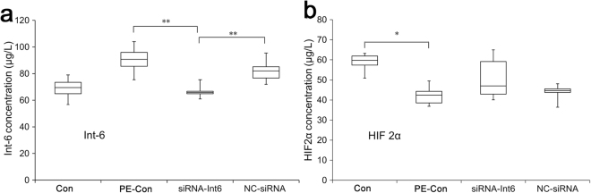
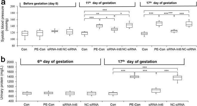
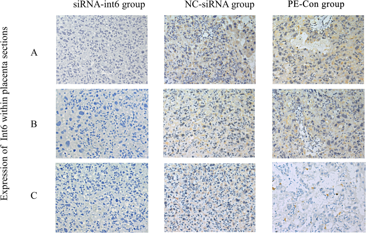
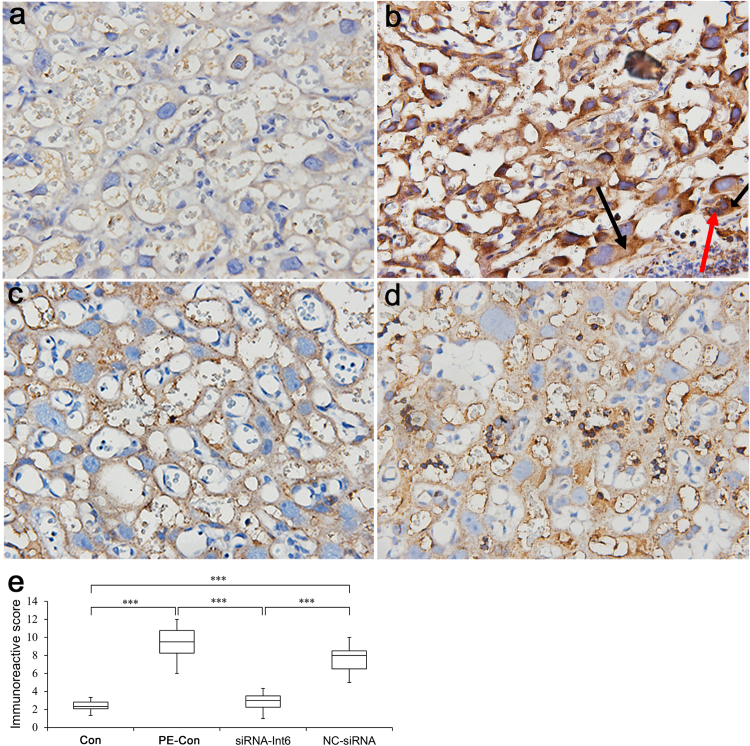
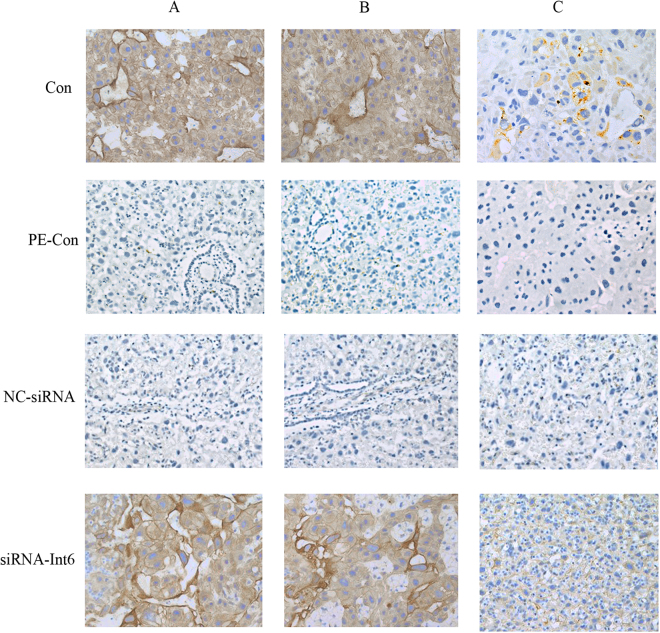
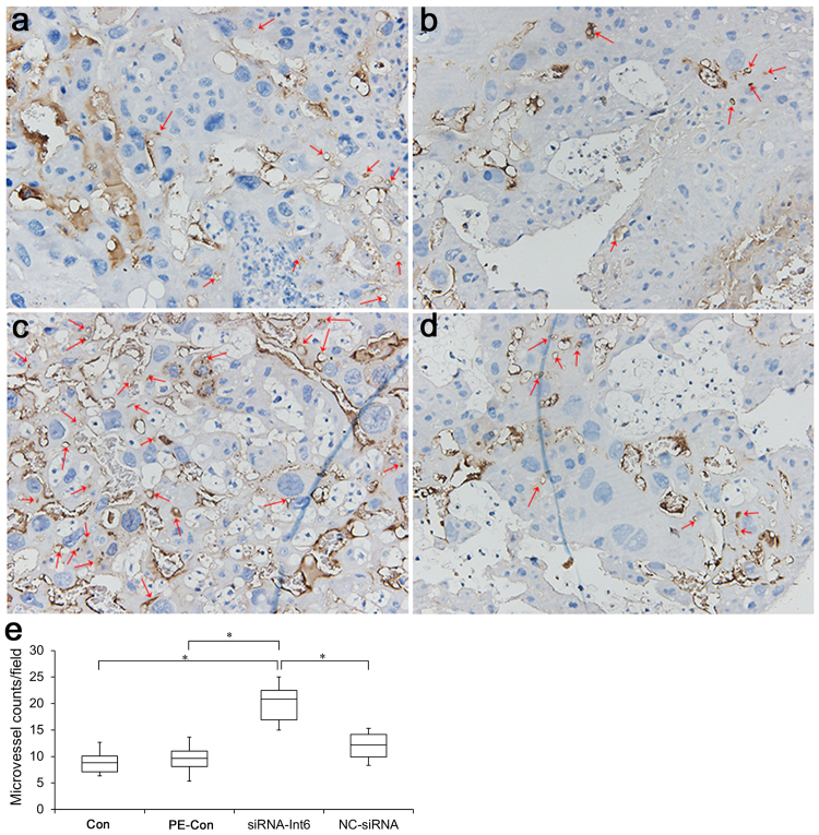
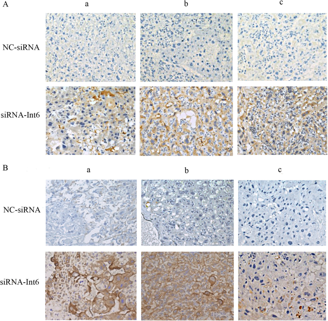
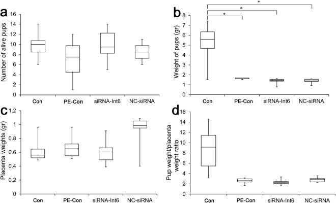
We investigated whether stable eukaryotic translation initiation factor 3e/inter 6 (eIF-3e/Int6) RNA-silencing (siRNA-Int6) can ameliorate pre-eclampsia (PE) by promoting angiogenesis in an N-nitro-L-arginine methyl ester (L-NAME)-induced rat pre-eclampsia (PE) model. Twenty-four pregnant female Sprague-Dawley rats were allocated into 4 groups, including controls (Con) without any treatment, and 18 from gestational day (GD) 7 to GD17 L-NAME-treated rats, which were divided into stable siRNA-Int6 transfected (siRNA-Int6), negative vector control siRNA (NC-siRNA) and PE control (PE-Con) groups. All adenovirus siRNA transfections were performed on GD7 via intravenous tail injection. On GD0, GD11 and GD17, blood pressure, and on GD6 and GD17, protein estimations in 24 h urine samples were conducted. All animals were sacrificed on GD18. In the PE-Con group, placental Int6 was expressed to a significantly greater level than in the Con group, which was reversed by the application of siRNA-Int6. Blood pressure and proteinuria were significantly lower in the siRNA-Int6 group than in the PRE-Con group. As shown by CD31 and IB4 expression, placental micro-vascular density (MVD) was significantly higher in the siRNA-Int6 group than in the PE-Con and NC-siRNA groups, which has accompanied by enhanced trophoblast invasion. Int6 silencing alleviated the maternal clinical manifestations of pre-eclampsia and promoted placental angiogenesis in pregnant L-NAME-treated rats.
Conflict of interest statement
The authors declare no competing interests.
Figures

Similar articles
-
Int6/eIF3e silencing promotes functional blood vessel outgrowth and enhances wound healing by upregulating hypoxia-induced factor 2alpha expression.Circulation. 2010 Aug 31;122(9):910-9. doi: 10.1161/CIRCULATIONAHA.109.931931. Epub 2010 Aug 16. Circulation. 2010. PMID: 20713899
-
[Regulation of pravastatin on long-chain fatty acid oxidative enzyme in pre-eclampsia-like mouse model].Zhonghua Fu Chan Ke Za Zhi. 2018 Mar 25;53(3):183-189. doi: 10.3760/cma.j.issn.0529-567X.2018.03.008. Zhonghua Fu Chan Ke Za Zhi. 2018. PMID: 29609233 Chinese.
-
Protective effect of metformin on the NG-nitro-l-arginine methyl ester (l-NAME)-induced rat models of preeclampsia.Biochem Biophys Res Commun. 2024 Dec 20;739:150996. doi: 10.1016/j.bbrc.2024.150996. Epub 2024 Nov 13. Biochem Biophys Res Commun. 2024. PMID: 39550868
-
Ferulic acid ameliorated placental inflammation and apoptosis in rat with preeclampsia.Clin Exp Hypertens. 2019;41(6):524-530. doi: 10.1080/10641963.2018.1516773. Epub 2018 Sep 5. Clin Exp Hypertens. 2019. PMID: 30183401 Review.
-
The Bad, the Good and eIF3e/INT6.Front Biosci (Landmark Ed). 2017 Jan 1;22(1):1-20. doi: 10.2741/4469. Front Biosci (Landmark Ed). 2017. PMID: 27814599 Review.
Cited by
-
FOXP2 overexpression upregulates LAMA4 expression and thereby alleviates preeclampsia by regulating trophoblast behavior.Commun Biol. 2024 Nov 1;7(1):1427. doi: 10.1038/s42003-024-07149-7. Commun Biol. 2024. PMID: 39487340 Free PMC article.
-
Comparative proteomic analysis identifies differentially expressed proteins and reveals potential mechanisms of traumatic heterotopic ossification progression.J Orthop Translat. 2022 May 14;34:42-59. doi: 10.1016/j.jot.2022.04.003. eCollection 2022 May. J Orthop Translat. 2022. PMID: 35615641 Free PMC article.
-
The Impact of Oxidative Stress of Environmental Origin on the Onset of Placental Diseases.Antioxidants (Basel). 2022 Jan 1;11(1):106. doi: 10.3390/antiox11010106. Antioxidants (Basel). 2022. PMID: 35052610 Free PMC article. Review.
References
Publication types
MeSH terms
Substances
LinkOut - more resources
Full Text Sources
Other Literature Sources
Molecular Biology Databases
Research Materials
Miscellaneous
