. 2018 Jun 12;8(1):8927.
doi: 10.1038/s41598-018-27182-x.

Evaluation of an artificial vertebral body fabricated by a tantalum-coated porous titanium scaffold for lumbar vertebral defect repair in rabbits

Affiliations

Evaluation of an artificial vertebral body fabricated by a tantalum-coated porous titanium scaffold for lumbar vertebral defect repair in rabbits

Faqi Wang et al. Sci Rep. .

Abstract

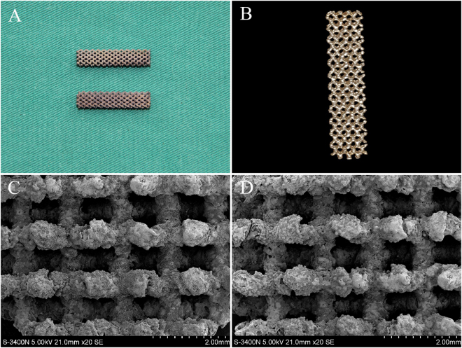
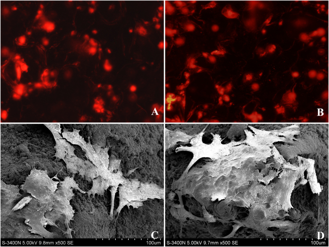
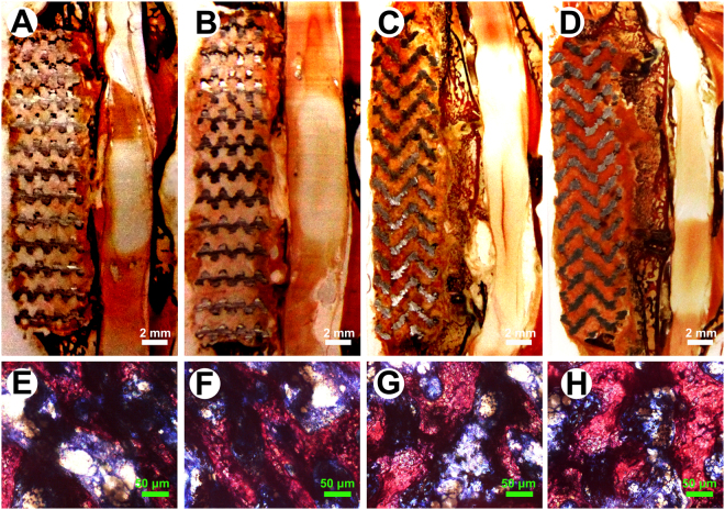
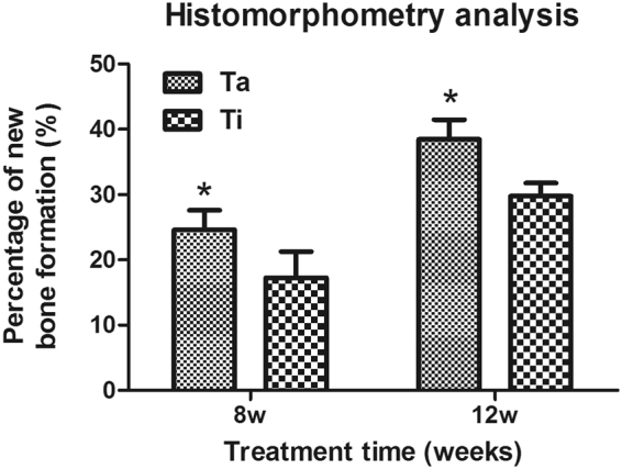
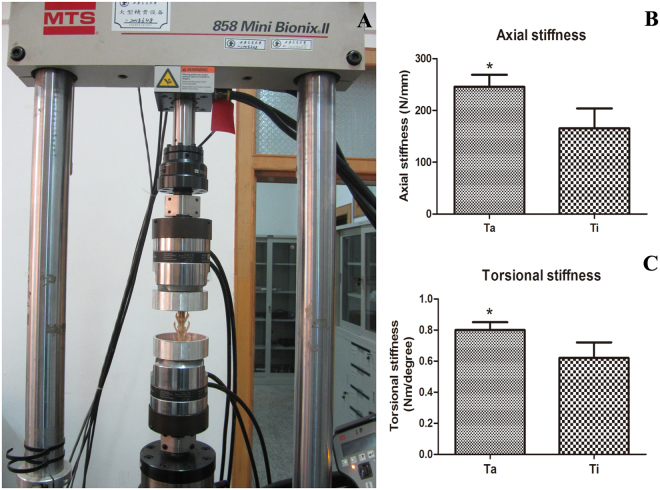
Tantalum (Ta)-coated porous Ti-6A1-4V scaffolds have better bioactivity than Ti-6A1-4V scaffolds; however, their bioperformance as an artificial vertebral body (AVB) is unknown. In the present study, we combined a Ta-coated Ti-6A1-4V scaffold with rabbit bone marrow stromal cells (BMSCs) for tissue-engineered AVB (TEAVB) construction and evaluated the healing and fusion efficacy of this scaffold in lumbar vertebral defects after corpectomy in rabbits. The results showed that BMSCs on the surface of the Ta-coated Ti scaffolds proliferated better than BMSCs on Ti scaffolds. Histomorphometry showed better bone formation when using Ta-coated TEAVBs than that with Ti TEAVBs at both 8 and 12 weeks after implantation. In addition, the vertical and rotational stiffness results showed that, compared with uncoated TEAVBs, Ta-coated TEAVBs enhanced rabbit lumbar vertebral defect repair. Our findings demonstrate that Ta-coated TEAVBs have better healing and fusion efficacy than Ti TEAVBs in rabbit lumbar vertebral defects, which indicates their good prospects for clinical application.


Conflict of interest statement

The authors declare no competing interests.

Figures

Figure 1
Figure 1
Porous Ti6Al4V scaffolds with and without Ta coating. (A) The general view of the scaffolds. Top: Ti6Al4V scaffold; bottom: Ta-coated Ti6Al4V scaffold. (B) Characteristic micro-CT image of uncoated and Ta-coated scaffolds. (C,D) The morphological structures of uncoated and Ta-coated scaffolds, respectively.
Figure 2
Figure 2
Morphological evaluation of BMSCs on the scaffolds. CM-DiI-labeled BMSCs attached to uncoated (A) and Ta-coated scaffolds (B) and SEM morphology of BMSCs attached to uncoated (C) and Ta-coated scaffolds (D) after 72 h of culture.
Figure 3
Figure 3
Attachment, proliferation and ALP activity of BMSCs on the scaffolds. (A) Adhesion of BMSCs cultured on uncoated and Ta-coated scaffolds at 2, 4, and 8 h. (B) Proliferation of BMSCs cultured on uncoated and Ta-coated scaffolds at 1, 4, and 7 d. (C) ALP activity of BMSCs cultured on uncoated and Ta-coated scaffolds at 1, 4, and 7 d. All data are expressed as the mean ± SD. *P < 0.05, **P < 0.01.
Figure 4
Figure 4
Micro-CT 3D reconstruction and bone tissue metrology analysis of scaffolds postoperation. (A,D) Micro-CT images of Ta-coated TEAVBs and Ti TEAVBs at 8 w, respectively. (B,E) Micro-CT images of Ta-coated TEAVBs and Ti TEAVBs at 12 w, respectively. (C,F) Micro-CT 3D reconstruction images of Ta-coated TEAVBs and Ti TEAVBs at 12 w, respectively. (G,K) Analysis of tissue mass density, bone volume fraction, number of bone trabeculae, thickness of bone trabeculae, and bone trabecular space at 12 w, respectively. All data are expressed as the mean ± SD. *P < 0.05.
Figure 5
Figure 5
Representative histological analysis of scaffolds with VG staining postoperation in the L6 of rabbits. (A,C) General view of longitudinal sections of uncoated TEAVBs at 8 and 12 w of cell culture postoperatively. (E,G) Representative images of uncoated, VG-stained TEAVBs at 8 and 12 w of cell culture postoperatively. (B,D) General view of longitudinal sections of Ta-coated TEAVBs at 8 and 12 w of cell culture postoperatively. (F,H) Representative images of VG-stained, Ta-coated TEAVBs at 8 and 12 w of cell culture postoperatively. The tissue stained red was the newly formed bone with visible cell nuclei. The tissue stained blue was fibrous tissue. Scale bar: 2 mm (white), 50 μm (green).
Figure 6
Figure 6
Histomorphometric analysis of Ta-coated TEAVBs and uncoated TEAVBs in defects in the L6 of rabbits. All data are expressed as the mean ± SD. *P < 0.05.
Figure 7
Figure 7
Biomechanical evaluation of scaffolds. (A) Overview of the biomechanical testing process. (B) Axial stiffness and (C) torsional stiffness of the TEAVBs constructed from uncoated and Ta-coated scaffolds at 12 w postoperation. All data are expressed as the mean ± SD. *P < 0.05.
Figure 8
Figure 8
The surgical procedure of the implantation of a Ta-coated scaffold into defects in the L6 of a rabbit. (A) The exposure of the forepart of the lumbar vertebral body. (B) L6 corpectomy. (C) The implantation of the scaffold into the defect site. (D) X-ray examination during the operation to check the positions of TEAVBs. Top: lateral radiograph; bottom: anteroposterior image.

Similar articles

Cited by

References

    1. Tomita K, et al. Total en bloc spondylectomy. A new surgical technique for primary malignant vertebral tumors. Spine (Phila Pa 1976) 1997;22:324–333. doi: 10.1097/00007632-199702010-00018. - DOI - PubMed
    1. Dai LY, Jiang LS, Wang W, Cui YM. Single-stage anterior autogenous bone grafting and instrumentation in the surgical management of spinal tuberculosis. Spine (Phila Pa 1976) 2005;30:2342–2349. doi: 10.1097/01.brs.0000182109.36973.93. - DOI - PubMed
    1. McDonough, P. W., Davis, R., Tribus, C. & Zdeblick, T. A. The management of acute thoracolumbar burst fractures with anterior corpectomy and Z-plate fixation. Spine (Phila Pa 1976)29, 1901–1908; discussion 1909 (2004). - PubMed
    1. Silber JS, et al. Donor site morbidity after anterior iliac crest bone harvest for single-level anterior cervical discectomy and fusion. Spine (Phila Pa 1976) 2003;28:134–139. doi: 10.1097/00007632-200301150-00008. - DOI - PubMed
    1. Macdonald RL, et al. Multilevel anterior cervical corpectomy and fibular allograft fusion for cervical myelopathy. J. Neurosurg. 1997;86:990–997. doi: 10.3171/jns.1997.86.6.0990. - DOI - PubMed

Publication types

MeSH terms

LinkOut - more resources