. 2018 Jun 12;8(1):8930.
doi: 10.1038/s41598-018-27208-4.

Multimodality cellular and molecular imaging of concomitant tumour enhancement in a syngeneic mouse model of breast cancer metastasis

Affiliations

Multimodality cellular and molecular imaging of concomitant tumour enhancement in a syngeneic mouse model of breast cancer metastasis

Katie M Parkins et al. Sci Rep. .

Abstract

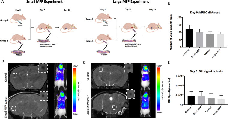
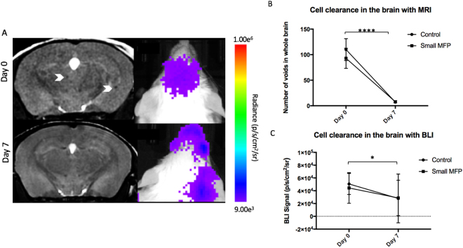
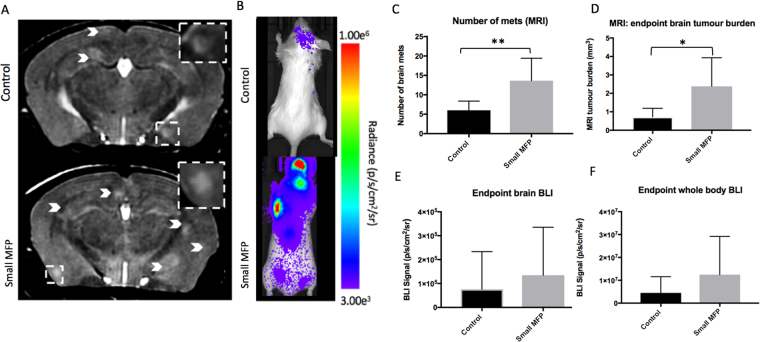
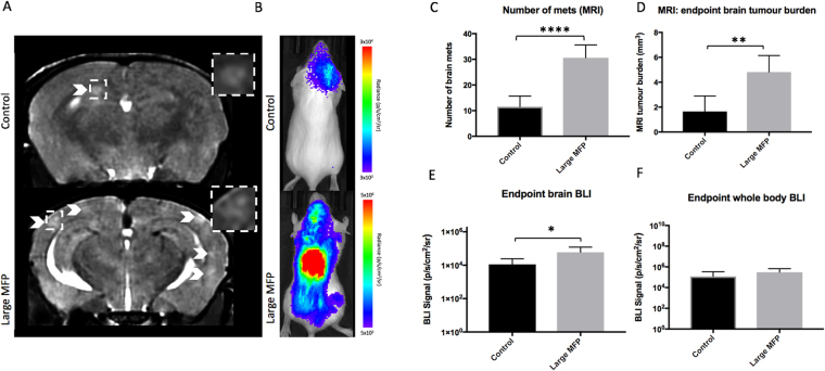
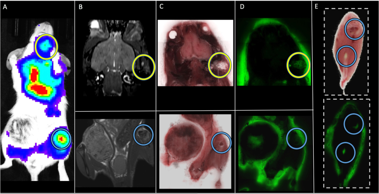
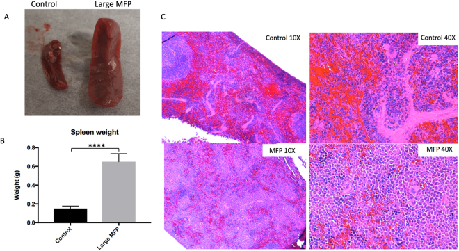
The mechanisms that influence metastatic growth rates are poorly understood. One mechanism of interest known as concomitant tumour resistance (CTR) can be defined as the inhibition of metastasis by existing tumour mass. Conversely, the presence of a primary tumour has also been shown to increase metastatic outgrowth, termed concomitant tumour enhancement (CTE). The majority of studies evaluating CTR/CTE in preclinical models have relied on endpoint histological evaluation of tumour burden. The goal of this research was to use conventional magnetic resonance imaging (MRI), cellular MRI, and bioluminescence imaging to study the impact of a primary tumour on the development of brain metastases in a syngeneic mouse model. Here, we report that the presence of a 4T1 primary tumour significantly enhances total brain tumour burden in Balb/C mice. Using in vivo BLI/MRI we could determine this was not related to differences in initial arrest or clearance of viable cells in the brain, which suggests that the presence of a primary tumour can increase the proliferative growth of brain metastases in this model. The continued application of our longitudinal cellular and molecular imaging tools will yield a better understanding of the mechanism(s) by which this physiological inhibition (CTR) and/or enhancement (CTE) occurs.


Conflict of interest statement

The authors declare no competing interests.

Figures

Figure 1
Figure 1
(A) Experimental design of animal model used for Small and Large MFP experiments (B) On day 0, the number of discrete signal voids, representing iron-labeled cells, that arrested in the brain on day 0 as well as brain BLI signal was not significantly different between mice with and without a small MFP tumour. (C) There were also no significant differences in cell arrest for mice with large MFP tumours. (D,E) All four groups of mice were not significantly different from each other in MRI cell arrest at day 0 as well as BLI signal in the brain at day 0. Data is presented as mean +/− SD.
Figure 2
Figure 2
(A) BLI and MR imaging data illustrates the signal loss over time with cell clearance in the brain of small MFP and control mice. (B) The number of signal voids in the brain decreases from days 0 to 7 as cancer cells are cleared from the brain. (C) BLI signal also decreases from day 0 to 7 as cancer cells are cleared from the brain. Data is presented as mean +/− SD. *Indicates p < 0.05.
Figure 3
Figure 3
(A) At day 14, brain metastases appeared as regions of hyperintensity by MRI, and (B) regions of BLI signal in the brain and body. (C) Mice with a primary small MFP tumor had significantly more brain metastases than mice without a primary tumor. (D) Mice with a primary tumor had significantly more total brain tumor burden than mice without a primary small MFP tumor. (E,F) There were no significant differences in BLI signal in the brain or body between mice with and without a primary small MFP tumour. Data is presented as mean +/− SD. *indicates p < 0.05; **Indicates p < 0.01.
Figure 4
Figure 4
(A) At day 14, brain metastases appeared as regions of hyperintensity by MRI, and (B) regions of BLI signal in the brain and body. (C) Mice with a large MFP tumor had significantly more brain metastases than mice without a primary tumor. (D) Mice with a large MFP tumour had significantly more total brain tumor burden than mice without a primary tumor. (E,F) Similarly, mice with a large MFP tumour had significantly more BLI signal in both the brain and the body compared to control mice. Data is presented as mean +/− SD. *Indicates p < 0.05; **indicates p < 0.01.
Figure 5
Figure 5
In a mouse from the large MFP group we were able to detect distant metastases (head circled in yellow; bone circled in blue) with BLI (A) and match them to corresponding MR (B) and brightfield (C,E) and fluorescence (D,E) cryoviz images.
Figure 6
Figure 6
(A,B) Spleens from large MFP tumor-bearing mice were enlarged and significantly higher in weight(g) than spleens collected from control mice. (C) Normal spleen histology was observed in control mice (Top) but it was found that the spleens isolated from large MFP mice showed a reduction in red pulp (bottom) compared to control mice and normal spleen histology. Data is presented as mean +/− SD. ****Indicates p < 0.0001.

Similar articles

Cited by

References

    1. Siegel R, Ma J, Zou Z, Jemal A. CA: a cancer journal for clinicians. Cancer Statistics. 2014;64(1):9–29. - PubMed
    1. Scully OJ, Bay BH, Yip G, Yu Y. Breast cancer metastasis. Cancer Genomics Proteomics. 2012;9(5):311–320. - PubMed
    1. Chiarella, P., Bruzzo, J., Meiss, R. P., Ruggiero, R. A. Concomitant tumor resistance. Cancer Lett. 28, 324(2): 133–41 (2012). - PubMed
    1. Prehn RT. The inhibition of tumor growth by tumor mass. Cancer Res. 1991;51:2–4. - PubMed
    1. Demicheli R, et al. Breast cancer recurrence dynamics following adjuvant CMF is consistent with tumour dormancy and mastectomy-driven acceleration of the metastatic process. Ann Oncol. 2005;16:1449–1457. doi: 10.1093/annonc/mdi280. - DOI - PubMed

Substances