. 2018 Jun 12;8(1):8996.
doi: 10.1038/s41598-018-27064-2.

An essential role for Wnt/β-catenin signaling in mediating hypertensive heart disease

Affiliations

An essential role for Wnt/β-catenin signaling in mediating hypertensive heart disease

Yue Zhao et al. Sci Rep. .

Abstract

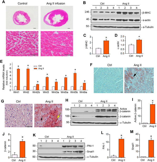
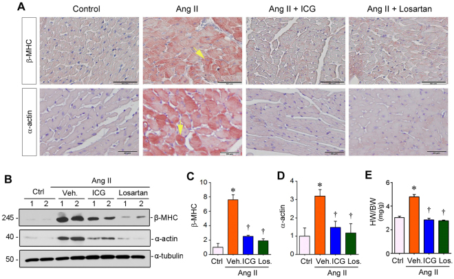
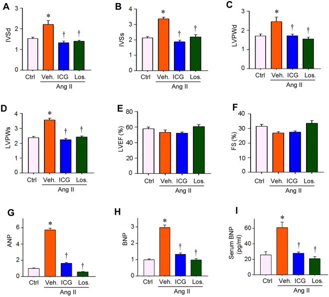
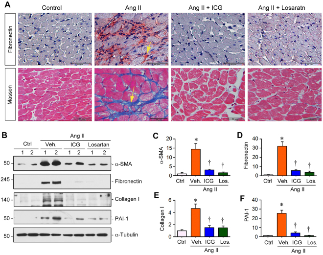
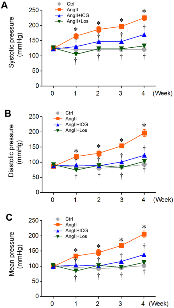
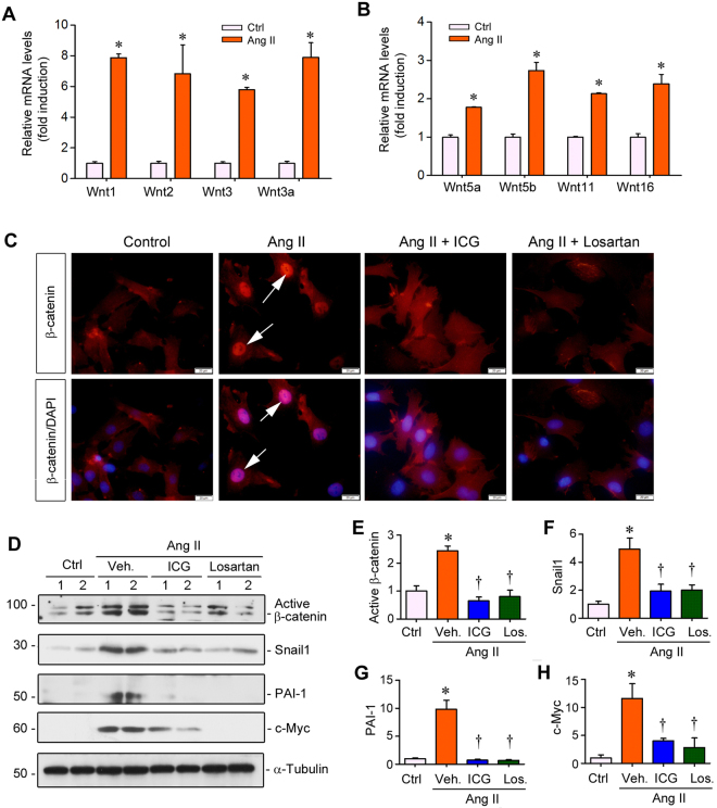
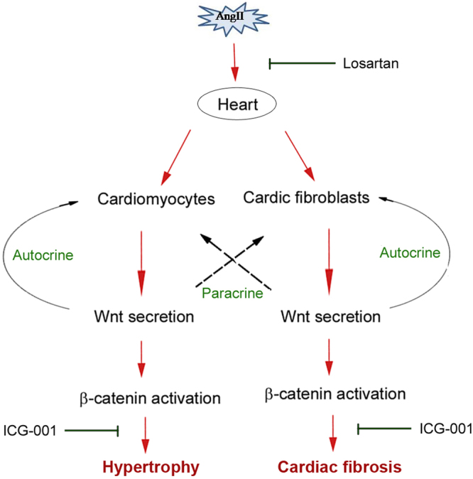
Activation of the renin-angiotensin system (RAS) is associated with hypertension and heart disease. However, how RAS activation causes cardiac lesions remains elusive. Here we report the involvement of Wnt/β-catenin signaling in this process. In rats with chronic infusion of angiotensin II (Ang II), eight Wnt ligands were induced and β-catenin activated in both cardiomyocytes and cardiac fibroblasts. Blockade of Wnt/β-catenin signaling by small molecule inhibitor ICG-001 restrained Ang II-induced cardiac hypertrophy by normalizing heart size and inhibiting hypertrophic marker genes. ICG-001 also attenuated myocardial fibrosis and inhibited α-smooth muscle actin, fibronectin and collagen I expression. These changes were accompanied by a reduced expression of atrial natriuretic peptide and B-type natriuretic peptide. Interestingly, ICG-001 also lowered blood pressure induced by Ang II. In vitro, Ang II induced multiple Wnt ligands and activated β-catenin in rat primary cardiomyocytes and fibroblasts. ICG-001 inhibited myocyte hypertrophy and Snail1, c-Myc and atrial natriuretic peptide expression, and abolished the fibrogenic effect of Ang II in cardiac fibroblasts. Finally, recombinant Wnt3a was sufficient to induce cardiomyocyte injury and fibroblast activation in vitro. Taken together, these results illustrate an essential role for Wnt/β-catenin in mediating hypertension, cardiac hypertrophy and myocardial fibrosis. Therefore, blockade of this pathway may be a novel strategy for ameliorating hypertensive heart disease.


Conflict of interest statement

The authors declare no competing interests.

Figures

Figure 1
Figure 1
Chronic Ang II infusion induces cardiac hypertrophy and activates Wnt/β-catenin signaling. (A) Histological staining (H.E.) revealed overt cardiac hypertrophy in SD rats after 4 weeks of Ang II infusion. Scale bar, 50 µm. (B) Western blot analysis showed an increased expression of hypertrophic markers such as β-MHC and α-actin in the heart after Ang II infusion. Whole cardiac lysates were immunoblotted with antibodies against β-MHC, α-actin and α-tubulin, respectively. (C and D) Quantitative data on β-MHC and α-actin levels in different groups as indicated. *P < 0.05 versus controls (n = 4). (E) Quantitative, real-time RT-PCR (qRT-PCR) showed mRNA induction of multiple Wnt ligands in the heart of SD rat after 4 weeks of Ang II infusion. *P < 0.05 versus controls (n = 4). (F) Immunohistochemical staining for Wnt3a protein in the heart after Ang II infusion. Arrow indicates positive staining. Scale bar, 20 µm. (G) Representative micrographs showed induction of β-catenin protein in the heart after Ang II infusion. Yellow arrow indicates positive cardiomyocyte, whereas black arrow denotes positive cardiac fibroblast. Scale bar, 50 µm. (HJ) Western blot analysis showed cardiac β-catenin and active β-catenin protein expression in the heart after Ang II infusion. Representative Western blot (H) and quantitative data (I,J) are presented. (KM) Western blot analysis showed the protein expression of β-catenin downstream target genes such as PAI-1 and Snail1. Representative Western blot (K) and quantitative data (L,M) are presented. *P < 0.05 versus controls (n = 4). Ctrl, controls. Ang II, angiotensin II.
Figure 2
Figure 2
Inhibition of β-catenin signaling ameliorates Ang II-induced cardiac hypertrophy. (A) Representative micrographs showed immunohistochemical staining for cardiac β-MHC and α-actin proteins in various groups as indicated. Arrows indicate positive staining. Scale bar, 50 µm (top panel) and 20 µm (bottom panel). (BD) Western blot analysis of β-MHC and α-actin in the heart after various treatments as indicated. Representative Western blots (B) and quantitative data for β-MHC (C) and α-actin (D) are presented. *P < 0.05 versus controls; P < 0.05 versus Ang II alone (n = 5). (E) Ratio of heart weight/body weight of SD rats in various groups as indicated. HW, heart weight. BW, body weight. Ctrl, sham controls; Veh., vehicle; ICG, ICG-001; Los., losartan. *P < 0.05 versus sham controls; P < 0.05 versus Ang II alone (n = 5).
Figure 3
Figure 3
ICG-001 prevents geometric abnormalities and inhibits cardiac injury after Ang II infusion in rats. (AD) Echocardiography showed cardiac geometric changes in various groups as indicated. Parameters of cardiac structure measured by echocardiography were as follows, interventricular septum depth at end-diastole (IVSd) (A), interventricular septum depth at end-systole (IVSs) (B), left ventricular posterior wall depth at end-diastole (LVPWd) (C), and left ventricular posterior wall depth at end-systole (LVPWs) (D), at 4 weeks after Ang II infusion in SD rats. (E,F) Echocardiography showed left ventricular ejection fraction (LVEF) and fractional shortening (FS) in rats at 4 weeks after various treatments. (G,H) qRT-PCR showed the mRNA expression of cardiac ANP and BNP in various groups as indicated. *P < 0.05 versus controls; P < 0.05 versus Ang II alone (n = 5). (I) Serum BNP levels in SD rats after various treatments as indicated. Serum BNP protein level was assessed by a specific ELISA. Ctrl, sham controls. Veh, vehicle. ICG, ICG-001. Los, Losartan. *P < 0.05 versus controls; P < 0.05 versus Ang II alone (n = 5).
Figure 4
Figure 4
Inhibition of β-catenin signaling by ICG-001 suppresses Ang II-triggered cardiac fibrosis. (A) Representative micrographs showed fibronectin protein and collagen deposition in the heart after various treatments as indicated. Immunohistochemical staining for fibronectin protein (upper panel) and Masson’s trichrome staining for collagen deposition (lower panel) are shown. Arrows indicate positive staining. Scale bar, 50 µm (top panel) and 20 µm (bottom panel). (B) Representative Western blot showed the protein levels of α-SMA, fibronectin, collagen I and PAI-1 in various groups as indicated. (CF) Quantitative data on the protein levels of α-SMA, fibronectin, collagen I and PAI-1 in various groups as indicated. Relative protein levels over the controls (fold induction) are presented. Ctrl, sham controls. ICG, ICG-001. Los, Losartan. *P < 0.05 versus controls; P < 0.05 versus Ang II alone (n = 5).
Figure 5
Figure 5
ICG-001 lowers blood pressure after Ang II infusion in rats. (A) Graphic presentation of the systolic arterial pressure of SD rats during 4 weeks in various treatment groups as indicated. (B) Graphic presentation of the diastolic arterial pressure of SD rats during 4 weeks. (C) Graphic presentation of the mean arterial pressure of SD rats during 4 weeks. Data are presented as mean ± SEM (n = 5). Ctrl, sham controls. ICG, ICG-001. Los, Losartan. *P < 0.05 versus controls; P < 0.05 versus Ang II alone.
Figure 6
Figure 6
Ang II induces Wnt/β-catenin activation in primary cardiomyocytes. (A,B) qRT-PCR showed that a battery of Wnts mRNA was induced by Ang II. Primary cardiomyocytes were incubated with Ang II (10−6 M) for 12 hours. (C) Immunofluorescent staining for β-catenin in cardiomyocytes treated with Ang II in the absence or presence of ICG-001 or losartan. Arrows indicate nuclear localization of β-catenin after treatment with Ang II in primary cardiomyocytes. (D) Representative Western blot showed protein levels of active β-catenin, Snail1, PAI-1 and c-Myc in primary cardiomyocytes after various treatments for 24 hours as indicated. (EH) Graphic presentation showed the relative protein levels of active β-catenin (E), Snail1 (F), PAI-1 (G) and c-Myc (H) in cardiomyocytes after various treatments. Ctrl, controls. Veh, Vehicle. ICG, ICG-001. Los, Losartan. *P < 0.05 versus controls; P < 0.05 versus Ang II alone (n = 5).
Figure 7
Figure 7
Inhibition of β-catenin by ICG-001 prevents Ang II-induced cardiomyocyte hypertrophy in vitro. (A) Representative micrographs showed rhodamine staining of cardiomyocytes stimulated by Ang II in the absence or presence of ICG-001 or losartan. Scale bar, 50 µm. (B) Graphic presentation showed the relative sizes of cardiomyocytes in various groups as indicated. *P < 0.05 versus controls; P < 0.05 versus Ang II alone (n = 15). (C) Representative Western blot analysis showed the protein levels of α-actin and β-MHC in cardiomyocytes after various treatments as indicated. (D,E) Graphic presentation showed the relative levels of α-actin and β-MHC proteins. *P < 0.05 versus controls; P < 0.05 versus Ang II alone (n = 5). (FH) qRT-PCR demonstrated the mRNA levels of ANP, BNP and β-MHC in cardiomyocytes after various treatments as indicated. (IK) Western blots showed that recombinant Wnt3a induced α-actin and β-MHC expression in primary cardiomyocytes. Rat cardiomyocytes were treated with Wnt3a (100 ng/ml) for 24 hours. Western blot (I) and quantitative data (J and K) are presented. Ctrl, controls. Veh, Vehicle. ICG, ICG-001. Los, Losartan. *P < 0.05 versus controls; P < 0.05 versus Ang II alone (n = 5).
Figure 8
Figure 8
Inhibition of β-catenin by ICG-001 suppresses Ang II-induced fibroblast activation in vitro. (A) Representative phase-contrast micrographs of primary cardiac fibroblasts in the absence or presence of Ang II in vitro. Scale bar, 100 µm. (B) qRT-PCR showed that Ang II induced mRNA expression of a group of Wnt ligands in cultured cardiac fibroblasts. *P < 0.05 versus controls. (C) Representative Western blot showed the protein level of active β-catenin, α-SMA, fibronectin and collagen I in cardiac fibroblasts in various groups as indicated. (DG) Graphic presentation showed the relative abundances of active β-catenin (D), α-SMA (E), fibronectin (F) and collagen I (G). *P < 0.05 versus controls; P < 0.05 versus angiotensin II alone (n = 5). (H) Immunofluorescent staining showed fibronectin protein deposition by fibroblasts after various treatments as indicated. Scale bar, 20 µm. Ctrl, controls. ICG, ICG-001. (I,J) Recombinant Wnt3a induced α-SMA and fibronectin expression in primary cardiac fibroblasts. Rat cardiac fibroblasts were treated with Wnt3a (100 ng/ml) for 24 hours. Western blot (I) and quantitative data (J) are presented.
Figure 9
Figure 9
Schematic diagram depicts the role of Wnt/β-catenin signaling in mediating Ang II-induced cardiac hypertrophy and fibrosis. Ang II induces de novo expression of a variety of Wnt ligands in both cardiomyocytes and cardiac fibroblasts, and then stimulates β-catenin activation by autocrine and paracrine mechanism. As expected, blockade of Ang II action by losartan represses Wnt/β-catenin signaling. Similarly, inhibition of β-catenin signaling by ICG-001 also blocks cardiomyocyte hypertrophy and fibroblast activation, thereby preventing Ang II-induced hypertension, cardiac injury, hypertrophy and fibrosis.

Similar articles

Cited by

References

    1. Alpert CM, Smith MA, Hummel SL, Hummel EK. Symptom burden in heart failure: assessment, impact on outcomes, and management. Heart Fail Rev. 2017;22:25–39. doi: 10.1007/s10741-016-9581-4. - DOI - PMC - PubMed
    1. Reyes EB, et al. Heart failure across Asia: Same healthcare burden but differences in organization of care. Int J Cardiol. 2016;223:163–167. doi: 10.1016/j.ijcard.2016.07.256. - DOI - PubMed
    1. Shimokawa H, Miura M, Nochioka K, Sakata Y. Heart failure as a general pandemic in Asia. Eur J Heart Fail. 2015;17:884–892. doi: 10.1002/ejhf.319. - DOI - PubMed
    1. Shimizu I, Minamino T. Physiological and pathological cardiac hypertrophy. J Mol Cell Cardiol. 2016;97:245–262. doi: 10.1016/j.yjmcc.2016.06.001. - DOI - PubMed
    1. Larstorp AC, et al. Regression of ECG-LVH is associated with lower risk of new-onset heart failure and mortality in patients with isolated systolic hypertension; The LIFE study. American journal of hypertension. 2012;25:1101–1109. doi: 10.1038/ajh.2012.86. - DOI - PubMed

Publication types

MeSH terms