

Expression of Notch receptors and their ligands in pancreatic ductal adenocarcinoma
- PMID: 29896227
- PMCID: PMC5995048
- DOI: 10.3892/etm.2018.6172
Expression of Notch receptors and their ligands in pancreatic ductal adenocarcinoma
Abstract
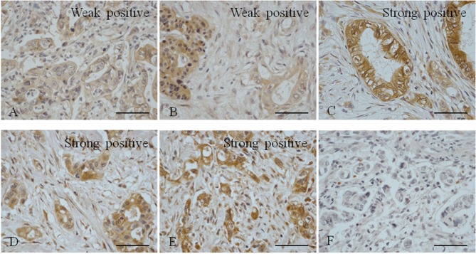
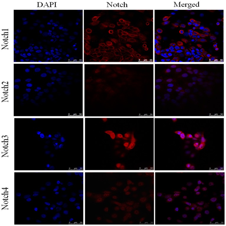
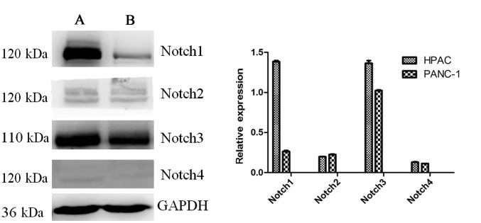
Pancreatic cancer is the fourth leading cause of cancer-associated mortality in developed countries. Pancreatic ductal adenocarcinoma (PDAC) accounts for ~90% of all pancreatic cancer cases. The Notch signaling pathway serves a crucial role in embryonic development, as well as during the tumorigenesis of different types of cancer. However, Notch signaling serves either oncogenic or tumor suppressor roles depending on the tissue type. There are four Notch receptors (Notch1-4) and five ligands [Jagged1, Jagged2, δ-like ligand protein (DLL)1, DLL3 and DLL4]; therefore, it has been suggested that the different Notch receptors serve distinct roles in the same type of tissue. To determine whether this is the case, the present study measured the expression of all Notch receptors and their ligands in PDAC tissue samples and cells. Immunohistochemistry was performed to measure the expression of Notch receptors and their ligands in paraffin-embedded PDAC tissue samples. Immunofluorescence was used to detect the expression of Notch receptors in the pancreatic cancer cell lines human pancreatic adenocarcinoma (HPAC) and PANC-1. In addition, levels of Notch receptors and ligands in HPAC and PANC-1 cells were analyzed by western blot analysis. The results revealed that levels of Notch1 and Notch3 were increased in PDAC tissues, whereas levels of Notch2 and Notch3 were not. The expression of Notch receptors in the pancreatic cancer cell lines HPAC and PANC-1 was consistent with their expression in PDAC tissues. Additionally, levels of the ligands DLL1, DLL3 and DLL4 were increased in HPAC and PANC-1 cells, as well as PDAC tissue samples. However, the expression of Jagged1 and 2 remained low. These results indicate that Notch1, Notch3, DLL1, DLL3 and DLL4 are upregulated in PDAC, a positive correlation was observed between the expression of Notch1 and Notch3, and between Notch1 and the ligands DLL1, DLL3 and DLL4. whereas Notch2, Notch4, Jagged1 and Jagged2 are not. The interaction of Notch1 and Notch3 with Notch ligands DLL1, DLL3 and DLL4 may be important in maintaining the tumor phenotype of pancreatic cancer.
Keywords: Notch ligands; Notch receptors; cancer biomarkers; expression; pancreatic cancer.
Figures


Similar articles
-
Differential expression of the Notch1 receptor, and its ligands Dll1, Dll3 and Dll4 in distinct human pituitary adenoma subtypes.Oncol Lett. 2017 Jun;13(6):4533-4539. doi: 10.3892/ol.2017.5997. Epub 2017 Apr 5. Oncol Lett. 2017. PMID: 28599454 Free PMC article.
-
[Expressions of Notch signaling-associated proteins in esophageal squamous cell carcinoma].Zhonghua Wei Chang Wai Ke Za Zhi. 2015 Sep;18(9):909-13. Zhonghua Wei Chang Wai Ke Za Zhi. 2015. PMID: 26404689 Chinese.
-
Notch signaling dynamics in the adult healthy prostate and in prostatic tumor development.Prostate. 2016 Jan;76(1):80-96. doi: 10.1002/pros.23102. Epub 2015 Sep 30. Prostate. 2016. PMID: 26419726
-
Possible roles of DLK1 in the Notch pathway during development and disease.Biochim Biophys Acta. 2012 Jun;1822(6):988-95. doi: 10.1016/j.bbadis.2012.02.003. Epub 2012 Feb 12. Biochim Biophys Acta. 2012. PMID: 22353464 Review.
-
Precision medicine for human cancers with Notch signaling dysregulation (Review).Int J Mol Med. 2020 Feb;45(2):279-297. doi: 10.3892/ijmm.2019.4418. Epub 2019 Dec 4. Int J Mol Med. 2020. PMID: 31894255 Free PMC article. Review.
Cited by
-
Are ENT1/ENT1, NOTCH3, and miR-21 Reliable Prognostic Biomarkers in Patients with Resected Pancreatic Adenocarcinoma Treated with Adjuvant Gemcitabine Monotherapy?Cancers (Basel). 2019 Oct 23;11(11):1621. doi: 10.3390/cancers11111621. Cancers (Basel). 2019. PMID: 31652721 Free PMC article.
-
Potential ceRNA networks involved in autophagy suppression of pancreatic cancer caused by chloroquine diphosphate: A study based on differentially‑expressed circRNAs, lncRNAs, miRNAs and mRNAs.Int J Oncol. 2019 Feb;54(2):600-626. doi: 10.3892/ijo.2018.4660. Epub 2018 Dec 10. Int J Oncol. 2019. PMID: 30570107 Free PMC article.
-
GenomeForest: An Ensemble Machine Learning Classifier for Endometriosis.AMIA Jt Summits Transl Sci Proc. 2020 May 30;2020:33-42. eCollection 2020. AMIA Jt Summits Transl Sci Proc. 2020. PMID: 32477621 Free PMC article.
-
Krüppel-like factor 10 modulates stem cell phenotypes of pancreatic adenocarcinoma by transcriptionally regulating notch receptors.J Biomed Sci. 2023 Jun 12;30(1):39. doi: 10.1186/s12929-023-00937-z. J Biomed Sci. 2023. PMID: 37308977 Free PMC article.
-
Therapeutic Status and Available Strategies in Pancreatic Ductal Adenocarcinoma.Biomedicines. 2021 Feb 11;9(2):178. doi: 10.3390/biomedicines9020178. Biomedicines. 2021. PMID: 33670230 Free PMC article. Review.
References
LinkOut - more resources
Full Text Sources
Other Literature Sources
Miscellaneous