Changes of lysosomal membrane permeabilization and lipid metabolism in sidt2 deficient mice
- PMID: 29896245
- PMCID: PMC5995057
- DOI: 10.3892/etm.2018.6187
Changes of lysosomal membrane permeabilization and lipid metabolism in sidt2 deficient mice
Abstract
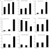
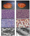
The SID1 transmembrane family member 2 (sidt2) deficient mouse model was used to investigate the function of sidt2 in lysosomal membrane permeabilization and lipid metabolism of liver tissue. The mouse model was established by Cre/LoxP technology. Enzymatic methods were used to analyze the sidt2-/- mouse serum lipids, aspartate transaminase, alanine transaminase and serum bilirubin, compared with sidt2+/+ mice. Defective lipid metabolism and damaged liver functions were observed in the sidt2-/- mice. By using hematoxylin and eosin and Oil Red O staining, changes of morphology were observed in sidt2-/- mice with optical microscopy. Transmission electron microscopy was also used. Hepatic steatosis and partial liver tissue apoptosis were observed. The tissue distribution of sidt2 protein and mRNA was measured in knockout mice. The results indicated that negligible sidt2 mRNA and protein expression were observed in sidt2-/- mice, and that sidt2-/- mice had abnormal liver functions. Transmission electron microscopy revealed membrane lipid droplets in the liver cell cytoplasm, and some apoptotic body formation. These results demonstrated that absence of the lysosomal membrane protein sidt2 led to changes in lysosomal membrane permeabilization and lipid metabolism.
Keywords: SID1 transmembrane family member 2−/− mice; lipid metabolism; lysosomal membrane permeabilization.
Figures


Similar articles
-
Sidt2 regulates hepatocellular lipid metabolism through autophagy.J Lipid Res. 2018 Mar;59(3):404-415. doi: 10.1194/jlr.M073817. Epub 2018 Jan 23. J Lipid Res. 2018. PMID: 29363559 Free PMC article.
-
Spontaneous nonalcoholic fatty liver disease and ER stress in Sidt2 deficiency mice.Biochem Biophys Res Commun. 2016 Aug 5;476(4):326-332. doi: 10.1016/j.bbrc.2016.05.122. Epub 2016 May 24. Biochem Biophys Res Commun. 2016. PMID: 27233614
-
The functions of SID1 transmembrane family, member 2 (Sidt2).FEBS J. 2023 Oct;290(19):4626-4637. doi: 10.1111/febs.16641. Epub 2022 Oct 14. FEBS J. 2023. PMID: 36176242 Review.
-
SID1 transmembrane family, member 2 (Sidt2): a novel lysosomal membrane protein.Biochem Biophys Res Commun. 2010 Nov 26;402(4):588-94. doi: 10.1016/j.bbrc.2010.09.133. Epub 2010 Oct 20. Biochem Biophys Res Commun. 2010. PMID: 20965152
-
The role of lysosomal membrane proteins in glucose and lipid metabolism.FASEB J. 2021 Oct;35(10):e21848. doi: 10.1096/fj.202002602R. FASEB J. 2021. PMID: 34582051 Review.
Cited by
-
Structural basis for double-stranded RNA recognition by SID1.Nucleic Acids Res. 2024 Jun 24;52(11):6718-6727. doi: 10.1093/nar/gkae395. Nucleic Acids Res. 2024. PMID: 38742627 Free PMC article.
-
Lysosome biology in autophagy.Cell Discov. 2020 Feb 11;6:6. doi: 10.1038/s41421-020-0141-7. eCollection 2020. Cell Discov. 2020. PMID: 32047650 Free PMC article. Review.
-
Structural insight into the human SID1 transmembrane family member 2 reveals its lipid hydrolytic activity.Nat Commun. 2023 Jun 15;14(1):3568. doi: 10.1038/s41467-023-39335-2. Nat Commun. 2023. PMID: 37322007 Free PMC article.
-
Interaction between SIDT2 and ABCA1 Variants with Nutrients on HDL-c Levels in Mexican Adults.Nutrients. 2023 Jan 11;15(2):370. doi: 10.3390/nu15020370. Nutrients. 2023. PMID: 36678241 Free PMC article.
-
Genome-Wide Association Study Identifies a Functional SIDT2 Variant Associated With HDL-C (High-Density Lipoprotein Cholesterol) Levels and Premature Coronary Artery Disease.Arterioscler Thromb Vasc Biol. 2021 Sep;41(9):2494-2508. doi: 10.1161/ATVBAHA.120.315391. Epub 2021 Jul 8. Arterioscler Thromb Vasc Biol. 2021. PMID: 34233476 Free PMC article.
References
-
- Oberle C, Huai J, Reinheckel T, Tacke M, Rassner M, Ekert PG, Buellesbach J, Borner C. Lysosomal membrane permeabilization and cathepsin release is a Bax/Bak-dependent, amplifying event of apoptosis in fibroblasts and monocytes. Cell Death Differ. 2010;17:1167–1178. doi: 10.1038/cdd.2009.214. - DOI - PubMed
LinkOut - more resources
Full Text Sources
Other Literature Sources
