

Thoracic manifestation of Wegener's granulomatosis: Computed tomography findings and analysis of misdiagnosis
- PMID: 29896268
- PMCID: PMC5995034
- DOI: 10.3892/etm.2018.6154
Thoracic manifestation of Wegener's granulomatosis: Computed tomography findings and analysis of misdiagnosis
Abstract
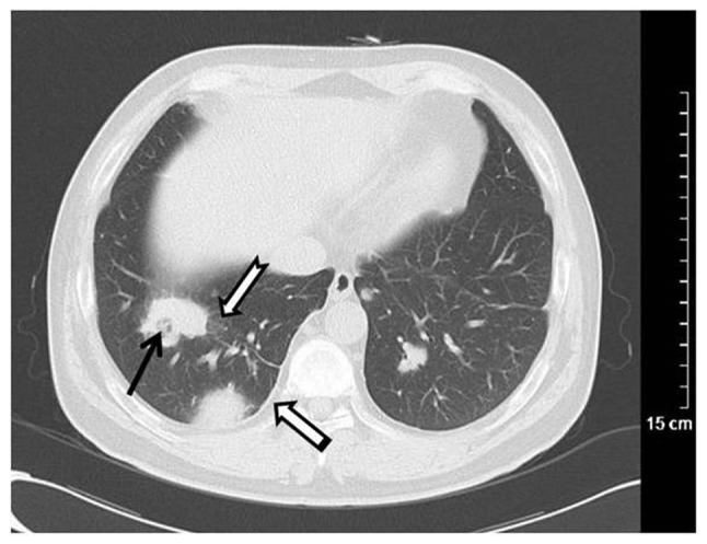
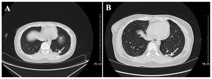
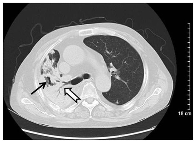
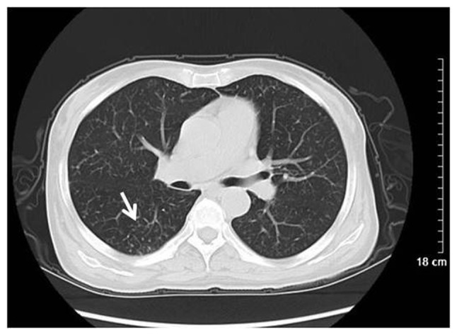
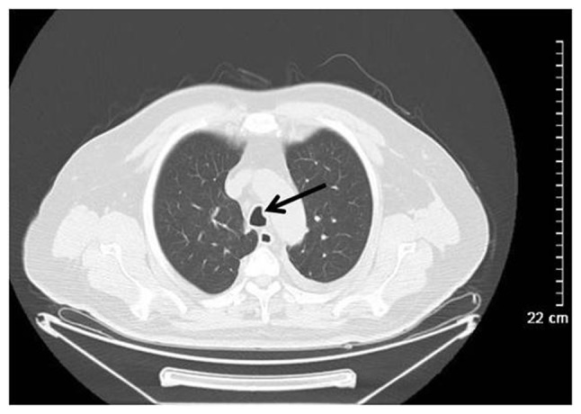
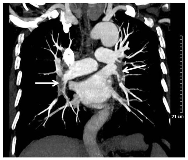
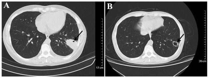
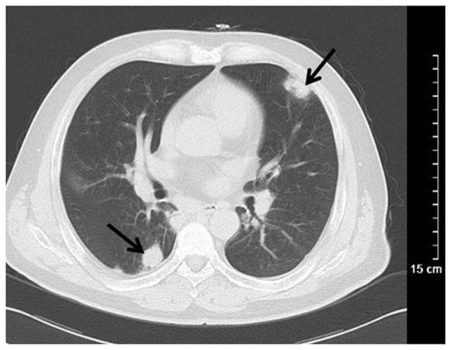
The aim of the present study was to investigate the computed tomography (CT) manifestations of Wegener granulomatosis (WG) in the chest and potential reasons for misdiagnosis. Conventional CT scans and clinical data of 45 patients with WG were retrospectively analyzed. Patients typically presented with multiple system involvement, primarily in the upper and lower respiratory tract. The incidence of thoracic involvement was 75.56% (34/45). Radiographic features were varied between cases in the present study, with the most common features being numerous cavitary nodules and masses in the lungs. Cavitations were usually irregular, with uneven wall thickness, partial centrality, fuzzy inner edges and piecemeal necrosis. These results indicate that WG typically has multiple system involvement, with the chest being most prominent. Multiple variable-sized cavitary nodules with irregular edges and piecemeal necrosis were the most notable features revealed using CT scanning; however, in order to give a definitive diagnosis, biopsies should be performed.
Keywords: Wagener's granulomatosis; X-ray computed; diagnostic errors; tomography.
Figures
Similar articles
-
[Clinical data analysis and chest radiographic features of Wegener's granulomatosis with pulmonary involvement].Nan Fang Yi Ke Da Xue Xue Bao. 2010 Apr;30(4):786-8. Nan Fang Yi Ke Da Xue Xue Bao. 2010. PMID: 20423851 Chinese.
-
Wegener's granulomatosis in childhood.Pediatr Radiol. 1991;21(8):552-5. doi: 10.1007/BF02012595. Pediatr Radiol. 1991. PMID: 1815173
-
Imaging of thoracic Wegener's granulomatosis: the computed tomographic appearance.Am J Med. 1992 Nov;93(5):529-36. doi: 10.1016/0002-9343(92)90581-u. Am J Med. 1992. PMID: 1442856
-
[Computed tomography findings in pulmonary involvement of Wegener's granulomatosis: pictorial review].Tuberk Toraks. 2015;63(4):257-64. doi: 10.5578/tt.9986. Tuberk Toraks. 2015. PMID: 26963309 Review. Turkish.
-
Wegener's granulomatosis in the chest: high-resolution CT findings.AJR Am J Roentgenol. 2009 Mar;192(3):676-82. doi: 10.2214/AJR.08.1837. AJR Am J Roentgenol. 2009. PMID: 19234263 Review.
Cited by
-
Tips and Tricks in Thoracic Radiology for Beginners: A Findings-Based Approach.Tomography. 2023 Jun 14;9(3):1153-1186. doi: 10.3390/tomography9030095. Tomography. 2023. PMID: 37368547 Free PMC article. Review.
-
Noninfectious Granulomatous Diseases of the Chest.Radiographics. 2020 Jul-Aug;40(4):1003-1019. doi: 10.1148/rg.2020190180. Epub 2020 Jun 5. Radiographics. 2020. PMID: 32501738 Free PMC article. Review.
-
Granulomatosis with polyangiitis presenting as exudative pleural effusion: A rare case report.Lung India. 2023 Mar-Apr;40(2):177-179. doi: 10.4103/lungindia.lungindia_263_22. Lung India. 2023. PMID: 37006106 Free PMC article. No abstract available.
-
Noninfectious Granulomatous Lung Disease: Radiological Findings and Differential Diagnosis.J Pers Med. 2024 Jan 23;14(2):134. doi: 10.3390/jpm14020134. J Pers Med. 2024. PMID: 38392568 Free PMC article. Review.
-
Massive, life-threatening hemoptysis due to localized granulomatosis with polyangiitis.J Cardiothorac Surg. 2023 Jul 26;18(1):239. doi: 10.1186/s13019-023-02302-5. J Cardiothorac Surg. 2023. PMID: 37496070 Free PMC article.
References
-
- Wegener F. Über eine eigenartige rhinogene Granulomatose mit besonderer Beteiligung des Arterien systems und der Nieren. Beiträge Zur Pathologie. 1976;158:127–143. doi: 10.1016/S0005-8165(76)80082-0. - DOI
-
- Bişkin S, Yazici ZM, Kayhan FT, Erdur Ö. Wegener Granülomatoziste KBB Tutulumu. KBB ve BBC Dergisi. 2009;17:62–65.
-
- Scalcon MRR, Pereira IA, Filho AR, Paiva EDS. Manifestação otológica localizada em paciente com granulomatose de Wegener Localized otologic manifestation in a patient with Wegener's granulomatosis. Rev Bras Reumatol. 2008;48:253–255. doi: 10.1590/S0482-50042008000400011. - DOI
LinkOut - more resources
Full Text Sources
Other Literature Sources