

Cascade-amplifying synergistic effects of chemo-photodynamic therapy using ROS-responsive polymeric nanocarriers
- PMID: 29896295
- PMCID: PMC5996363
- DOI: 10.7150/thno.24015
Cascade-amplifying synergistic effects of chemo-photodynamic therapy using ROS-responsive polymeric nanocarriers
Abstract
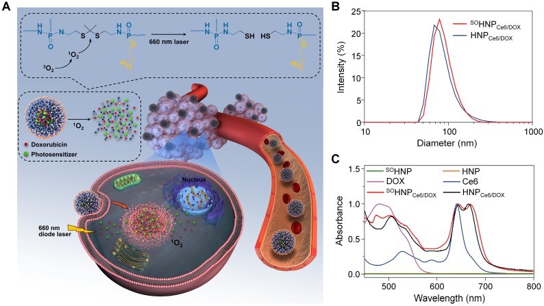
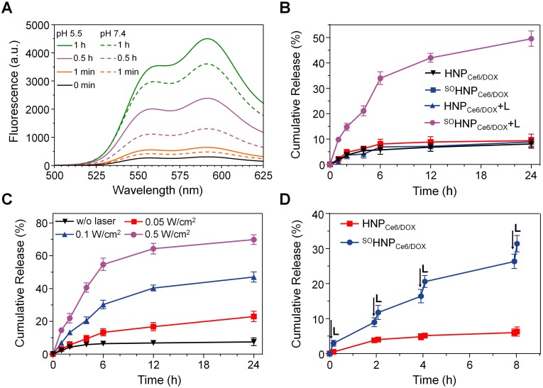
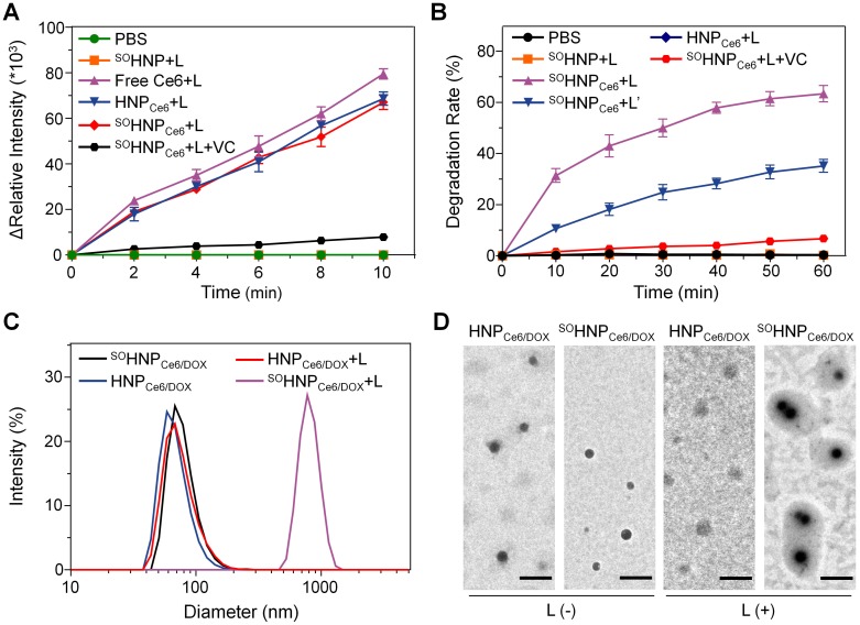
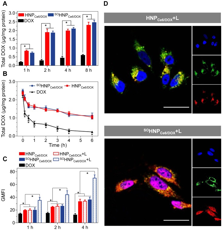
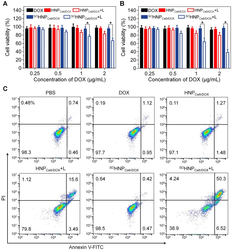
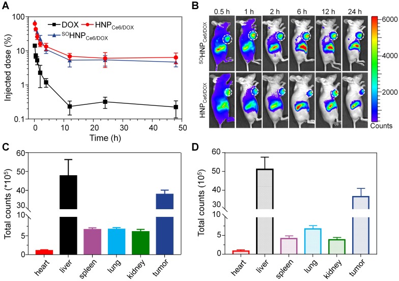
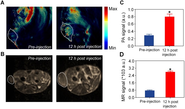
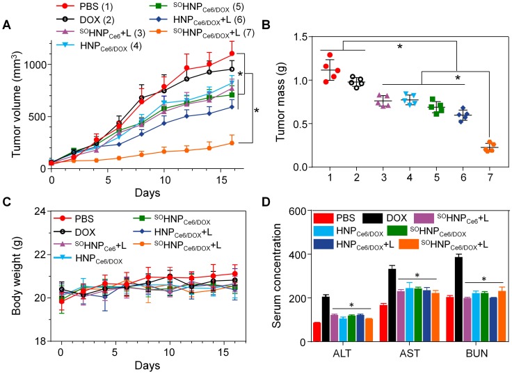
The simple integration of chemotherapeutic drugs and photosensitizers (PSs) into the same nanocarriers only achieves a combination of chemo-photodynamic therapy but may not confer synergistic effects. The boosted intracellular release of chemotherapeutic drugs during the photodynamic therapy (PDT) process is necessary to achieve a cascade of amplified synergistic therapeutic effects of chemo-photodynamic therapy. Methods: In this study, we explored an innovative hyperbranched polyphosphate (RHPPE) containing a singlet oxygen (SO)-labile crosslinker to boost drug release during the PDT process. The photosensitizer chlorin e6 (Ce6) and doxorubicin (DOX) were simultaneously loaded into RHPPE nanoparticles (denoted as SOHNPCe6/DOX). The therapeutic efficacy of SOHNPCe6/DOX against drug-resistant cancer was evaluated in vitro and in vivo. Results: Under 660-nm light irradiation, SOHNPCe6/DOX can produce SO, which not only induces PDT against cancer but also cleaves the thioketal linkers to destroy the nanoparticles. Subsequently, boosted DOX release can be achieved, activating a chemotherapy cascade to synergistically destroy the remaining tumor cells after the initial round of PDT. Furthermore, SOHNPCe6/DOX also efficiently detected the tumor area by photoacoustic/magnetic resonance bimodal imaging. Under the guidance of bimodal imaging, the laser beam was precisely focused on the tumor areas, and subsequently, SOHNPCe6/DOX realized a cascade of amplified synergistic chemo-photodynamic therapeutic effects. High antitumor efficacy was achieved even in a drug-resistant tumor model. Conclusion: The designed SOHNPCe6/DOX with great biocompatibility is promising for use as a co-delivery carrier for combined chemo-photodynamic therapy, providing an alternative avenue to achieve a cascade of amplified synergistic effects of chemo-photodynamic therapy for cancer treatment.
Keywords: ROS responsive; chemo-photodynamic therapy; drug-resistant cancer; on-demand drug release; synergistic therapy.
Conflict of interest statement
Competing Interests: The authors have declared that no competing interest exists.
Figures
Similar articles
-
Doxorubicin-loaded pH-responsive nanoparticles coated with chlorin e6 for drug delivery and synergetic chemo-photodynamic therapy.Nanotechnology. 2020 May 8;31(19):195103. doi: 10.1088/1361-6528/ab6fd5. Epub 2020 Jan 24. Nanotechnology. 2020. PMID: 31978912
-
Synergistic chemo-photodynamic therapy mediated by light-activated ROS-degradable nanocarriers.J Mater Chem B. 2019 Jan 21;7(3):460-468. doi: 10.1039/c8tb03030h. Epub 2018 Dec 19. J Mater Chem B. 2019. PMID: 32254733
-
pH-responsive and self-targeting assembly from hyaluronic acid-based conjugate toward all-in-one chemo-photodynamic therapy.J Colloid Interface Sci. 2019 Jul 1;547:30-39. doi: 10.1016/j.jcis.2019.03.087. Epub 2019 Mar 26. J Colloid Interface Sci. 2019. PMID: 30933691
-
A comprehensive review on singlet oxygen generation in nanomaterials and conjugated polymers for photodynamic therapy in the treatment of cancer.Nanoscale. 2024 Feb 15;16(7):3243-3268. doi: 10.1039/d3nr05801h. Nanoscale. 2024. PMID: 38265094 Review.
-
Natural-product-based, carrier-free, noncovalent nanoparticles for tumor chemo-photodynamic combination therapy.Pharmacol Res. 2024 May;203:107150. doi: 10.1016/j.phrs.2024.107150. Epub 2024 Mar 21. Pharmacol Res. 2024. PMID: 38521285 Review.
Cited by
-
ROS-responsive nano-drug delivery system combining mitochondria-targeting ceria nanoparticles with atorvastatin for acute kidney injury.Theranostics. 2020 Jan 16;10(5):2342-2357. doi: 10.7150/thno.40395. eCollection 2020. Theranostics. 2020. PMID: 32104507 Free PMC article.
-
Photoactivated HPPH-Liposomal therapy for the treatment of HPV-Negative head and neck cancer.Oral Oncol. 2023 Sep;144:106487. doi: 10.1016/j.oraloncology.2023.106487. Epub 2023 Jul 7. Oral Oncol. 2023. PMID: 37423200 Free PMC article.
-
A ROS-responsive polymeric prodrug nanosystem with self-amplified drug release for PSMA (-) prostate cancer specific therapy.J Nanobiotechnology. 2019 Aug 26;17(1):91. doi: 10.1186/s12951-019-0521-z. J Nanobiotechnology. 2019. PMID: 31451114 Free PMC article.
-
TRPM8-regulated calcium mobilization plays a critical role in synergistic chemosensitization of Borneol on Doxorubicin.Theranostics. 2020 Aug 13;10(22):10154-10170. doi: 10.7150/thno.45861. eCollection 2020. Theranostics. 2020. PMID: 32929340 Free PMC article.
-
Tumor microenvironment-responsive hyperbranched polymers for controlled drug delivery.J Pharm Anal. 2024 Dec;14(12):101003. doi: 10.1016/j.jpha.2024.101003. Epub 2024 May 22. J Pharm Anal. 2024. PMID: 39831051 Free PMC article. Review.
References
-
- Lucky SS, Soo KC, Zhang Y. Nanoparticles in photodynamic therapy. Chem Rev. 2015;115:1990–2042. - PubMed
-
- Dolmans D, Fukumura D, Jain RK. Photodynamic therapy for cancer. Nat Rev Cancer. 2003;3:380–7. - PubMed
-
- Fan WP, Huang P, Chen XY. Overcoming the Achilles' heel of photodynamic therapy. Chem Soc Rev. 2016;45:6488–519. - PubMed
-
- Liu K, Xing RR, Zhou QL, Ma GH, Möhwald H, Yan XH. Simple peptide-tuned self-assembly of photosensitizers towards anticancer photodynamic therapy. Angew Chem Int Ed. 2016;55:3036–9. - PubMed
Publication types
MeSH terms
Substances
LinkOut - more resources
Full Text Sources
Other Literature Sources
