. 2018 Jun 1;9(3):334-345.
doi: 10.14336/AD.2017.0809. eCollection 2018 Jun.

Anserine/Carnosine Supplementation Preserves Blood Flow in the Prefrontal Brain of Elderly People Carrying APOE e4

Affiliations

Anserine/Carnosine Supplementation Preserves Blood Flow in the Prefrontal Brain of Elderly People Carrying APOE e4

Qiong Ding et al. Aging Dis. .

Abstract

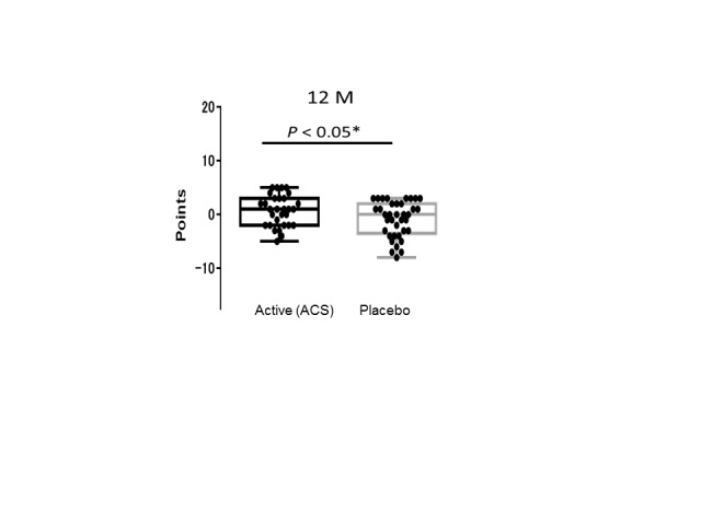
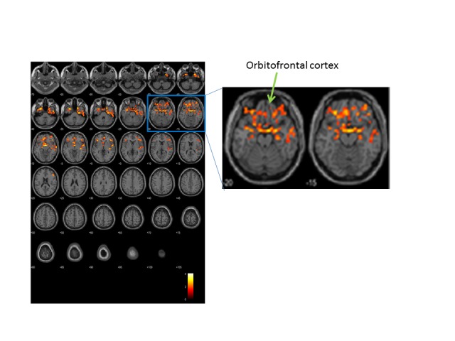
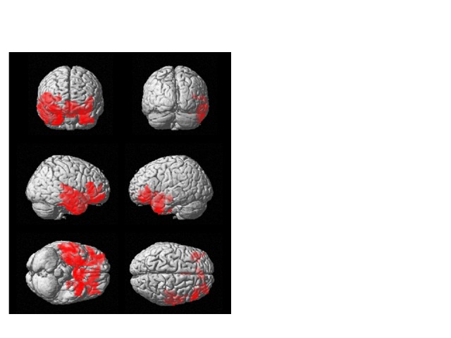
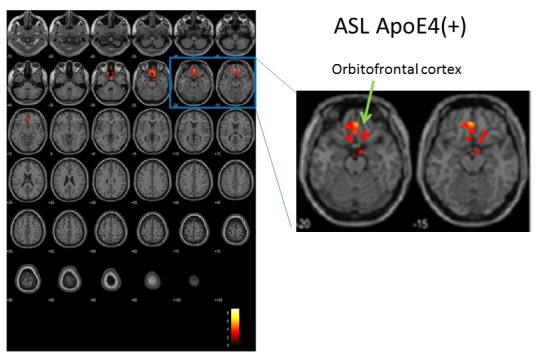
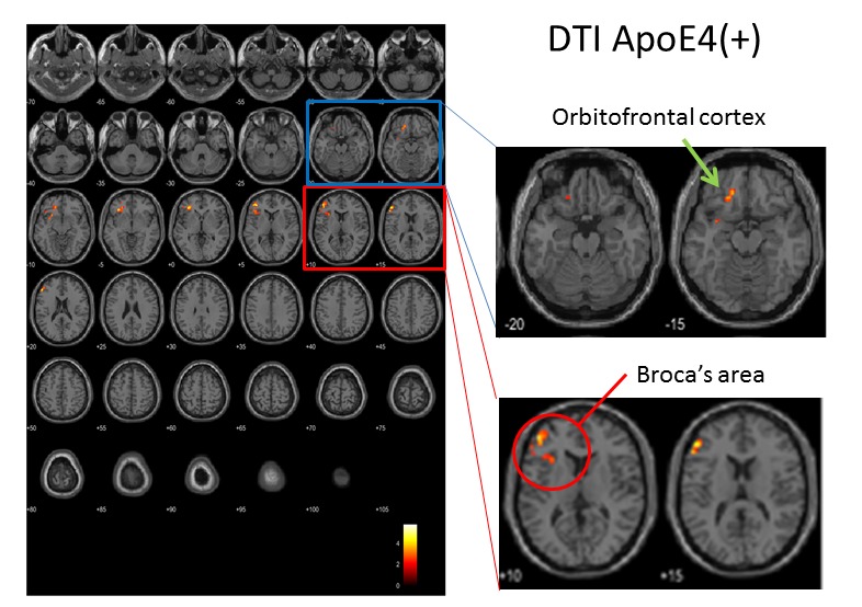
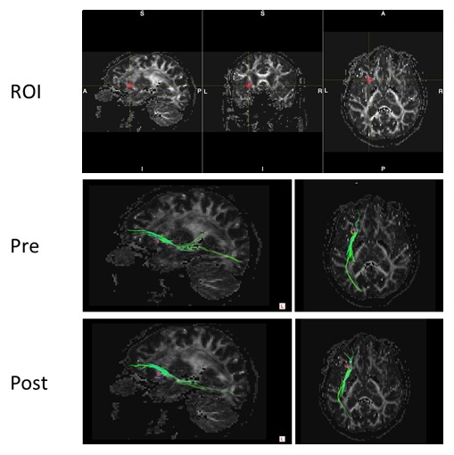
In a previously reported double-blind, randomized controlled trial (RCT), we demonstrated that daily supplementation with anserine (750 mg) and carnosine (250 mg) improves brain blood flow and memory function in elderly people. Here, we conducted a sub-analysis of MRI data and test scores from the same RCT to determine whether anserine/carnosine supplementation specifically benefits elderly people carrying the APOE e4 allele, which is a risk gene for accelerated brain aging and for the onset of Alzheimer's Disease. We collected data from 68 participants aged 65 years or older who received anserine/carnosine supplementation (ACS) or placebo for 12 months. Subjects were assessed at the start and end of the trial using several neuropsychological tests, including the Wechsler Memory Scale-Logical Memory (WMS-LM). We also collected two types of MRI data, arterial spin labeling (ASL) and diffusion tensor imaging (DTI) at the start and end of the trial. We found that ACS significantly preserved verbal memory (WMS-LM, F[1,65] = 4.2003, p = 0.0445) and blood flow at frontal areas of the brain (FWEcluster level, p < 0.001). Sub-analysis based on the APOE4 genotype showed a significant preservation of blood flow (p = 0.002, by ASL analysis) and white-matter microstructure (p = 0.003, by DTI analysis) at prefrontal areas in APOE4+ subjects in the active group, while there was no significant difference between APOE4- subjects in the active and placebo groups. The effect of ACS in preserving brain structure and function in elderly people carrying APOE4 should be verified by further studies.

Keywords: APOE e4; ASL; Alzheimer’s Disease; DTI; RCT; verbal memory.


Conflict of interest statement

Conflict of Interest NH Food Ltd., provided the supplements free of charge. The authors declare that the research was conducted in the absence of any commercial or financial relationships that could be construed as a potential conflict of interest.

Figures

Figure 1.
Figure 1.
Longitudinal changes in WMS-LM2 scores. A box plot of the WMS-LM2 data in Table 2 for subjects in the active (n = 30) and placebo (n = 37) groups. Each black dot indicates the difference between the first and final test scores for one volunteer. Box shows the 25-75 percentile, and solid bar shows the median. (One subject in the active group could not complete the final WMS-LM2 test.)
Figure 2.
Figure 2.
Longitudinal changes in WMS-LM2 scores at the mid-term test. A box plot of the change of WMS-LM2 story A data (the score of 6-month test - the score of the initial test) in the active and placebo groups. Each black dot indicates the difference between the first and final test scores for one volunteer. Box shows the 25-75 percentile, and solid bar shows the median.
Figure 3.
Figure 3.
Longitudinal changes in brain perfusion. Brain blood flow was analyzed by ASL. Color indicates regions where changes in brain perfusion differed between the two groups (n = 31 active, 37 placebo). After repeated two-way ANOVA in SPM, the biggest difference was at (x, y, z) = (3, -1, -10) in Montreal Neurological Institute (MNI) coordinate, T=4.09; other two locations were (x, y, z) = (-30, 11, -37) and (24, 11, -43). SPM statistics showed significance between Active and Placebo (Active > Placebo). P(FWE-corr) < 0.001, KE=2857 voxels. Note that the brain locations of the preservation of blood flow by ACS included both sides of temporal lobes, orbitofrontal cortices, dorsolateral prefrontal cortices and anterior cingulate cortices.
Figure 4.
Figure 4.
ACS preserves blood flow in the prefrontal brain of elderly people. Location of differential longitudinal changes in brain perfusion (red) in the active and placebo groups, on a brain montage from the SPM platform based on equivalent calculations to those in Fig. 3. In the active group, blood flow in these areas was elevated in the follow-up MRI scan.
Figure 5.
Figure 5.
Longitudinal changes in brain perfusion in APOE4+ subjects, assessed by ASL. Color indicates brain regions with differences in longitudinal changes in brain perfusion between the active (n = 8) and placebo (n = 4) groups. After repeated two-way ANOVA in SPM, the biggest difference was at (x, y, z) = (-3, 44, -16), T=9.03. SPM statistics showed significance between Active and Placebo (Active > Placebo). P(FWE-corr) = 0.002, KE=378 voxels.
Figure 6.
Figure 6.
Longitudinal changes in FA (fraction anisotropy) values in APOE4+ subjects, assessed by DTI. Color indicates areas of the brain where FA values differed between the active (n = 8) and placebo (n = 4) groups. After repeated two-way ANOVA in SPM, the biggest difference was at (x, y, z) = (-32, 32, -4), T=5.75; other two locations were (x, y, z) = (-38, 44, 8) and (-50, 36, 18). SPM statistics showed significance between Active and Placebo (Active > Placebo). P(FWE-corr) = 0.003, KE=754 voxels.
Figure 7.
Figure 7.
Tract-graph analysis of APOE4/E4 subject (#75) in the active group. Top panel shows a seed ROI (x, y, z) = (-30, 30, -4), Sphere Radius = 3.00 mm. Middle panel: a tract-graph using this spherical seed ROI at the start-up scan (Pre). Bottom panel: a tract-graph using this spherical seed ROI at the follow-up scan (Post). Note the longer tract (green) toward the frontal pole seemed to be stronger at the follow-up scan.

References

    1. Ritchie K, Lovestone S (2002). The dementias. The Lancet, 360:1759-66. - PubMed
    1. Lindenberger U (2014). Human cognitive aging: corriger la fortune? Science, 346:572-8. - PubMed
    1. Cooper JK (2014). Nutrition and the brain: what advice should we give? Neurobiol Aging, (Suppl 2) 35: S79-83. - PubMed
    1. Rokicki J, Li L, Imabayashi E, Kaneko J, Hisatsune T, Matsuda H (2015). Daily Carnosine and Anserine Supplementation Alters Verbal Episodic Memory and Resting State Network Connectivity in Healthy Elderly Adults. Front Aging Neurosci, 7:219. - PMC - PubMed
    1. Hisatsune T, Kaneko J, Kurashige H, Cao Y, Satsu H, Totsuka M, Katakura Y, Imabayashi E, Matsuda H (2016). Effect of Anserine/Carnosine Supplementation on Verbal Episodic Memory in Elderly People. J Alzheimers Dis, 50:149-59. - PMC - PubMed

LinkOut - more resources