. 2019 Apr;107(3):479-489.
doi: 10.1002/jbm.b.34138. Epub 2018 Jun 13.

In vivo biocompatibility and time-dependent changes in mechanical properties of woven collagen meshes: A comparison to xenograft and synthetic mid-urethral sling materials

Affiliations

In vivo biocompatibility and time-dependent changes in mechanical properties of woven collagen meshes: A comparison to xenograft and synthetic mid-urethral sling materials

Katherine Chapin et al. J Biomed Mater Res B Appl Biomater. 2019 Apr.

Abstract

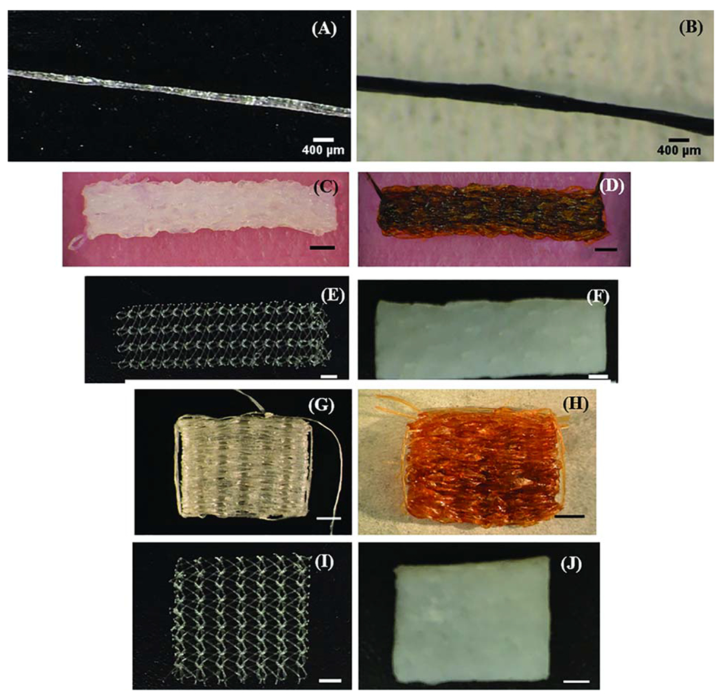
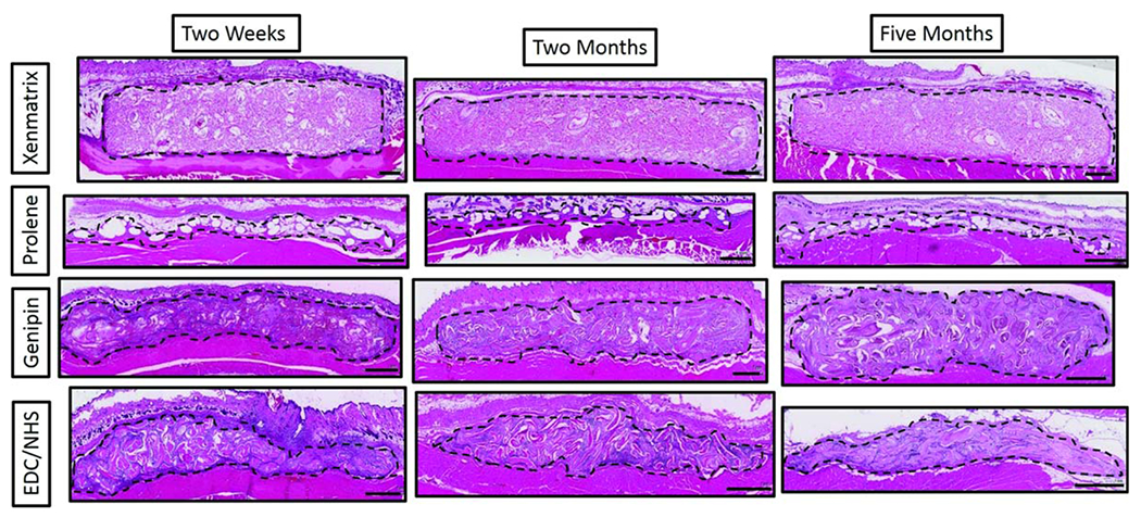
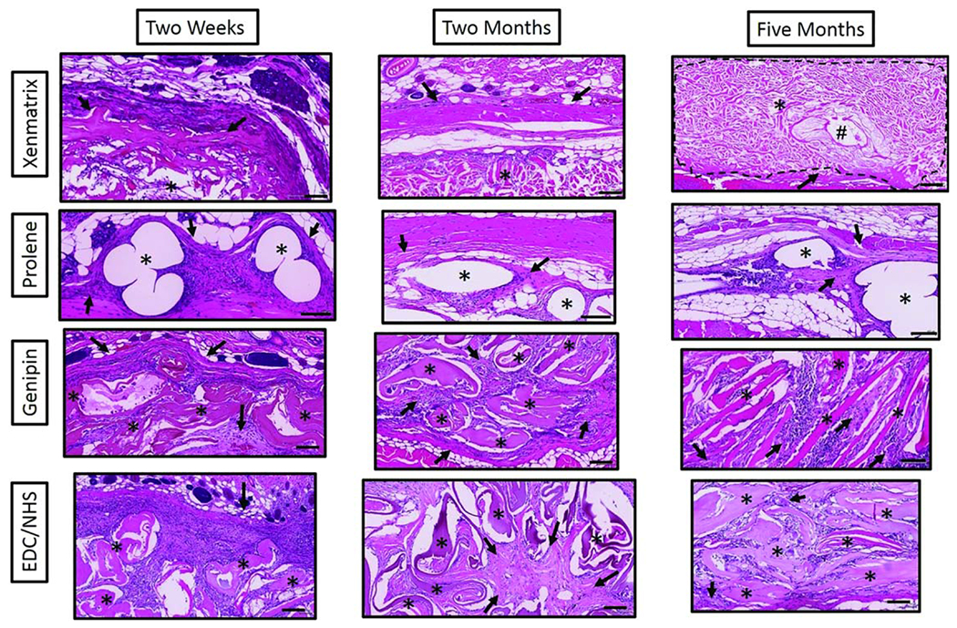
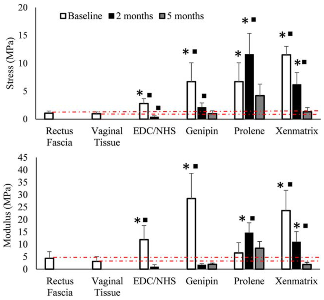
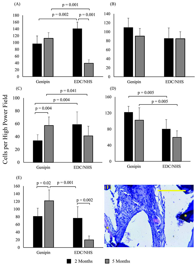
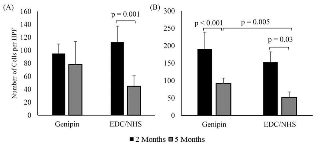
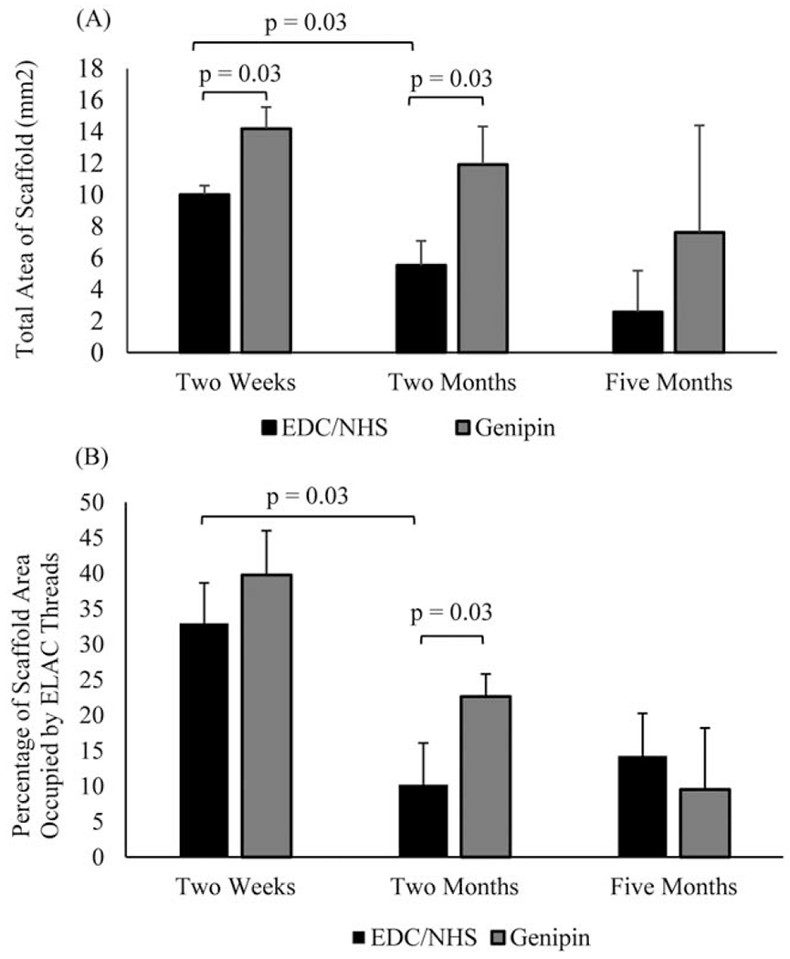
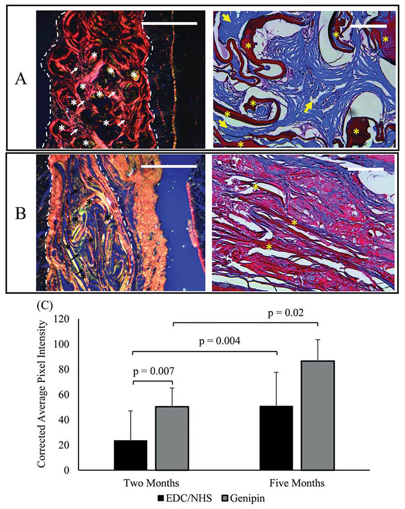
Meshes woven from highly aligned collagen threads crosslinked using either genipin or 1-ethyl-3-(3-dimethylaminopropyl) carboiimide and N-hydroxy succinimide (EDC/NHS) were implanted in a subcutaneous rat model to evaluate their biocompatibility (at 2 weeks, 2 months, and 5 months), mechanical properties (at baseline, 2 months, and 5 months) and ultimately their suitability for use as mid-urethral slings (MUS) for management of stress urinary incontinence. Porcine dermal (Xenmatrix) and monofilament polypropylene (Prolene) meshes were also implanted to provide comparison to clinically used materials. Quantitative histological scoring showed tissue integration in Xenmatrix was almost absent, while the open network of woven collagen and Prolene meshes allowed for cellular and tissue integration. However, strength and stiffness of genipin-crosslinked collagen (GCC), Prolene, and Xenmatrix meshes were not significantly different from those of native rectus fascia and vaginal tissues of animals at 5 months. EDC/NHS-crosslinked collagen (ECC) meshes were degraded so extensively at five months that samples could only be used for histological staining. Picrosirius red and Masson's trichrome staining revealed that integrated tissue within GCC meshes was more aligned (p = 0.02) and appeared more concentrated than ECC meshes at 5 months. Furthermore, immunohistochemical staining showed that GCC meshes attracted a greater number of cells expressing markers for M2 macrophages, those associated with regeneration, than ECC meshes (p = 0.01 for CD206+ cells, p = 0.001 CD163+ cells) at 5 months. As such, GCC meshes hold promise as a new MUS biomaterial based on favorable induction of fibrous tissue resulting in mechanical stiffness matching that of native tissue. © 2018 Wiley Periodicals, Inc. J Biomed Mater Res Part B: Appl Biomater, 2018. © 2018 Wiley Periodicals, Inc. J Biomed Mater Res Part B: Appl Biomater 107B: 479-489, 2019.

Keywords: collagen; stress urinary incontinence; tissue engineering.


Figures

FIGURE 1.
FIGURE 1.
Light microscopy images of (A) Two-ply ECC yarn (B) Two-ply GCC yarn (C–F) ECC, GCC, Prolene and Xenmatrix meshes implanted to evaluate mechanical properties (G–J) ECC, GCC, Prolene and Xenmatrix meshes for assessing mechanical properties. Scale bars for meshes are 2 mm.
FIGURE 2.
FIGURE 2.
Low magnification light microscopy images of H&E stained mesh sections at 2 weeks, 2 months and 5 months. Meshes were collected along with overlying skin and underlying abdominal muscle. Sections of meshes (within dotted outline) are oriented here with the skin at the top of images and the abdominal muscle at the bottom of the images. Scale bars are 1 mm.
FIGURE 3.
FIGURE 3.
High magnification light microscopy images of H&E stained mesh sections at 2 weeks, 2 months and 5 months. Stars denote the respective mesh materials and arrows indicate areas of new collagen deposition in an around mesh materials. The pound symbol (#) denotes hair follicles in Xenmatrix mesh sections. Scale bars are 100 μm.
FIGURE 4.
FIGURE 4.
Average (A) modulus and (B) stress to failure for meshes over time (n = 5 per time point). Squares indicate a value is significantly different (p<0.05) from the proceeding value (intra-group comparison) and stars indicate that a given value is significantly different (p<0.05) from native tissue at that time point (inter-group comparison). Dashed red lines are meant to show how meshes compare to native tissues over time. Error bars indicate the SD for test groups.
FIGURE 5.
FIGURE 5.
Immunohistochemical identification of macrophage markers in woven collagen scaffolds. (A) General macrophage marker CD68, (B) pro-inflammatory macrophage markers B7, and (C) IL6, anti-inflammatory macrophage markers (D) CD163, and (E) CD206. In all bar graphs n = 15 per time point and horizontal connecting bars indicate a significant difference (p <0.05) between the number of expressive cells for groups. Error bars indicate the SD for test groups (F) Immunohistochemical image of CD206 positive macrophages in genipin crosslinked collagen meshes. Asterisks highlight collagen threads. The scale bar is 0.25 mm.
FIGURE 6.
FIGURE 6.
Immunohistochemical markers for endothelium in woven collagen meshes at 2 and 5 months (n = 15 per time point). (A) CD31 and (B) α-SMA. Horizontal connecting bars indicate a significant difference (p< 0.05) between the number of expressive cells for groups, and error bars indicate the SD for test groups.
FIGURE 7.
FIGURE 7.
Changes in the (A) morphology and (B) thread area density of woven collagen scaffolds over time. Significant differences are indicated by connecting bars (p<0.05) and error bars indicate the SD for test groups.
FIGURE 8.
FIGURE 8.
Five months explanted GCC (A) and ECC (B) meshes stained using picrosirius red (left) and Masson’s trichrome (right). Picrosirius red stained slides were imaged under cross polarized conditions. Highly aligned collagen is predominant in GCC samples as manifested by red/orange polarization patterns and loosely aligned collagen is predominant in ECC as manifested by yellow/green polarization in picrosirius stained sections. Masson’s trichrome images show ample amount of de novo collagen deposition (arrows) in genipin crosslinked threads (asterisk). On the other hand, there is minimal collagen deposition in EDC/NHS crosslinked threads. (C) Degree of alignment of newly deposited collagen from picrosirius stained sections was measured by the corrected pixel intensity of images of scaffolds at 2 and 5 months (n = 15 per time point). Significant differences are indicated by connecting bars (p<0.05), and error bars indicate SD for test groups. Scale bars for picrosirius and Masson’s trichrome images are 1 mm and 0.25 mm in length, respectively.

References

    1. Nitti VW. The prevalence of urinary incontinence. Rev Urol 2001; 3:2–6. - PMC - PubMed
    1. Sangster P, Roland M. Biomaterials in urinary incontinence and treatment of their complications. Indian J Urol 2010;26:221–229. - PMC - PubMed
    1. Gandhi S, Kubba LM, Abramov Y, Botros SM, Goldberg RP, Victor TA, Sand PK. Histopathologic changes of porcine dermis xenografts for transvaginal suburethral slings. Am J Obstet Gynecol 2005;192:1643–1648. - PubMed
    1. Khan ZA, Nambiar A, Morley R, Chapple CR, Emery SJ, Lucas MG. Long-term follow-up of a multicentre randomised controlled trial comparing tension-free vaginal tape, xenograft and autologous fascial slings for the treatment of stress urinary incontinence in women. BJU Int 2015;115:968–977. - PubMed
    1. Nilsson C, Palva K, Aarnio R, Morcos E, Falconer C. Seventeen years’ follow-up of the tension-free vaginal tape procedure for female stress urinary incontinence. Int Urogynecol J 2013;24: 1265–1269. - PubMed