. 2018 Jul 5;10(26):21825-21835.
doi: 10.1021/acsami.8b03962. Epub 2018 Jun 22.

Establishment of a Human iPSC- and Nanofiber-Based Microphysiological Blood-Brain Barrier System

Affiliations

Establishment of a Human iPSC- and Nanofiber-Based Microphysiological Blood-Brain Barrier System

Dianjun Qi et al. ACS Appl Mater Interfaces. .

Abstract

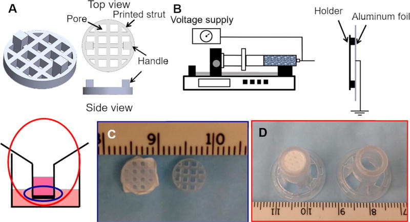
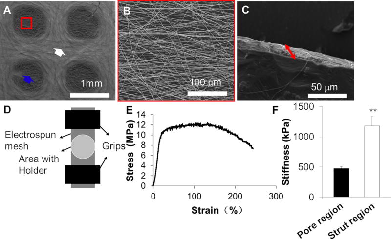
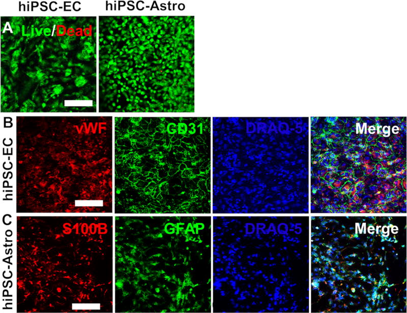
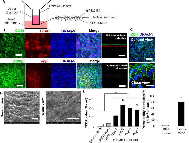
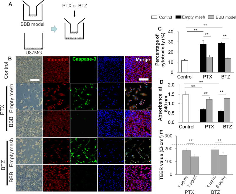
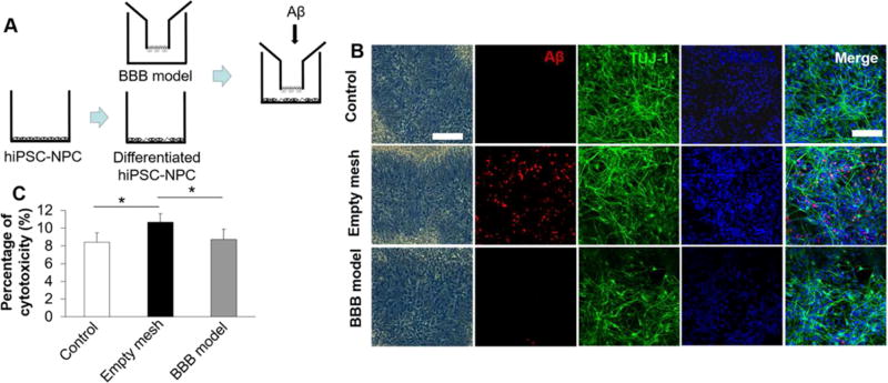
The blood-brain barrier (BBB) is an active and complex diffusion barrier that separates the circulating blood from the brain and extracellular fluid, regulates nutrient transportation, and provides protection against various toxic compounds and pathogens. Creating an in vitro microphysiological BBB system, particularly with relevant human cell types, will significantly facilitate the research of neuropharmaceutical drug delivery, screening, and transport, as well as improve our understanding of pathologies that are due to BBB damage. Currently, most of the in vitro BBB models are generated by culturing rodent astrocytes and endothelial cells, using commercially available transwell membranes. Those membranes are made of plastic biopolymers that are nonbiodegradable, porous, and stiff. In addition, distinct from rodent astrocytes, human astrocytes possess unique cell complexity and physiology, which are among the few characteristics that differentiate human brains from rodent brains. In this study, we established a novel human BBB microphysiologocal system, consisting of a three-dimensionally printed holder with a electrospun poly(lactic- co-glycolic) acid (PLGA) nanofibrous mesh, a bilayer coculture of human astrocytes, and endothelial cells, derived from human induced pluripotent stem cells (hiPSCs), on the electrospun PLGA mesh. This human BBB model achieved significant barrier integrity with tight junction protein expression, an effective permeability to sodium fluorescein, and higher transendothelial electrical resistance (TEER) comparing to electrospun mesh-based counterparts. Moreover, the coculture of hiPSC-derived astrocytes and endothielial cells promoted the tight junction protein expression and the TEER value. We further verified the barrier functions of our BBB model with antibrain tumor drugs (paclitaxel and bortezomib) and a neurotoxic peptide (amyloid β 1-42). The human microphysiological system generated in this study will potentially provide a new, powerful tool for research on human BBB physiology and pathology.

Keywords: 3D printing; drug screening; electrospinning; human induced pluripotent stem cells; transepithelial electrical resistance.


Conflict of interest statement

The authors declare no competing financial interest.

Figures

Figure 1
Figure 1
Fabrication of the insert consisting of a 3D-printed holder and an electrospun PLGA mesh for the BBB model: (A) design of a holder that can fit into a commercial transwell frame. The holder has pore and strut areas with two handles for handling; (B) schematic of electrospinning PLGA nanofibers onto the holder. The holder is fixed onto the aluminum foil on the collector; (C) the insert with the PLGA mesh (left) and an empty holder without the mesh (right); and (D) the inserts were placed into the commercial transwell frame after removing the plastic membranes. Left: insert with the holder and PLGA mesh; Right: insert only.
Figure 2
Figure 2
Characterization of nanofibrous PLGA meshes electrospun onto the holders: (A–C) typical SEM micrographs of the PLGA mesh. (A) Overview of the morphology. Two areas with similar fibrous morphology but different thicknesses were observed. The white arrow indicates the strut region with a thicker thickness, and the red frame and blue arrow indicate the pore region with thinner thickness; (B) close view of the pore region; (C) side view of the pore region, showing the thickness of the PLGA mesh; (D) schematic of the tensile test of PLGA meshes peeled from holders; (E) typical tensile stress–strain curve; and (F) stiffness of the pore region and strut region tested by nanoindentation (~50 points from three different samples, **p < 0.01).
Figure 3
Figure 3
Cell viability and phenotypic marker expression of hiPSC-ECs and hiPSC-Astro on nanofibrous PLGA meshes: (A) Live/Dead assay images (scale bar: 100 µm) and (B,C) typical IF images, showing the expression of vWF (red), CD31 (green) for hiPSC-EC and S100B (red), GFAP (green) for hiPSC-Astro (scale bar: 100 µm).
Figure 4
Figure 4
Generation and characterization of the in vitro BBB model: (A) schematic of a coculture of hiPSC-ECs and hiPSC-Astro on the PLGA mesh that was electrospun onto the 3D-printed holder and fit into the transwell frame. Human iPSC-ECs were cultured on the top side of the PLGA mesh and conditioned in EGM in the upper chamber, whereas hiPSC-Astro were cultured in the bottom side in astrocyte medium in the lower chamber; (B) IF staining of cocultured hiPSC-ECs and hiPSC-Astro after 7 day culture. In the upper panel, the BBB model was costained with CD31 for hiPSC-ECs and GFAP for hiPSC-Astro, whereas in the lower panel, S100B and vWF were stained for hiPSC-Astro and hiPSC-ECs, respectively. Volume-rendered side views of the bilayer cells were also presented (scale bar: 100 µm); (C) expression of the tight junction protein ZO-1 in the cocultured hiPSC-EC (scale bar: 100 µm for general view, and 20 µm for close view); (D) hiPSC-EC morphology on the electrospun PLGA mesh after 7 day coculture (scale bar: 50 µm); (E) TEER value of the empty mesh, without cells, in the transwell insert (white), BBB model with hiPSC-Astro alone (gray), and with coculture of hiPSC-ECs and hiPSC-Astro at different time points (black) (n = 3–5, **p < 0.01, $ indicates a significant difference between days 1 and 3, % indicates a significant difference between days 3 and 7); and (F) permeability coefficient of sodium fluorescein through the in vitro BBB model (n = 3–5, **p < 0.01).
Figure 5
Figure 5
Barrier functions of the in vitro BBB model to PTX and BTZ: (A) schematic of a triculture of hiPSC-ECs and hiPSC-Astro on the PLGA mesh in the upper chamber with U87MG cells in the lower chamber of a transwell insert and the addition of PTX and BTZ. The upper chamber was filled with EGM and the lower chamber was filled with astrocyte medium; (B) typical images of U87MG response to the treatment of PTX (1 µg/mL) and BTZ (4 µg/mL) with or without the BBB model. Left one panel: optical images (scale bar: 100 µm); right four panels: IF staining images (scale bar: 100 µm); (C) LDH assay showed cytotoxicity (n = 3, **p < 0.01); (D) MTT assay showed cell metabolic activity (n = 3, **p < 0.01); and (E) TEER value with changing the dose of PTX and BTZ (n = 3, **p < 0.01). The dashed line indicates the average TEER value without the addition of anti-GBM drugs.
Figure 6
Figure 6
Barrier functions of the in vitro BBB model to Aβ: (A) schematic of a triculture of hiPSC-ECs and hiPSC-Astro on the PLGA mesh in the upper chamber with hiPSC–NPCs in the lower chamber of a transwell insert and the addition of Aβ; hiPSC–NPCs were induced in neuronal differentiation medium for 14 days before triculture. The upper chamber was filled with EGM and the lower chamber was filled with neuronal differentiation medium; (B) typical images of hiPSC–NPC response to the treatment of Aβ with or without the BBB model. Left one panel: optical images (scale bar: 100 µm); right four panels: IF staining images (scale bar: 100 µm); and (C) LDH assay showed cytotoxicity (n = 3, *p < 0.05). Control indicates the group without electrospun meshes, hiPSC-ECs, hiPSC-Astro, and without Aβ treatment; the empty mesh indicates the group with electrospun meshes but without hiPSC-ECs, hiPSC-Astro, and with Aβ treatment; and the BBB model indicates the group with electrospun meshes, hiPSC-ECs, hiPSC-Astro, and with Aβ treatment.

Similar articles

Cited by

References

    1. Abbott NJ, Rönnbäck L, Hansson E. Astrocyte-Endothelial Interactions at the Blood-Brain Barrier. Nat. Rev. Neurosci. 2006;7:41–53. - PubMed
    1. Armulik A, Genové G, Mäe M, Nisancioglu MH, Wallgard E, Niaudet C, He L, Norlin J, Lindblom P, Strittmatter K, Johansson BR, Betsholtz C. Pericytes Regulate the Blood-Brain Barrier. Nature. 2010;468:557–561. - PubMed
    1. Daneman R, Zhou L, Kebede AA, Barres BA. Pericytes Are Required for Blood-Brain Barrier Integrity during Embryogenesis. Nature. 2010;468:562–566. - PMC - PubMed
    1. Stamatovic SM, Keep RF, Andjelkovic AV. Brain Endothelial Cell-Cell Junctions: How to ″Open″ the Blood Brain Barrier. Curr. Neuropharmacol. 2008;6:179–192. - PMC - PubMed
    1. Deeken JF, Loscher W. The Blood-Brain Barrier and Cancer: Transporters, Treatment, and Trojan Horses. Clin. Cancer Res. 2007;13:1663–1674. - PubMed

Substances

LinkOut - more resources