. 2018 Oct 1;315(4):F1006-F1018.
doi: 10.1152/ajprenal.00602.2017. Epub 2018 Jun 13.

Dual gain and loss of cullin 3 function mediates familial hyperkalemic hypertension

Affiliations

Dual gain and loss of cullin 3 function mediates familial hyperkalemic hypertension

Ryan J Cornelius et al. Am J Physiol Renal Physiol. .

Abstract

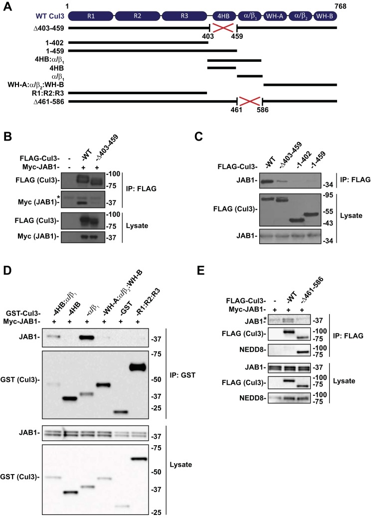
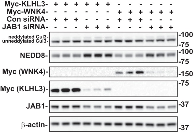
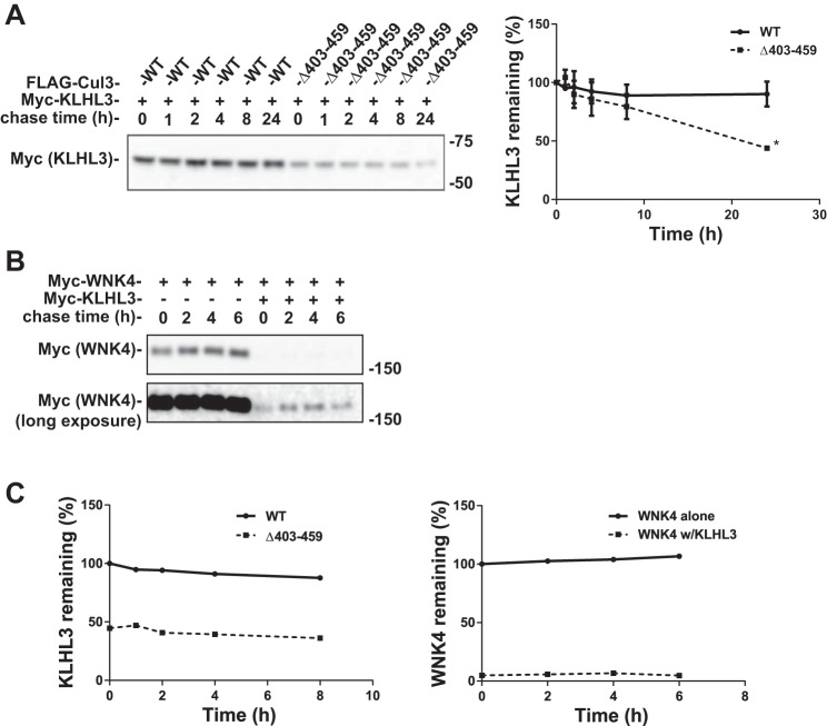
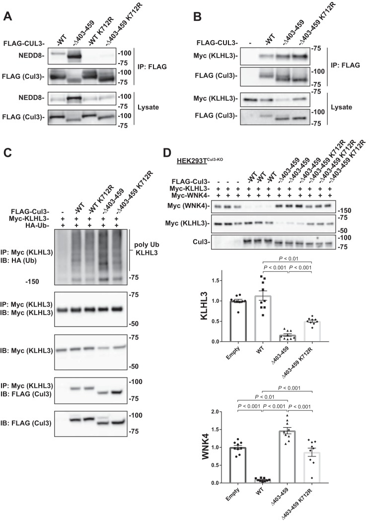
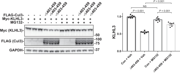
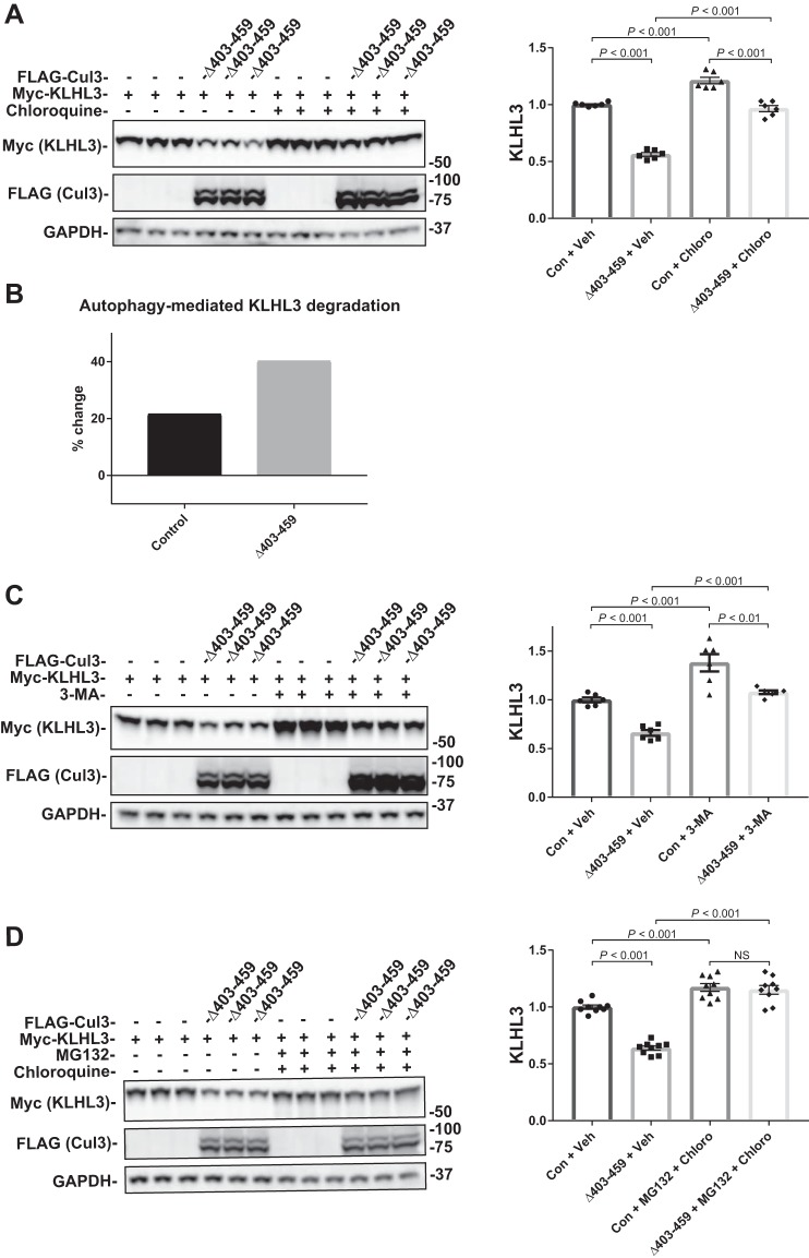
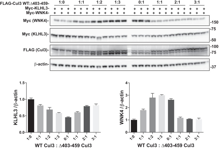
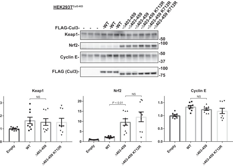
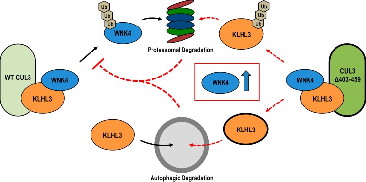
Familial hyperkalemic hypertension is caused by mutations in with-no-lysine kinases (WNKs) or in proteins that mediate their degradation, kelch-like 3 (KLHL3) and cullin 3 (CUL3). Although the mechanisms by which WNK and KLHL3 mutations cause the disease are now clear, the effects of the disease-causing CUL3Δ403-459 mutation remain controversial. Possible mechanisms, including hyperneddylation, altered ubiquitin ligase activity, decreased association with the COP9 signalosome (CSN), and increased association with and degradation of KLHL3 have all been postulated. Here, we systematically evaluated the effects of Cul3Δ403-459 using cultured kidney cells. We first identified that the catalytically active CSN subunit jun activation domain-binding protein-1 (JAB1) does not associate with the deleted Cul3 4-helix bundle domain but instead with the adjacent α/β1 domain, suggesting that altered protein folding underlies the impaired binding. Inhibition of deneddylation with JAB1 siRNA increased Cul3 neddylation and decreased KLHL3 abundance, similar to the Cul3 mutant. We next determined that KLHL3 degradation has both ubiquitin ligase-dependent and -independent components. Proteasomal KLHL3 degradation was enhanced by Cul3Δ403-459; however, autophagic degradation was also upregulated by this Cul3 mutant. Finally, to evaluate whether deficient substrate adaptor was responsible for the disease, we restored KLHL3 to wild-type (WT) Cul3 levels. In the absence of WT Cul3, WNK4 was not degraded, demonstrating that Cul3Δ403-459 itself cannot degrade WNK4; conversely, when WT Cul3 was present, as in diseased humans, WNK4 degradation was restored. In conclusion, deletion of exon 9 from Cul3 generates a protein that is itself ubiquitin-ligase defective but also capable of enhanced autophagocytic KLHL3 degradation, thereby exerting dominant-negative effects on the WT allele.

Keywords: JAB1; cullin-RING ubiquitin ligase; deneddylation; neddylation.


Figures

Fig. 1.
Fig. 1.
CSN binds to Cul3 at the α/β1 domain. A: Diagram of Cul3 domain structure and schematic of the Cul3 constructs. B: Coimmunoprecipitation was performed with HEK293 cells transfected with myc-JAB1 and FLAG-tagged WT Cul3 or Cul3Δ403–459 and analyzed by immunoblot. Cul3Δ403–459 exhibited a decreased interaction with JAB1 compared with WT Cul3. C: Effects of Cul3Δ403–459 on JAB1 binding was determined by coimmunoprecipitation of HEK293 cells with N-terminal domain Cul3 constructs using anti-FLAG and analyzed by immunoblot. Coimmunoprecipitation of N-terminal domain Cul3 constructs with (1–459) and without (1–402) the 4HB domain showed no binding to JAB1. D: Segments of the Cul3 protein were generated with a GST tag and cotransfected with myc-tagged JAB1 in HEK293 cells. Coimmunoprecipitation was performed using glutathione sepharose beads. Immunoblotting for JAB1 showed binding to 4HB:α/β1 and α/β1 Cul3 constructs but not to 4HB, WH-A:α/β:WH-B, or R1:R2:R3 Cul3 constructs. E: Coimmunoprecipitation was performed in HEK293 cells with myc-JAB1 and FLAG-tagged WT Cul3 or Cul3Δ461–586 constructs. Cul3Δ461–586 demonstrated less binding to JAB1 protein compared with WT Cul3. Immunoblotting for NEDD8 showed enhanced neddylation of the Cul3Δ461–586 construct compared with WT Cul3. *Nonspecific band. 4HB, 4-helix bundle; Cul3, cullin 3; CSN, COP9 signalosome; GST, glutathione S-transferase; HEK, human embryonic kidney; IP, immunoprecipitation; JAB1, jun activation domain-binding protein-1; NEDD8, neuronal precursor cell expressed developmentally downregulated protein 8; WT, wild type.
Fig. 2.
Fig. 2.
Effects of JAB1 inhibition on Cul3 neddylation and substrate protein abundance. Myc-tagged KLHL3 or WNK4 was cotransfected into HEK293 cells with either JAB1 siRNA or control siRNA. The proteins were examined by immunoblot in cells with endogenous WT Cul3. JAB1 siRNA decreased JAB1, KLHL3, and WNK4 abundance and increased NEDD8 abundance and the neddylated form of Cul3 (top band). β-actin was used as a loading control. Con, control; Cul3, cullin 3; HEK, human embryonic kidney; JAB1, jun activation domain-binding protein-1; KLHL3, kelch-like 3; NEDD8, neuronal precursor cell expressed developmentally downregulated protein 8; WNK, with-no-lysine kinase; WT, wild type.
Fig. 3.
Fig. 3.
Cul3Δ403–459 decreases the stability of KLHL3. A: Cycloheximide chase assay was performed with HEK293 cells cotransfected with myc-tagged KLHL3 and either FLAG-WT Cul3 or FLAG-Cul3Δ403–459. Because of a robust decrease in KLHL3 by the Cul3Δ403–459 that prevented quantification, the amount of Cul3Δ403–459 transfected was reduced to half of WT Cul3. Cycloheximide (100 µg/ml) was added 36 h posttransfection, and cells were lysed at 0, 1, 2, 4, 8, and 24 h time points. KLHL3 protein abundance was more rapidly degraded in cells coexpressing Cul3Δ403–459. Right, quantitative analysis of KLHL3 protein abundance. Stain-free imaging was used as a loading control. Linear regression was used to determine the slope of each group. The differences between the slopes were significantly different (P < 0.001). Data represent mean values ± SE relative to the 0 h time point. Statistical differences were examined using two-tailed unpaired Student’s t-test. *P = 0.01 vs. WT. B: Cycloheximide chase assay was performed with HEK293 cells cotransfected with myc-tagged WNK4 in the presence or absence of KLHL3. Cycloheximide (100 µg/ml) was added 36 h posttransfection and cells were lysed at 0, 2, 4, and 6 h time points. Stain-free imaging was used as a loading control. C: Left, quantitative analysis of KLHL3; all data points are relative to WT Cul3 0 h time point. Right, quantitative analysis of WNK4 protein abundance; all data points are relative to WNK4 without KLHL3 0 h time point. The effects of Cul3Δ403–459 on KLHL3 abundance is similar to the effects of KLHL3 on WNK4 abundance. Cul3, cullin 3; KLHL3, kelch-like 3; WNK, with-no-lysine kinase; WT, wild type.
Fig. 4.
Fig. 4.
Ligase-deficient Cul3Δ403–459 K712R double mutant blunts the effects of Cul3Δ403–459 on KLHL3 and WNK4. A: FLAG-tagged Cul3 constructs were cotransfected into HEK293 cells and immunoprecipitated using FLAG antibody. Immunoblotting for NEDD8 showed no neddylation of the K712R mutant for both WT Cul3 and Cul3Δ403–459. B: Coimmunoprecipitation was performed with HEK293 cells transfected with myc-KLHL3 and FLAG-tagged WT Cul3, Cul3Δ403–459, Cul3Δ403–459 K712R, or empty vector. Pull-down with FLAG antibodies showed that KLHL3 had more binding to Cul3Δ403–459 and Cul3Δ403–459 K712R proteins. C: Ubiquitin assay was performed for KLHL3 in HEK293 cells by cotransfecting FLAG-tagged Cul3 constructs with myc-KLHL3 and HA-tagged ubiquitin. Immunoprecipitation was performed using anti-myc antibody and polyubiquitylation of KLHL3 was visualized by immunoblotting for anti-HA. Cul3Δ403–459 K712R double mutant attenuated the higher abundance of KLHL3 ubiquitylation shown with Cul3Δ403–459. D: Top, abundance of myc-tagged KLHL3 and WNK4 protein was examined by immunoblot in Cul3 knockdown HEK293T (HEK293TCul3-KO) cells cotransfected with different FLAG-tagged Cul3 constructs. KLHL3 and WNK4 expression was higher and lower, respectively, in Cul3Δ403–459 K712R compared with Cul3Δ403–459. Bottom, quantitative analysis of KLHL3 and WNK4 protein abundance. Stain-free imaging was used as a loading control. Data represent individual values as well as means ± SE relative to control. Statistical differences were examined by one-way ANOVA with Tukey’s post hoc analysis. Cul3, cullin 3; HA, hemagglutinin; HEK, human embryonic kidney; IB, immunoblot; IP, immunoprecipitation; KLHL3, kelch-like 3; Ub, ubiquitin; WNK, with-no-lysine kinase; WT, wild type.
Fig. 5.
Fig. 5.
Effects of proteasome inhibition on Cul3Δ403–459-mediated KLHL3 degradation. The pathway for degradation of KLHL3 by the Cul3Δ403–459 mutant was examined by inhibiting the proteasomal pathway with the drug MG132. HEK293 cells were cotransfected with myc-KLHL3 and either no Cul3 or FLAG-Cul3Δ403–459. The cells were incubated with vehicle or 10 μM MG132 for 18 h before harvesting. Immunoblot analysis showed that inhibition of the proteasomal pathway partially blocked Cul3Δ403–459-mediated KLHL3 degradation. Right, quantitative analysis of KLHL3 protein abundance. GAPDH was used as a loading control. Data represent individual values as well as means ± SE relative to control. Statistical differences were examined by one-way ANOVA with Tukey’s post hoc analysis. Con, control; Cul3, cullin 3; HEK, human embryonic kidney; KLHL3, kelch-like 3; NS, not significant; Veh, vehicle.
Fig. 6.
Fig. 6.
Effects of autophagy inhibition on Cul3Δ403–459-mediated KLHL3 degradation. The pathway for degradation of KLHL3 by the Cul3Δ403–459 mutant was examined by inhibiting the autophagy pathway with the drugs chloroquine or 3-methyladenine (3-MA). HEK293 cells were cotransfected with myc-KLHL3 and either no Cul3 or FLAG-Cul3Δ403–459. The cells were incubated with vehicle or 100 μM chloroquine (A) or 5 mM 3-MA (C) for 18 h before harvesting. Immunoblot analysis showed that inhibition of autophagy with chloroquine or 3-MA partially blocked Cul3Δ403–459-mediated KLHL3 degradation, whereas administration of the drugs together completely eliminated KLHL3 degradation. Right, quantitative analysis of KLHL3 protein abundance. B: Bar graph depicting the percent change in KLHL3 protein abundance caused by autophagy inhibition from chloroquine administration between control and Cul3Δ403–459 groups. D: HEK293 cells were incubated with both the proteasomal inhibitor MG132 and autophagy inhibitor chloroquine, simultaneously. Administration of the drugs together completely eliminated KLHL3 degradation. GAPDH was used as a loading control. Data represent individual values as well as means ± SE relative to control. Statistical differences were examined by one-way ANOVA with Tukey’s post hoc analysis. Con, control; Cul3, cullin 3; HEK, human embryonic kidney; KLHL3, kelch-like 3; Veh, vehicle.
Fig. 7.
Fig. 7.
Increased expression of KLHL3 can overcome effects of Cul3Δ403–459 on WNK4 in the presence of WT Cul3. HEK293TCul3-KO cells (A) or HEK293 cells (B) were transfected with myc-WNK4 and either FLAG-tagged WT Cul3 or Cul3Δ403–459 along with increasing amounts of myc-tagged KLHL3 and analyzed by immunoblot. The increased KLHL3 expression only slightly decreased WNK4 protein abundance in HEK293TCul3-KO cells, however, HEK293 cells had a larger decrease in WNK4 which was not significantly different from WT Cul3. Bar graphs depict quantification of KLHL3 and WNK4 protein abundance. Stain-free imaging was used as a loading control. Data represent relative individual values as well as means ± SE. Statistical differences were examined by one-way ANOVA with Tukey’s post hoc analysis. Cul3, cullin 3; HEK, human embryonic kidney; KLHL3, kelch-like 3; WNK, with-no-lysine kinase; WT, wild type.
Fig. 8.
Fig. 8.
WT Cul3 and Cul3Δ403–459 compete for KLHL3. Myc-tagged KLHL3 and WNK4 were co-transfected with different amounts of FLAG-tagged WT Cul3 and Cul3Δ403–459 into HEK293 cells. The ratio of FLAG-WT Cul3 to Cul3Δ403–459 was adjusted as labeled and analyzed by immunoblot. β-actin was used as a loading control. Increasing the ratio of Cul3Δ403–459 to WT Cul3 decreased KLHL3 and increased WNK4 protein expression. The opposite was observed when increasing the ratio of WT Cul3 to Cul3Δ403–459. Bar graphs are a summary of the densitometry analysis of the blot. Cul3, cullin 3; KLHL3, kelch-like 3; WNK, with-no-lysine kinase; WT, wild type.
Fig. 9.
Fig. 9.
Effects of Cul3Δ403–459 on Keap1, Nrf2, and cyclin E. Top, abundance of endogenous Keap1, Nrf2, and cyclin E protein was examined in HEK293TCul3-KO cells cotransfected with different FLAG-tagged Cul3 constructs and analyzed by immunoblot. Keap1 and cyclin E showed no difference in protein abundance between the groups. Nrf2 protein levels were higher in Cul3Δ403–459 and Cul3Δ403–459 K712R transfected cells. Stain-free imaging was used as a loading control. Bottom, quantitative analysis of Keap1, Nrf2, and cyclin E protein abundance. Data represent relative individual values as well as means ± SE. Statistical differences were examined by one-way ANOVA with Tukey’s post hoc analysis. Cul3, cullin 3; HEK, human embryonic kidney; Keap1, kelch-like ECH-associated protein-1; Nrf2, nuclear factor erythroid 2-related factor 2.
Fig. 10.
Fig. 10.
Simplified model of Cul3Δ403–459 effects on KLHL3 and WNK4. KLHL3 is degraded by two separate pathways. Under normal conditions, the WT Cul3-KLHL3 ubiquitin ligase complex (left) ubiquitylates WNK4 targeting it for degradation via the proteasome. Separate from cullin-RING-ligase activity, KLHL3 is also degraded through selective autophagy. The Cul3Δ403–459 FHHt mutant (right) targets KLHL3 instead of WNK4 for ubiquitylation; causing proteasomal degradation of KLHL3 while preventing WNK4 turnover. Additionally, expression of the Cul3 mutant causes enhanced autophagic-mediated degradation of KLHL3. The lower levels of KLHL3 through both proteasomal and autophagic degradation prevent WT Cul3 from interacting with WNK4, leading to an increase in WNK4 protein abundance. Cul3, cullin 3; FHHt, familial hyperkalemic hypertension; KLHL3, kelch-like 3; WNK, with-no-lysine kinase.

Similar articles

Cited by

References

    1. Agbor LN, Ibeawuchi S, Hu C, Wu J, Davis DR, Keen HL, Quelle FW, Sigmund CD. Cullin-3 mutation causes arterial stiffness and hypertension through a vascular smooth muscle mechanism. JCI Insight 1: e91015, 2016. doi:10.1172/jci.insight.91015. - DOI - PMC - PubMed
    1. Araki Y, Rai T, Sohara E, Mori T, Inoue Y, Isobe K, Kikuchi E, Ohta A, Sasaki S, Uchida S. Generation and analysis of knock-in mice carrying pseudohypoaldosteronism type II-causing mutations in the cullin 3 gene. Biol Open 4: 1509–1517, 2015. doi:10.1242/bio.013276. - DOI - PMC - PubMed
    1. Boh BK, Smith PG, Hagen T. Neddylation-induced conformational control regulates cullin RING ligase activity in vivo. J Mol Biol 409: 136–145, 2011. doi:10.1016/j.jmb.2011.03.023. - DOI - PubMed
    1. Boyden LM, Choi M, Choate KA, Nelson-Williams CJ, Farhi A, Toka HR, Tikhonova IR, Bjornson R, Mane SM, Colussi G, Lebel M, Gordon RD, Semmekrot BA, Poujol A, Välimäki MJ, De Ferrari ME, Sanjad SA, Gutkin M, Karet FE, Tucci JR, Stockigt JR, Keppler-Noreuil KM, Porter CC, Anand SK, Whiteford ML, Davis ID, Dewar SB, Bettinelli A, Fadrowski JJ, Belsha CW, Hunley TE, Nelson RD, Trachtman H, Cole TRP, Pinsk M, Bockenhauer D, Shenoy M, Vaidyanathan P, Foreman JW, Rasoulpour M, Thameem F, Al-Shahrouri HZ, Radhakrishnan J, Gharavi AG, Goilav B, Lifton RP. Mutations in kelch-like 3 and cullin 3 cause hypertension and electrolyte abnormalities. Nature 482: 98–102, 2012. doi:10.1038/nature10814. - DOI - PMC - PubMed
    1. Cavadini S, Fischer ES, Bunker RD, Potenza A, Lingaraju GM, Goldie KN, Mohamed WI, Faty M, Petzold G, Beckwith REJ, Tichkule RB, Hassiepen U, Abdulrahman W, Pantelic RS, Matsumoto S, Sugasawa K, Stahlberg H, Thomä NH. Cullin-RING ubiquitin E3 ligase regulation by the COP9 signalosome. Nature 531: 598–603, 2016. doi:10.1038/nature17416. - DOI - PubMed

Publication types

MeSH terms