Review
. 2018 Jul 1;98(3):1765-1803.
doi: 10.1152/physrev.00064.2017.

DEPTOR at the Nexus of Cancer, Metabolism, and Immunity

Affiliations
Review

DEPTOR at the Nexus of Cancer, Metabolism, and Immunity

Alexandre Caron et al. Physiol Rev. .

Abstract

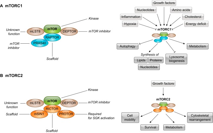
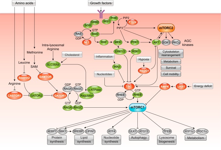
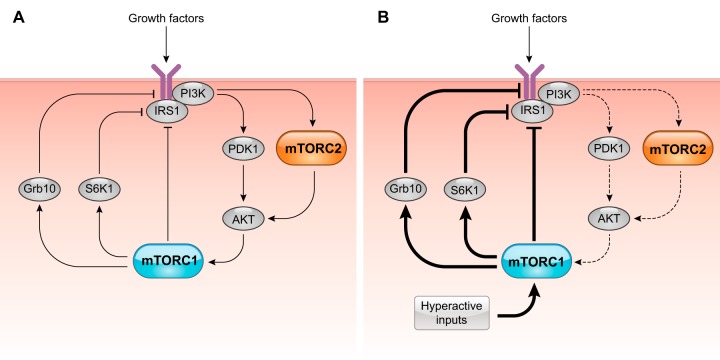
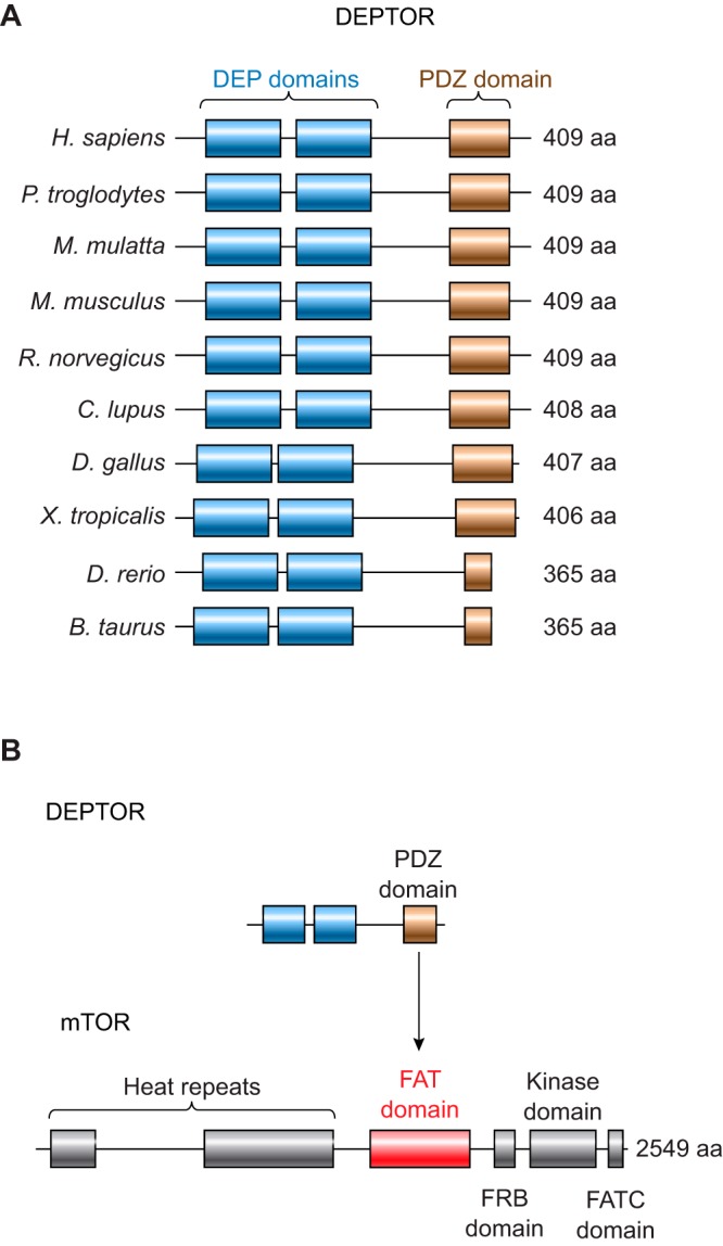
DEP domain-containing mechanistic target of rapamycin (mTOR)-interacting protein (DEPTOR) is an important modulator of mTOR, a kinase at the center of two important protein complexes named mTORC1 and mTORC2. These highly studied complexes play essential roles in regulating growth, metabolism, and immunity in response to mitogens, nutrients, and cytokines. Defects in mTOR signaling have been associated with the development of many diseases, including cancer and diabetes, and approaches aiming at modulating mTOR activity are envisioned as an attractive strategy to improve human health. DEPTOR interaction with mTOR represses its kinase activity and rewires the mTOR signaling pathway. Over the last years, several studies have revealed key roles for DEPTOR in numerous biological and pathological processes. Here, we provide the current state of the knowledge regarding the cellular and physiological functions of DEPTOR by focusing on its impact on the mTOR pathway and its role in promoting health and disease.


Figures

FIGURE 1.
FIGURE 1.
Protein composition and functions of mechanistic target of rapamycin complex 1 (mTORC1) and mTORC2. A, left: mTORC1 is composed of several proteins that are essential for its assembly and its activation. Right: the activity of mTORC1 is regulated by various inputs, including hypoxia, inflammation, energy deficit, growth factors, amino acids, cholesterol, and nucleotides. When active, mTORC1 controls several biological processes, including autophagy, macromolecule biosynthesis, lysosome biogenesis, and metabolism. B, left: mTORC2 is composed of several proteins that are essential for its assembly and its activation. Right: the activity of mTORC2 is primarily activated by growth factors. When active, mTORC2 controls various biological processes, including cell mobility, survival, metabolism, and cytoskeletal rearrangement. DEPTOR, DEP domain-containing mTOR-interacting protein; mLST8, mammalian lethal with Sec13 protein 8; mSIN1, mammalian stress-activated protein kinase interacting protein; PRAS40, proline-rich AKT substrate 40 kDa; PROTOR, protein observed with Rictor; RAPTOR, regulatory-associated protein of mTOR; RICTOR, rapamycin-insensitive companion of mTOR; SGK, serum- and glucocorticoid-induced protein kinase.
FIGURE 2.
FIGURE 2.
The mechanistic target of rapamycin (mTOR) signaling network. The mTOR signaling pathway responds to various inputs to regulate biological processes controlling cell growth, proliferation, and metabolism. In the figure, factors that contribute to the activation of mTOR are presented in green, whereas negative regulators are shown in red. In response to growth factors, tyrosine kinase receptors autophosphorylate and recruit adaptor proteins, including growth factor receptor-bound protein 2 (GRB2) and insulin receptor substrate 1 (IRS1), which contribute to activate RAt sarcoma (RAS) and phosphoinositide 3-kinase (PI3K), respectively. Active RAS amplifies PI3K and turns on mitogen-activated protein (MAP) kinases, including RAF proto-oncogene serine/threonine-protein kinase (RAF), mitogen-activated kinase kinase (MEK), and extracellular-signal-regulated kinase 1/2 (ERK1/2). When active, PI3K promotes the production of phosphatidylinositol (3,5)-triphosphate (PIP3) from phosphatidylinositol (4,5)-bisphosphate (PIP2), an event that activates mTOR complex 2 (mTORC2). PIP3 production is also required for the activation of 3-phosphoinositide-dependent kinase 1 (PDK1) by growth factors. The activation of mTORC2 and PDK1 promotes protein kinase B (AKT) phosphorylation and activity. In addition to AKT, mTORC2 also activates other AGC [protein kinase A, G, and C families (PKA, PKC, PKG)] kinases, including serum- and glucocorticoid-induced protein kinase 1 (SGK1) and protein kinase C-α (PKC-α). The activation of these kinases by mTORC2 affects different biological processes, including cytoskeletal rearrangement, metabolism, survival, and cell mobility. A major hub integrating growth factor inputs to control the activity of mTORC1 is the protein complex tuberous sclerosis complex (TSC). Phosphorylation of TSC by ERK, ribosomal S6 kinase (RSK), and AKT inhibits TSC and promotes the guanosine triphosphate (GTP) loading of RHEB, a key activator of mTORC1. Inflammation, hypoxia, nucleotide levels, and energy deficits also signal to mTORC1, at least in part, by affecting the activity of TSC toward RHEB. Amino acids are well known to serve as a key activator of mTORC1. Amino acids promote the localization of mTORC1 and Ras homolog enriched in brain (Rheb) to the lysosome, where the complex can be fully activated. mTORC1 senses intralysosomal and cytosolic amino acids through distinct mechanisms. The lysosomal amino acid transporter solute carrier family 38 member 9 (SLC38A9) interacts with the RAG-Ragulator-v-ATPase (vacuolar H+-adenoside triphosphate) complex and is required for intralysosomal arginine to activate mTORC1. The binding of intralysosomal arginine to SLC38A9 also promotes the efflux of several essential amino acids to the cytosol, where they activate mTORC1. Cytosolic amino acids use various pathways to promote mTORC1. Arginine binds to its intracellular sensor CASTOR1 (cytosolic arginine sensor for mTORC1 subunit 1), an event that disrupts CASTOR interaction with GATOR2 (GAP activity toward Rags complex 2). In the CASTOR-free state, GATOR2 inhibits GATOR1, a complex that represses mTORC1 signaling by acting as a GAP for RAGA/B. Leucine similarly activates mTORC1 by activating GATOR2. The binding of leucine to its intracellular sensor Sestrin2 breaks up Sestrin2 interaction with GATOR2, allowing GATOR2 to repress GATOR1. Thus, in leucine- and arginine-rich conditions, RAGA is thought to be mostly GTP loaded, leading to recruitment and activation of mTORC1. Lastly, methionine controls mTORC1 via the methionine-derived metabolite S-adenosylmethionne (SAM). SAM directly binds to its intracellular sensor SAMTOR (SAM sensor upstream of mTORC1), an event that blocks SAMTOR interaction with GATOR1. The loss of interaction blocks GATOR1 action on Ras-related GTP-binding protein A/B (RagA/B) and activates mTORC1. A complete overview of regulatory processes by which amino acids control mTORC1 was recently reviewed by others (224, 282). The activity of mTORC1 can also be activated by cholesterol. Lysosomal cholesterol activates mTORC1 through a mechanism that depends on SLC38A9. When active, mTORC1 promotes metabolism and the synthesis of several molecules to support growth, including proteins, lipids, and nucleotides, through the phosphorylation of several effectors. mTORC1 also blocks catabolism by repressing autophagy and lysosome biogenesis. 4E-BP1/2, eukaryotic translation initiation factor 4E-binding protein 1 and 2; AMPK, adenosine monophosphate-activated protein kinase; ATG13, autophagy-related protein 13; ATF4, activating transcription factor 4; GDP, guanosine diphosphate; HIF-1α, hypoxia-inducible factor-1α; IKKB, inhibitor of nuclear factor kappa B; LKB1, liver kinase B1; NF1, neurofibromatosis type 1; PDPK1, 3-phosphoinositide-dependent kinase 1; PGC-1α, peroxisome proliferator-activated receptor-γ coactivator-1α; PTEN, phosphatase and tensin homolog deleted on chromosome 10; RagC/D, Ras-related GTP-binding protein C/D; REDD1, regulated in development and DNA damage responses 1; S6K1, ribosomal protein S6 kinase 1; SOS, son of sevenless; SREBP, sterol regulatory element-binding protein; TFEB, transcription factor EB; ULK1, unc-51 like autophagy activating kinase 1.
FIGURE 3.
FIGURE 3.
Negative feedback loops emerging from mechanistic target of rapamycin complex 1 (mTORC1) to regulate phosphoinositide 3-kinase (PI3K) signaling. A: presentation of the negative feedback loops that link mTORC1 to PI3K. B: high inputs to mTORC1 exacerbate the feedback loops from mTORC1 and impair PI3K signaling. AKT, protein kinase B; GRB10, growth factor receptor-bound protein 10; IRS1, insulin receptor substrate 1; PDK1, 3-phosphoinositide-dependent kinase 1; S6K1, ribosomal protein S6 kinase 1.
FIGURE 4.
FIGURE 4.
DEPTOR (DEP domain containing mTOR-interacting protein) is a conserved protein that interacts with the mechanistic target of rapamycin (mTOR) kinase. A: illustration of DEPTOR organization and conservation between species. B: DEPTOR interacts with the FAT (FAT-carboxy terminal) domain of mTOR. DEP domain, Dishevelled, EGL-10, and pleckstrin domain; FATC domain, FRAP-ATM-TTRAP domain; FRB domain, FKBP12-rapamycin binding domain; HEAT repeats, Huntingtin-Elongation factor 3-regulatory subunit A of PP2A-TOR1 repeats; PDZ domain, postsynaptic density protein (PSD95), Drosophila disc large tumor suppressor (Dlg1), Zonula occludens-1 protein domain.
FIGURE 5.
FIGURE 5.
DEP (Dishevelled, Egl-10, and Pleckstrin) domain-containing proteins. Overview of mammalian DEP domain-containing proteins is shown. cNMP, cyclic nucleotide-binding domain; CPN, carboxypeptidase N; DEPDC1–7, DEP domain containing 1–7; DEPTOR, DEP domain-containing mTOR-interacting protein; DUF, domain of unknown function; EPAC1/2, exchange factor directly activated by cAMP 1/2; RAPGEF3-5, Rap guanine nucleotide exchange factor 3-5; FYVE, Fab 1 (yeast orthologue of PIKfyve), YOTB, Vac 1 (vesicle transport protein) and EEA1; GPR155, G protein-coupled receptor 155; MTD, minimal transactivation domain; PDZ, postsynaptic density 95, discs large, zonula occludens-1; PH, pleckstrin homology; PLEK, pleckstrin; P-REX1-2, phosphatidylinositol-3,4,5-trisphosphate dependent rac exchange factor 1-2; RA, Ras-associating; ZFYVE1, zinc finger FYVE-type containing 1.
FIGURE 6.
FIGURE 6.
Models presenting the possible impact of DEPTOR [Dishevelled, Egl-10, and Pleckstrin (DEP) domain-containing mTOR-interacting protein] on mechanistic target of rapamycin (mTOR) and phosphoinositide 3-kinase (PI3K) signaling. A: the linear model. Left: in this model, depleting DEPTOR levels relieves the inhibition on mTOR complex 1 (mTORC1) and complex 2 (mTORC2) and promotes the action of these complexes toward their respective substrats. Elevated ribosomal protein S6 kinase 1 (S6K1) and protein kinase B (AKT) are observed in this context. Right: conversely, overexpression of DEPTOR inhibits mTORC1 and mTORC2 and reduces the action of these complexes toward their respective substrates. Reduced S6K1 and AKT are measured in this situation. In the models depicted in these panels, the contribution of the negative feedback loop to the regulation of PI3K is minimal. B: the feedback model. Left: in this model, depleting DEPTOR levels activates mTORC1 and turns on the negative feedback loops from mTORC1 to PI3K. Elevated S6K1, but reduced AKT, is observed in this context. Right: conversely, overexpression of DEPTOR inhibits mTORC1 and blocks the negative feedback loops from mTORC1 to PI3K. Reduced S6K1, but elevated AKT, is measured in this situation. In this model, the negative feedback loops from mTORC1 dominate over mTORC2 for the activation of AKT. In these panels, the green color indicates activation, whereas red indicates inhibition. Dashed lines are used to illustrate decreases in protein content or activation. IRS1, insulin receptor substrate 1.
FIGURE 7.
FIGURE 7.
Retrospective analysis presenting the reported impact of DEPTOR (DEP domain-containing mTOR-interacting protein) knockdown on mechanistic target of rapamycin (mTOR) and phosphoinositide 3-kinase (PI3K) signaling. All of the reports describing the impact of DEPTOR depletion (knockdown or knockout) on mTORC1, mTORC2, or PI3K activation were analyzed and categorized. The phosphorylation of ribosomal protein S6 kinase 1 (S6K1) (Thr389), S6 (Ser235/236; Ser240/244), and 4EBP1 (Thr36/46) were used as markers of mTORC1 activity. The phosphorylation state of protein kinase B (AKT) (AKT) (Ser473) and N-myc downstream regulated 1 (NDRG1) (Thr346), a surrogate marker of serum- and glucocorticoid-induced protein kinase 1 (SGK1) activation, were used as markers of mTORC2 activity. The phosphorylation of AKT (Thr308) and the total levels of insulin receptor substrate 1 (IRS1) were used as markers of PI3K activation. The phosphorylation of IRS1 (Ser636/639) was also used as an indicator of impaired PI3K signaling. Green indicates activation, whereas red indicates inhibition.
FIGURE 8.
FIGURE 8.
Retrospective analysis presenting the reported impact of DEPTOR (DEP domain-containing mTOR-interacting protein) overexpression on mechanistic target of rapamycin (mTOR) and phosphoinositide 3-kinase (PI3K) signaling. All of the reports describing the impact of DEPTOR overexpression on mTOR complex 1 (mTORC1), complex 2 (mTORC2), or PI3K activation were analyzed and categorized. The phosphorylation of ribosomal protein S6 kinase 1 (S6K1) (Thr389), S6 (Ser235/236; Ser240/244), and 4EBP1 (Thr36/46) were used as markers of mTORC1 activity. The phosphorylation state of protein kinase B (AKT) (Ser473) and N-myc downstream regulated 1 (NDRG1) (Thr346), a surrogate marker of serum- and glucocorticoid-induced protein kinase 1 (SGK1) activation, were used as markers of mTORC2 activity. The phosphorylation of AKT (Thr308) and the total levels of insulin receptor substrate 1 (IRS1) were used as markers of PI3K activation. The phosphorylation of IRS1 (Ser636/639) was also used as an indicator of impaired PI3K signaling. Green indicates activation, whereas red indicates inhibition.
FIGURE 9.
FIGURE 9.
Retrospective analysis presenting the impact of DEPTOR (DEP domain-containing mTOR-interacting protein) on mechanistic target of rapamycin (mTOR) and phosphoinositide 3-kinase (PI3K) signaling. Top: the impact of DEPTOR depletion on mTOR and PI3K signaling. Bottom: the impact of DEPTOR overexpression. The pie charts presented have been produced using the data presented in FIGURES 7 AND 8. mTORC1/2, mTOR complex 1 and 2.
FIGURE 10.
FIGURE 10.
Mechanisms controlling DEPTOR transcription. A: impact of growth factors on the control of DEPTOR [Dishevelled, Egl-10, and Pleckstrin (DEP) domain-containing mTOR-interacting protein] expression. Growth factor signaling represses the expression of DEPTOR via a mechanism that remains to be characterized. B: Notch signaling is a positive regulator of DEPTOR expression. Extracellular ligands trigger the cleavage of the Notch receptor by the enzyme γ-secretase, the release of Notch intracellular domain (NICD), and its translocation in the nucleus. NICD binds a conserved sequence close to the transcription initiation site in DEPTOR promoter to promote its expression. C: nuclear receptors control the expression of DEPTOR. Estrogen receptor (ER) α, glucocorticoid receptor (GR), and vitamin D receptor (VDR) all promote the expression of DEPTOR, whereas androgen receptor (AR) represses it. With the exception of GR, which directly binds the DEPTOR promoter to increase its expression, the mechanism by which other nuclear receptors control DEPTOR transcription still remains to be established. D: the MAF (MAF BZIP transcription factor) proteins control the expression of DEPTOR. MAF transcription factors are often amplified in multiple myelomas (MMs) and contribute to promote the expression of DEPTOR in these cells. How MAF proteins control DEPTOR expression is still unclear. E: Che-1 [also known as AATF (apoptosis antagonizing transcription factor)] controls the expression of DEPTOR in response to stress. In response to DNA damage, hypoxia, and glucose deprivation, Che-1 is activated and directly binds the DEPTOR promoter to activate its transcription. F: regulation of Deptor expression by the Baf60c [also known as SMARCD3 (SWI/SNF-related, matrix-associated, actin-dependent regulator of chromatin, subfamily D, member 3)]/Six complex. Baf60c physically interacts with the transcription factor Six4 (sine oculis homeobox homolog 4) to control the transcription of Deptor. Baf60c functions as a coactivator for Six4 and stimulates Deptor expression by altering the chromatin structure and the epigenetic landscape of Deptor locus. G: control of DEPTOR transcription by enhancer of zeste homolog (EZH) and argininosuccinate synthase 1 (ASS1). EZH interacts with MTA2 (metastasis associated 1 family member 2) to blocks DEPTOR expression by binding and promoting the methylation (H3K27me3) of the DEPTOR promoter. Conversely, ASS1 promotes DEPTOR transcription by repressing H3K27me3. H: control of DEPTOR expression by micro-RNAs (miRNAs). The miR155 and miR375 minds DEPTOR transcript to reduce DEPTOR protein levels. AKT, protein kinase B; ERK, extracellular-signal-regulated kinase; MEK, mitogen-activated kinase kinase; mIR, micro-RNA; mTORC1/2, mechanistic target of rapamycin complex 1 and 2; PI3K, phosphoinositide 3-kinase; PTEN, phosphatase and tensin homolog deleted on chromosome 10; RAF, RAF proto-oncogene serine/threonine-protein kinase; RAS, RAt sarcoma; SWI/SNF, switch/sucrose non-fermentable; TSC, tuberous sclerosis complex.
FIGURE 11.
FIGURE 11.
Mechanisms controlling DEPTOR protein stability. A: the activation of mechanistic target of rapamycin (mTOR) signaling promotes the degradation of DEP domain-containing mTOR-interacting protein (DEPTOR) by the proteasome. In response to growth factors, mTORC1 and mTORC2 are activated. Increased mTOR kinase activity promotes the phosphorylation of DEPTOR, an event that promotes the dissociation of DEPTOR from mTOR. In the panel, the phosphorylation sites regulated by mTOR are labeled in green. The phosphorylation of DEPTOR by mTOR primes its phosphorylation on a specific degron by casein kinase 1α (CK1α). The phosphorylation sites found inside this sequence are presented in grey. Other kinases have been suggested to control the phosphorylation of DEPTOR in the degron including ribosomal protein S6 kinase 1 (S6K1) and ribosomal S6 kinase 1 (RSK1). The hyperphosphorylation of DEPTOR favors the binding of Skp1/Cul1/F box protein (SCF)βTrcp, the ubiquitination, and the degradation of DEPTOR by the proteasome. B: overview of the specific phosphorylation sites that play roles in DEPTOR degradation by the proteasome. mLST8, mammalian lethal with Sec13 protein 8; mSIN1, mammalian stress-activated protein kinase interacting protein; PDZ domain, postsynaptic density protein (PSD95), Drosophila disc large tumor suppressor (Dlg1), zonula occludens-1 protein domain; PRAS40, proline-rich AKT substrate 40 kDa; PROTOR, protein observed with Rictor; RAPTOR, regulatory-associated protein of mTOR; RICTOR, rapamycin-insensitive companion of mTOR.
FIGURE 12.
FIGURE 12.
DEPTOR [Dishevelled, Egl-10, and Pleckstrin (DEP) domain-containing mTOR-interacting protein] as an oncogene. Oncogenic Notch activation, MAF (transcription factor MAF) amplification, and possibly other unknown oncogenic drivers promote the expression of DEPTOR in some cancers. DEPTOR increases cell proliferation and survival and blocks cell cycle arrest and apoptosis through various complementary mechanisms. DEPTOR overexpression blocks mechanistic target of rapamycin complex 1 (mTORC1) activity, which 1) relieves the negative feedback loops on phosphoinositide 3-kinase (PI3K) and promotes protein kinase B (AKT) activation; 2) reduces the expression of the cell cycle inhibitor p21; 3) reduces protein synthesis, endoplasmic reticulum (ER) stress, and unfolded protein response (UPR) activation; and 4) promotes autophagy. All of these effects contribute to increase the resistance of cancer cells to various cellular stress (genotoxic stress, ER stress, and energetic stress) and to promote drug resistance. The small-molecule NSC126405 binds to DEPTOR and impairs its binding to mTOR. When used in multiple myeloma (MM) cells, NSC126405 impairs cell survival by promoting mTORC1-mediated inhibition of autophagy. p21, Cyclin-dependent kinase inhibitor 1.
FIGURE 13.
FIGURE 13.
DEPTOR expression levels in cancer. All of the reports presenting DEPTOR expression in human cancer are presented. Protein or mRNA expression were considered in the analysis. In every case, comparisons were made vs. normal tissues or cell types. Green indicates high DEPTOR levels, whereas red indicates low DEPTOR levels.
FIGURE 14.
FIGURE 14.
DEPTOR [Dishevelled, Egl-10, and Pleckstrin (DEP) domain-containing mTOR-interacting protein] as a tumor suppressor. Oncogenic activation of phosphoinositide 3-kinase (PI3K) signaling activates the mechanistic target of rapamycin (mTOR) pathway and reduces the expression and the stability of DEPTOR. Low DEPTOR expression relieves inhibition on mTOR complex 1 (mTORC1) and complex 2 (mTORC2), which further amplifies their activity. The activation of mTORC1 linked to low DEPTOR expression stimulates anabolism, growth and proliferation. On the other hand, activation of mTORC2 promotes the phosphorylation of protein kinase B (AKT) and serum- and glucocorticoid-induced protein kinase 1 (SGK1), which increases cell survival and resistance to stress and reduces apoptosis. The reduction in DEPTOR expression and the activation of AKT signaling promotes cancer by activating epithelial-mesenchymal transition (EMT), cell migration, and metastasis.
FIGURE 15.
FIGURE 15.
DEPTOR [Dishevelled, Egl-10, and Pleckstrin (DEP) domain-containing mTOR-interacting protein] in metabolism. In adipose tissue, overexpression of DEPTOR promotes adipogenesis by partially reducing mechanistic target of rapamycin complex 1 (mTORC1) activity. Mechanistically, this relieves the negative feedback loop toward growth factors signaling and increases the pro-adipogenic functions of protein kinase B (AKT). As such, DEPTOR ensures an optimal balance between AKT and mTORC1 signaling in adipocytes. This balance is critical for the activation of the master regulator of adipogenesis, peroxisome proliferator-activated receptor γ (PPAR-γ). In skeletal muscle, Baf60c [also known as SMARCD3 (SWI/SNF-related, matrix-associated, actin-dependent regulator of chromatin, subfamily D, member 3)] stimulates a metabolic switch from oxidation to glycolysis by promoting DEPTOR expression and AKT activation. Forcing this pathway improves insulin sensitivity and promotes glucose uptake and glycolysis. In the liver, DEPTOR is key for an appropriate feeding-to-fasting transition. Fasting increases DEPTOR expression in order to potentiate the inhibition of mTORC1 during a state of low energy. This prevents the activation of oxidative metabolism. The deletion of DEPTOR in hepatocytes reduces blood glucose and increases liver oxidative metabolism by preventing an optimal inhibition of mTORC1 during fasting. In the mediobasal hypothalamus, overexpression of DEPTOR reduces the ability of mTORC1 to reduce insulin signaling, resulting in higher insulin sensitivity, through an AKT-FoxO1 (Forkhead box protein O1)-dependent mechanism. As such, hypothalamic DEPTOR regulates energy balance, and forcing its expression prevents the development of obesity. Although the identity of the neurons mediating the effects of DEPTOR on metabolism is still unknown, overexpressing DEPTOR in pro-opiomelanocortin (POMC) neurons affects liver lipid and glucose metabolism. TCA, tricarboxylic acid cycle.

Similar articles

Cited by

References

    1. Abbas AK, Murphy KM, Sher A. Functional diversity of helper T lymphocytes. Nature 383: 787–793, 1996. doi:10.1038/383787a0. - DOI - PubMed
    1. Agrawal P, Reynolds J, Chew S, Lamba DA, Hughes RE. DEPTOR is a stemness factor that regulates pluripotency of embryonic stem cells. J Biol Chem 289: 31818–31826, 2014. doi:10.1074/jbc.M114.565838. - DOI - PMC - PubMed
    1. Albert V, Cornu M, Hall MN. mTORC1 signaling in Agrp neurons mediates circadian expression of Agrp and NPY but is dispensable for regulation of feeding behavior. Biochem Biophys Res Commun 464: 480–486, 2015. doi:10.1016/j.bbrc.2015.06.161. - DOI - PubMed
    1. Altamirano F, Oyarce C, Silva P, Toyos M, Wilson C, Lavandero S, Uhlén P, Estrada M. Testosterone induces cardiomyocyte hypertrophy through mammalian target of rapamycin complex 1 pathway. J Endocrinol 202: 299–307, 2009. doi:10.1677/JOE-09-0044. - DOI - PubMed
    1. André C, Cota D. Coupling nutrient sensing to metabolic homoeostasis: the role of the mammalian target of rapamycin complex 1 pathway. Proc Nutr Soc 71: 502–510, 2012. doi:10.1017/S0029665112000754. - DOI - PubMed

Publication types

MeSH terms

Substances

LinkOut - more resources