

Review
doi: 10.1152/physiol.00009.2018.
Neurons and Glia in the Enteric Nervous System and Epithelial Barrier Function
Affiliations
- PMID: 29897300
- PMCID: PMC6088142
- DOI: 10.1152/physiol.00009.2018
Item in Clipboard
Review
Neurons and Glia in the Enteric Nervous System and Epithelial Barrier Function
Physiology (Bethesda).
.
Abstract
The intestinal epithelial barrier is the largest exchange surface between the body and the external environment. Its functions are regulated by luminal, and also internal, components including the enteric nervous system. This review summarizes current knowledge about the role of the digestive "neuronal-glial-epithelial unit" on epithelial barrier function.
Figures
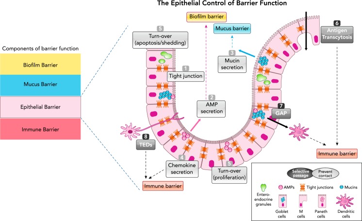
Schematic representation of the epithelial barrier in the GI tract Components of the intestinal barrier (left) and specifically the epithelial control of barrier function (right). The epithelial monolayer is composed of different cell types (represented here are Goblet cells, Paneth cells, and M cells), which exert barrier function either by preventing physical contact or by allowing selective passage of molecules. The physical barrier organized by the epithelium is composed of the tight-junction molecules (1), the secretion of antimicrobial peptides (AMPs; 2), the secretion of mucins (3), the secretion of chemokines that alert the immune barrier (4), cell turnover (equilibrium between shedding and proliferation; 5). Selective passage of antigens is ensured through M-cell transcytosis (6), Goblet cell antigen passages (GAP; 7), and transepithelial dendrite (TEDs) (8) pathways.
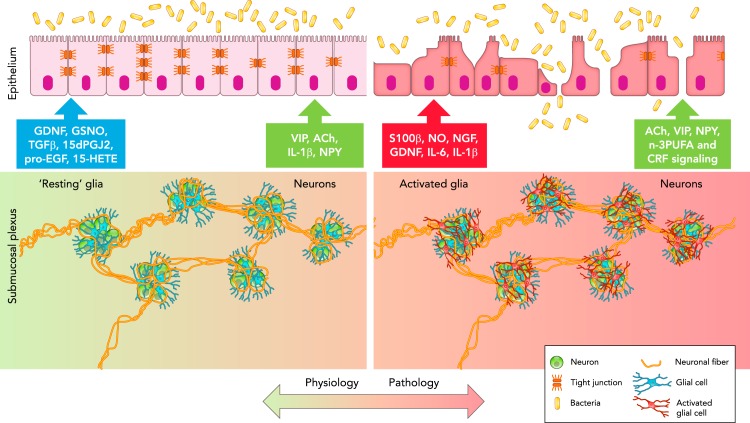
Schematic representation of the neuronal-glial-epithelial unit in the GI tract In physiological conditions (left), glial cells (blue) surround neurons (green) to form ganglia, which are interconnected through neuronal fibers (orange) to form the submucosal plexus. “Resting” glia communicate with epithelial cells via the release of GDNF, GSNO, TGFβ1, 15dPGJ2, pro-EGF, and 15-HETE to preserve barrier function, whereas submucosal neurons release VIP, ACh, IL-1β, and NPY. In pathological conditions (right), glial cells become activated (red) and release S100β, NO, NGF, GDNF, IL-6, and IL-1β, which alter epithelial barrier function, whereas submucosal neurons (green) release ACh, VIP, NPY, and n-3PUFA, and respond to CRF signaling, which affects epithelial barrier function.
References
-
- Abdo H, Mahé MM, Derkinderen P, Bach-Ngohou K, Neunlist M, Lardeux B. The omega-6 fatty acid derivative 15-deoxy-Δ12,14-prostaglandin J2 is involved in neuroprotection by enteric glial cells against oxidative stress. J Physiol 590: 2739–2750, 2012. doi: 10.1113/jphysiol.2011.222935. - DOI - PMC - PubMed
-
- Bishop AE, Carlei F, Lee V, Trojanowski J, Marangos PJ, Dahl D, Polak JM. Combined immunostaining of neurofilaments, neuron specific enolase, GFAP and S-100. A possible means for assessing the morphological and functional status of the enteric nervous system. Histochemistry 82: 93–97, 1985. doi: 10.1007/BF00502095. - DOI - PubMed
Publication types
MeSH terms
LinkOut - more resources
Full Text Sources
Other Literature Sources