. 2018 Jul 1;27(13):2344-2356.
doi: 10.1093/hmg/ddy143.

The c-Abl inhibitor, Radotinib HCl, is neuroprotective in a preclinical Parkinson's disease mouse model

Affiliations

The c-Abl inhibitor, Radotinib HCl, is neuroprotective in a preclinical Parkinson's disease mouse model

Saebom Lee et al. Hum Mol Genet. .

Abstract

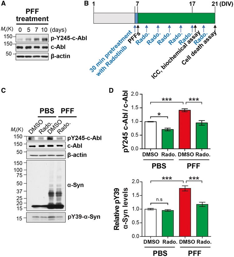
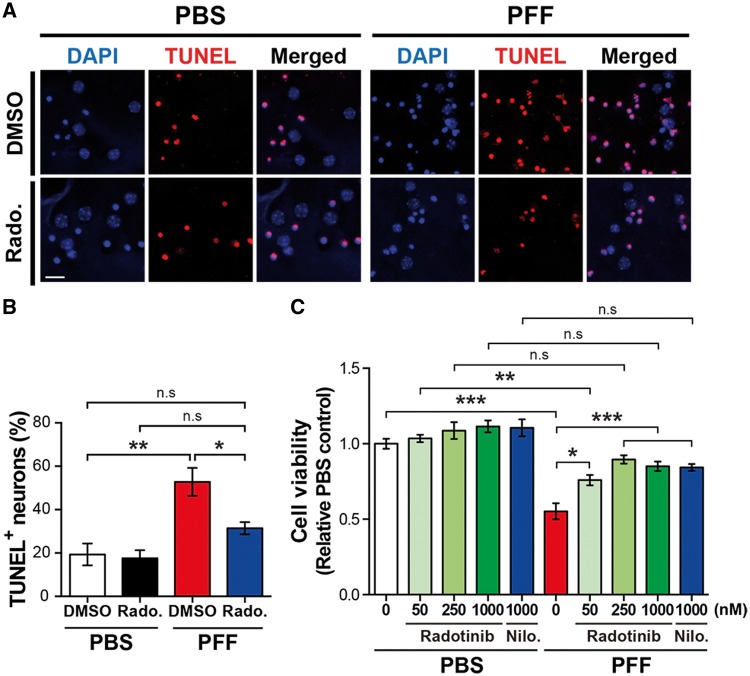
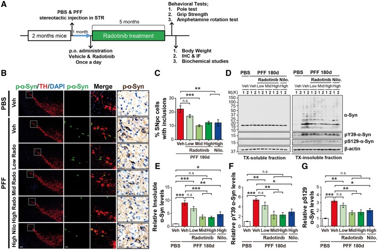
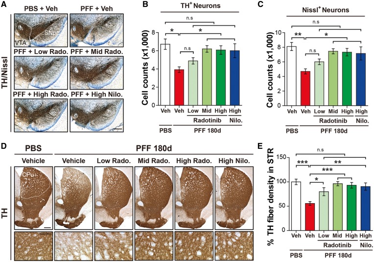
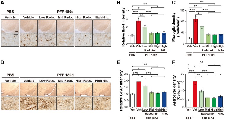
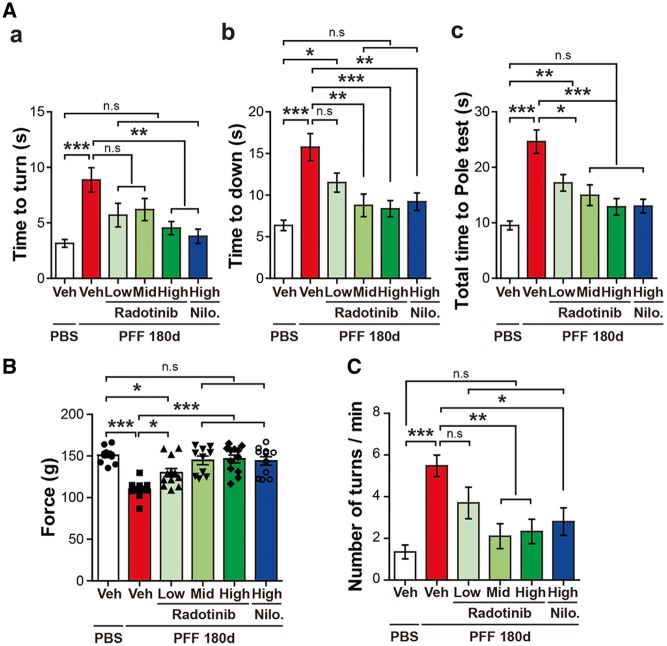
Accumulating evidence suggests that the non-receptor tyrosine kinase c-Abl plays an important role in the progression of Parkinson's disease (PD) and c-Abl inhibition could be neuroprotective in PD and related α-synucleinopathies. Nilotinib, a c-Abl inhibitor, has shown improved motor and cognitive symptoms in PD patients. However, issues concerning blood-brain barrier (BBB) penetration, lack of selectivity and safety still remain. Radotinib HCl is a selective Bcr-Abl kinase inhibitor that not only effectively access the brain, but also exhibits greater pharmacokinetic properties and safety profiles compared to Nilotinib and other c-Abl inhibitors. Here, we show the neuroprotective efficacy of Radotinib HCl, a brain penetrant c-Abl inhibitor, in a pre-clinical model of PD. Importantly, in vitro studies demonstrate that the treatment of Radotinib HCl protects the α-synuclein preformed fibrils (PFF)-induced neuronal toxicity, reduces the α-synuclein PFF-induced Lewy bodies (LB)/Lewy neurites (LN)-like pathology and inhibits the α-synuclein PFF-induced c-Abl activation in primary cortical neurons. Furthermore, administration of Radotinib HCl inhibits c-Abl activation and prevents dopaminergic neuron loss, neuroinflammation and behavioral deficits following α-synuclein PFF-induced toxicity in vivo. Taken together, our findings indicate that Radotinib HCl has beneficial neuroprotective effects in PD and provides an evidence that selective and brain permeable c-Abl inhibitors can be potential therapeutic agents for the treatment of PD and related α-synucleinopathies.


Figures

Figure 1.
Figure 1.
Radotinib HCl reduces α-synuclein PFF-induced c-Abl activation. (A) Western blot analysis of c-Abl activation by α-synuclein PFF treatment in cultured cortical neurons. (B) Experimental scheme. (C) Representative western blot data of reduced α-synuclein PFF-induced c-Abl activation by Radotinib HCl treatment (1 μM). (D) Quantification of pY245-c-Abl normalized to c-Abl. (E) Quantification pY39-α-synuclein normalized to β-actin. Bars indicate mean ± S.E.M. (n = 4). Statistical significance was determined by using one-way ANOVA with Tukey’s correction; *P < 0.05, ***P < 0.001. n.s., not significant. Radotinib, Radotinib HCl.
Figure 2.
Figure 2.
Radotinib HCl ameliorates α-synuclein PFF-induced α-synuclein aggregate formation and mitochondrial dysfunction. (A) Representative immunostaining images for p-α-synuclein (red) using pS129 antibody [MJF-R13 (8–8)] caused by α-synuclein PFF with or without Radotinib HCl (indicated concentrations) in primary cultured cortical neurons. The scale bar represents 50 μm. (B) Quantification of intensity of p-α-synuclein. Bars indicate mean ± S.E.M. (n = 3). (C) OCR was determined using the Seahorse XF24 Extracellular Flux Analyzer (Agilent Technologies, Santa Clara, CA). The basal respiratory rate (BRB) (D) and maximal respiratory rate (MRR) (E) were calculated (n = 8). Statistical significance was determined by using one-way ANOVA with Tukey’s correction; *P < 0.05, **P < 0.01, ***P < 0.001. n.s., not significant. Radotinib, Radotinib HCl.
Figure 3.
Figure 3.
Radotinib HCl protects against α-synuclein PFF-induced neuronal toxicity. (A) Representative images of TUNEL staining in primary cortical neurons. Radotinib HCl was added at 1 μm. The scale bar represents 20 μm. (B) Quantification of TUNEL positive neurons. Bars indicate mean ± S.E.M. (n = 3–5). (C) The neuronal cell death was measured by Alamar Blue assay. Bars indicate mean ± S.E.M. (n = 4). Statistical significance was determined by using one-way ANOVA with Tukey’s correction and two-way ANOVA with Bonferroni’s multiple comparisons test; *P < 0.05, **P < 0.01, ***P < 0.001. n.s., not significant. Radotinib, Radotinib HCl. Nilo., Nilotinib.
Figure 4.
Figure 4.
Radotinib HCl protects against α-synuclein PFF-induced pathology in vivo. (A) Preclinical study design of the α-synuclein PFF-induced PD mouse model. After 1 month, Radotinib HCl or Vehicle was delivered by oral administration at three different doses (3, 10 and 30 mg/kg) for 5 months. Nilotinib (30 mg/kg) was treated as a positive control. Mice were transcardially perfused after the last behavioral experiment. (B) Representative immunostaining for pS129-α-synuclein (green) and TH (red), immunohistological staining for pS129-α-synuclein (brown) in SNpc. The scale bars represent 100 μm (low magnification images) and 20 μm (high magnification images). (C) Quantification of SNpc regional neurons containing pS129-α-synuclein immunoreactive inclusions for each treatment group. Percentage of SNpc TH neurons with p-α-synuclein positive inclusion. Error bars represent the mean ± S.E.M, n = 4–6 mice per groups. (D) Representative immunoblots with sequential fractionation in 1% TX (TX-soluble) followed 2% SDS (TX-insoluble) in PBS and α-synuclein PFF stereotaxic injected mice treated with vehicle or Radotinib HCl and Nilotinib. (E) Quantification of levels of abnormal α-synuclein aggregates species, (F) pY39-α-synuclein and (G) pS129-α-synuclein in TX-insoluble fraction. Error bars represent the mean ± S.E.M, n = 4 mice per groups. One-way ANOVA was used to test for the statistical analysis, followed by Tukey’s correction; **P < 0.01, ***P < 0.001, n.s., not significant. Radotinib, Radotinib HCl. Nilo., Nilotinib.
Figure 5.
Figure 5.
Radotinib HCl protects against α-synuclein PFF-induced dopaminergic neurodegeneration and fiber density. (A) Representative photomicrographs from coronal mesencephalon sections containing TH-positive neurons in PBS and α-synuclein PFF stereotaxic injected mice treated with vehicle or Radotinib HCl and Nilotinib. The scale bar represents 500 μm. (B) Stereology counts of TH and (C) Nissl-positive neurons in the SNpc region. Unbiased sterologic counting was performed in SNpc region. Error bars represent the mean ± S.E.M, n = 4–6 mice per groups. (D) Representative photomicrograph of striatal sections stained for TH immunoreactivity. High power view of TH fiber density in the striatum (lower panels). The scale bars represent 100 μm (upper panles) and 50 μm (lower panels). CPu and STR, Striatum. (E) Quantification of dopaminergic nerve fiber densities in the striatum by using Image J software (NIH). Error bars represent the mean ± S.E.M, n = 4–6 mice per groups. One-way ANOVA was used to test for the statistical analysis, followed by Tukey’s correction; *P < 0.05, **P < 0.01, ***P < 0.001, n.s., not significant. Radotinib, Radotinib HCl. Nilo., Nilotinib.
Figure 6.
Figure 6.
Radotinib HCl restores α-synuclein PFF-induced neuroinflammation. (A) Representative images of immunohistochemistry data for Iba-1 in PBS and α-synuclein PFF stereotaxic injected mice treated with vehicle or Radotinib HCl and Nilotinib. The scale bars represent 500 μm (upper panels) and 50 μm (lower panels). (B) The intensity of Iba-1 positive signals in the SNpc and (C) quantification of Iba-1 positive cell number in SNpc. The intensity of Iba-1 immunoreactivity and densities of microglia in the SNpc region were measured with ImageJ software. Error bars represent the mean ± S.E.M, n = 4–6 mice per groups. (D) Representative images of immunohistochemistry data for GFAP in PBS and α-synuclein PFF stereotaxic injected mice treated with vehicle or Radotinib HCl and Nilotinib. The scale bars represent 500 μm (upper panles) and 50 μm (lower panels). (E) The intensity of GFAP positive signals in the SNpc and (F) quantification of GFAP positive cell number in SNpc. The intensity of GFAP immunoreactivity and densities of astrocyte in the SNpc region were measured with ImageJ software. Error bars represent the mean ± S.E.M, n = 4–6 mice per groups. One-way ANOVA was used to test for the statistical analysis, followed by by Tukey’s correction; *P < 0.05, **P < 0.01, ***P < 0.001, n.s., not significant. Radotinib, Radotinib HCl. Nilo., Nilotinib.
Figure 7.
Figure 7.
Radotinib HCl protects against α-synuclein PFF-induced behavioral deficits (A–C) 6 months after the α-synuclein PFF or PBS stereotaxic brain injection, the behavioral tests were performed in Vehicle and Radotinib HCl treated mice. Nilotinib-treated group as a positive control. Behavioral abnormalities were improved in mice administered with Radotinib HCl. Results of animals on the (A) Pole test, (B) grip strength, (C) amphetamine rotation test. Error bars represent the mean ± S.E.M (n = 9–12 mice per each group). One-way ANOVA was used for statistical analysis followed by Tukey’s correction; *P < 0.05, **P < 0.01, ***P < 0.001, n.s., not significant. Radotinib, Radotinib HCl. Nilo., Nilotinib.

Similar articles

Cited by

References

    1. Savitt J.M., Dawson V.L., Dawson T.M. (2006) Diagnosis and treatment of Parkinson disease: molecules to medicine. J. Clin. Investig., 116, 1744–1754. - PMC - PubMed
    1. Lang A.E., Lozano A.M. (1998) Parkinson's disease. First of two parts. N. Engl. J. Med., 339, 1044–1053. - PubMed
    1. Lang A.E., Lozano A.M. (1998) Parkinson's disease. Second of two parts. N. Engl. J. Med., 339, 1130–1143. - PubMed
    1. Dawson T.M., Dawson V.L. (2003) Molecular pathways of neurodegeneration in Parkinson's disease. Science, 302, 819–822. - PubMed
    1. Tsang A.H., Chung K.K. (2009) Oxidative and nitrosative stress in Parkinson's disease. Biochim. Biophys. Acta, 1792, 643–650. - PubMed

Publication types

MeSH terms