. 2018 Sep 6;46(15):e89.
doi: 10.1093/nar/gky423.

CancerDetector: ultrasensitive and non-invasive cancer detection at the resolution of individual reads using cell-free DNA methylation sequencing data

Affiliations

CancerDetector: ultrasensitive and non-invasive cancer detection at the resolution of individual reads using cell-free DNA methylation sequencing data

Wenyuan Li et al. Nucleic Acids Res. .

Abstract

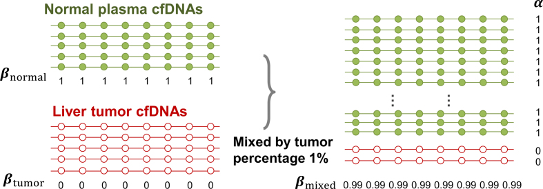
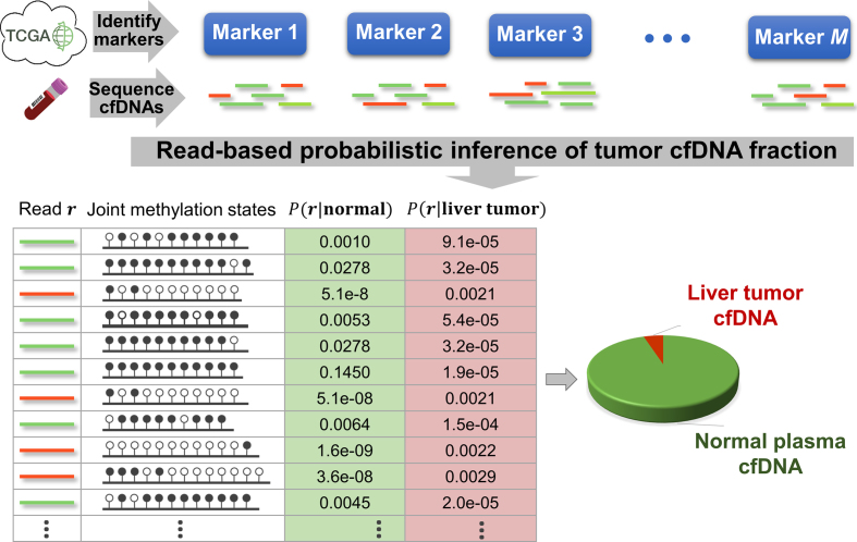
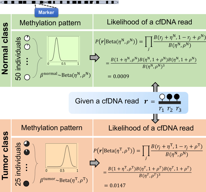
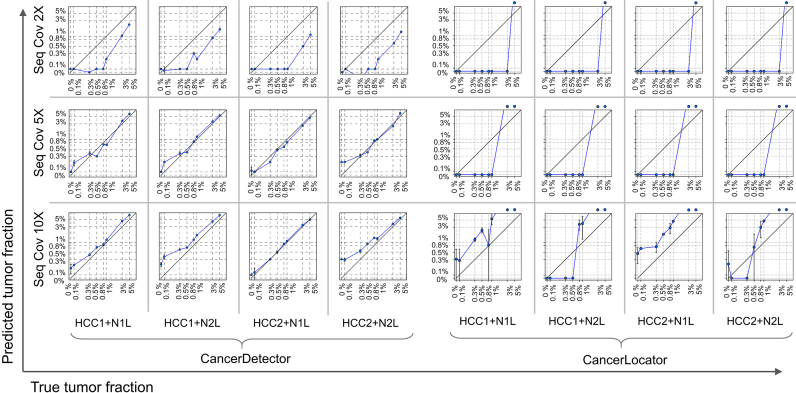
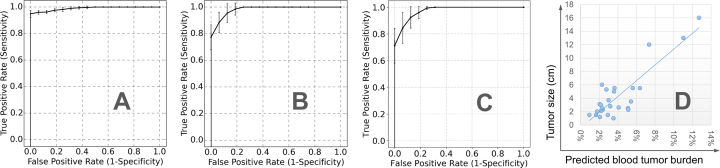
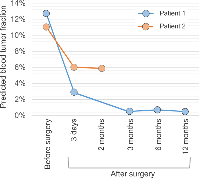
The detection of tumor-derived cell-free DNA in plasma is one of the most promising directions in cancer diagnosis. The major challenge in such an approach is how to identify the tiny amount of tumor DNAs out of total cell-free DNAs in blood. Here we propose an ultrasensitive cancer detection method, termed 'CancerDetector', using the DNA methylation profiles of cell-free DNAs. The key of our method is to probabilistically model the joint methylation states of multiple adjacent CpG sites on an individual sequencing read, in order to exploit the pervasive nature of DNA methylation for signal amplification. Therefore, CancerDetector can sensitively identify a trace amount of tumor cfDNAs in plasma, at the level of individual reads. We evaluated CancerDetector on the simulated data, and showed a high concordance of the predicted and true tumor fraction. Testing CancerDetector on real plasma data demonstrated its high sensitivity and specificity in detecting tumor cfDNAs. In addition, the predicted tumor fraction showed great consistency with tumor size and survival outcome. Note that all of those testing were performed on sequencing data at low to medium coverage (1× to 10×). Therefore, CancerDetector holds the great potential to detect cancer early and cost-effectively.


Figures

Figure 1.
Figure 1.
Illustration of the rationale why the methylation value averaged across all CpG sites in a sequencing read (α-value) is more sensitive at detecting tumor-derived cfDNAs than the traditional methylation level of a CpG site averaged across all reads (β-value). Each line represents a sequencing read and each dot represents a CpG site.
Figure 2.
Figure 2.
Overview of the CancerDetector method. The color of cfDNA sequencing reads represents their origin: red (green) reads are from tumor (normal plasma) cfDNA fragments. These reads are from a hypomethylated marker (chr2:4050595–4050945).
Figure 3.
Figure 3.
Illustration of calculating the likelihood of a cfDNA sequencing read in a marker, given the methylation patterns of normal and tumor classes.
Figure 4.
Figure 4.
Predicted blood tumor fractions (averaged over 10 runs) for the liver cancer cfDNA samples, simulated by subsampling and mixing sequencing reads from a real healthy cfDNA sample (N1L or N2L) and a solid liver tumor sample (HCC1 or HCC2) at eight different tumor fractions: 0, 0.1%, 0.3%, 0.5%, 0.8%, 1%, 3%, 5%, and at 3 different sequencing coverages (2×, 5× and 10×). In each log-log plot, a blue point represents a simulated sample with error bars (standard deviation of predicted tumor fraction), the x-axis is its true tumor fraction and the y-axis is its predicted tumor fraction. When the predicted tumor fraction is out of range (>5%), we draw the point above the box.
Figure 5.
Figure 5.
Predicted blood tumor fractions for the real data in all 10 runs: (A) average ROC curve with standard deviation bars for CancerDetector, (B) average ROC curve with standard deviation bars for our previous method CancerLocator, (C) average ROC curve with standard deviation bars for the methylated haplotype load based method (14) and (D) the relationship between the tumor size and average blood tumor fraction predicted by CancerDetector.
Figure 6.
Figure 6.
Average predicted blood tumor fractions for longitudinal data of two liver cancer patients before and after tumor resections in all 10 runs. The second patient passed away after surgery.

References

    1. Bettegowda C., Sausen M., Leary R.J., Kinde I., Wang Y., Agrawal N., Bartlett B.R., Wang H., Luber B., Alani R.M. et al. . Detection of circulating tumor DNA in early- and late-stage human malignancies. Sci. Transl. Med. 2014; 6:224ra24. - PMC - PubMed
    1. Newman A.M., Bratman S. V, To J., Wynne J.F., Eclov N.C.W., Modlin L.A., Liu C.L., Neal J.W., Wakelee H.A., Merritt R.E. et al. . An ultrasensitive method for quantitating circulating tumor DNA with broad patient coverage. Nat. Med. 2014; 20:548–554. - PMC - PubMed
    1. Newman A.M., Lovejoy A.F., Klass D.M., Kurtz D.M., Chabon J.J., Scherer F., Stehr H., Liu C.L., Bratman S. V, Say C. et al. . Integrated digital error suppression for improved detection of circulating tumor DNA. Nat. Biotechnol. 2016; 34:547–555. - PMC - PubMed
    1. Burrell R.A., McGranahan N., Bartek J., Swanton C.. The causes and consequences of genetic heterogeneity in cancer evolution. Nature. 2013; 501:338–345. - PubMed
    1. Greenman C., Stephens P., Smith R., Dalgliesh G.L., Hunter C., Bignell G., Davies H., Teague J., Butler A., Stevens C. et al. . Patterns of somatic mutation in human cancer genomes. Nature. 2007; 446:153–158. - PMC - PubMed

Publication types