Review
. 2018 Jun 1;365(12):fny123.
doi: 10.1093/femsle/fny123.

The Tat protein transport system: intriguing questions and conundrums

Affiliations
Review

The Tat protein transport system: intriguing questions and conundrums

Shruthi Hamsanathan et al. FEMS Microbiol Lett. .

Abstract

The Tat machinery catalyzes the transport of folded proteins across the cytoplasmic membrane in bacteria and the thylakoid membrane in plants. Transport occurs only in the presence of an electric field (Δψ) and/or a pH (ΔpH) gradient, and thus, Tat transport is considered to be dependent on the proton motive force (pmf). This presents a fundamental and major challenge, namely, that the Tat system catalyzes the movement of large folded protein cargos across a membrane without collapse of ion gradients. Current models argue that the active translocon assembles de novo for each cargo transported, thus providing an effective gating mechanism to minimize ion leakage. A limited structural understanding of the intermediates occurring during transport and the role of the pmf in stabilizing and/or driving this process have hindered the development of more detailed models. A fundamental question that remains unanswered is whether the pmf is actually 'consumed', providing an energetic driving force for transport, or alternatively, whether its presence is instead necessary to provide the appropriate environment for the translocon components to become active. Including addressing this issue in greater detail, we explore a series of additional questions that challenge current models, and, hopefully, motivate future work.


Figures
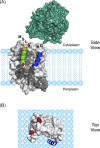
Figure 1.
Figure 1.
The receptor–substrate interaction. (A) Model of the interaction of the native substrate pre-SufI with TatBC. A signal peptide hairpin penetrates about halfway across the membrane in a deep groove on the side of TatC (gray; SWISS-MODEL ID: P69423) with both RR-motif (orange star) and mature domain (teal; PDB ID: 2UXV) on the cytoplasmic side of the membrane. Modeling suggests that the first part of the hairpin is helical (green), and the second half is disordered and interacts mostly with the TatB membrane domain (blue; residues L7-G21, Zhang et al.2014). Adapted from Hamsanathan et al. (2017). (B) Top view of TatBC. TatC residues E15 and E103 (red) interact with the RR-motif on Tat signal peptides (Rollauer et al.2012).
Figure 2.
Figure 2.
The Hairpin-Hinge model of Tat mature domain translocation. The Hairpin-Hinge model (Hamsanathan et al.2017) explains how the precursor protein remains continuously bound to the receptor complex while the C-terminal portion of the signal peptide hairpin and the mature domain migrate across the membrane. The membrane insertion of a signal peptide hairpin and the hairpin-hinge translocation mechanism are consistent with all of the receptor complex oligomerization models postulated thus far. For simplicity, a monomeric receptor complex is shown here. (A) The receptor complex inserts the signal peptide (black) of the precursor protein (mature domain in teal) into the membrane in a hairpin configuration that extends about halfway across the bilayer. Hairpin insertion is pmf-independent. The RR-motif (orange star) interacts with E15 and E103 (see Fig. 1) on the surface of TatC (gray) and the C-terminal end of the signal peptide after the hinge (red dot) interacts with TatB (dark blue) (see Fig. 1). (B) In the presence of a pmf, TatA (magenta) is recruited to the receptor–substrate complex, resulting in formation of a translocation conduit (dashed outline). (C) Unhinging of the signal peptide hairpin allows the mature domain to translocate through the pore across the membrane. (D) The translocation conduit disassembles after transport. The mature domain is released to the periplasm upon signal peptide cleavage.
Figure 3.
Figure 3.
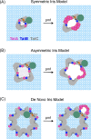
Models of the role of TatA in the Tat translocation mechanism. The number and arrangement of the TatA (magenta), TatB (dark blue) and TatC (gray) proteins in the receptor complex is unknown, though it is generally agreed that the signal peptide (black) of the precursor protein (orange star, RR-motif; teal, mature domain) binds in the groove on the side of TatC (see also Fig. 1). Three general types of current models are depicted. In all cases, the receptor complex oligomerization state shown is one of multiple reasonable possibilities, and TatA is recruited to the receptor–substrate complex in the presence of a pmf. Substantially different conformational rearrangements of the receptor complex and/or different TatA recruitment sites are feasible. The primary features distinguishing the different models is whether TatA is recruited to form one or multiple oligomeric structures and whether the signal peptide binding site is located within or outside of the receptor complex oligomer. (A) Symmetric iris model: this model assumes that the signal peptide binding pockets (dark gray) on TatC face inside the oligomer (face to face), and TatA is recruited symmetrically to the interfaces between protomers. A dimeric receptor arrangement is depicted here, as was originally postulated (Aldridge et al.2014), although a trimeric or tetrameric stoichiometry is also reasonable (Blummel et al.; Alcock et al.2016). (B) Asymmetric iris model: similar to the symmetric iris model, although in this case, the translocation conduit grows asymmetrically. While a tetrameric receptor arrangement is depicted here, either a tetrameric or trimeric stoichiometry was initially postulated (Alcock et al.2016), although a dimeric stochiometry is also reasonable. The central hole in the iris models (A and B) is likely blocked in some manner (indicated by the dotted shading) to prevent leakage before recruitment of additional TatA, most likely by the cytoplasmic domains of TatA and/or TatB in the receptor complex, or via lipids. (C) De novo iris model: for this model, the signal peptide binding sites of the receptor complex face outside, and recruited TatA forms a translocation conduit around the precursor bound to the receptor complex. An octomeric arrangement is shown here, based on biochemical evidence (Celedon and Cline 2012), though, in principle, any receptor complex oligomerization state is feasible.

Similar articles

Cited by

References

    1. Alami M, Luke I, Deitermann S et al. . Differential interactions between a twin-arginine signal peptide and its translocase in Escherichia coli. Mol Cell 2003;12:937–46. - PubMed
    1. Alcock F, Baker MAB, Green NP et al. . Live cell imaging shows reversible assembly of the TatA component of the twin-arginine protein transport system. Proc Natl Acad Sci USA 2013;110:E3650–9. - PMC - PubMed
    1. Alcock F, Damen MP, Levring J et al. . In vivo experiments do not support the charge zipper model for Tat translocase assembly. Elife 2017;6:e30127. - PMC - PubMed
    1. Alcock F, Stansfeld PJ, Basit H et al. . Assembling the Tat protein translocase. Elife 2016;5:e20718. - PMC - PubMed
    1. Alder NN, Theg SM. Energetics of protein transport across biological membranes. a study of the thylakoid DeltapH-dependent/cpTat pathway Cell 2003;112:231–42. - PubMed

Publication types

Substances

LinkOut - more resources